
15 mark essay on myocardial infarction
Reading File
Finding Sources
Finding Sources
Searching PubMed
"myocardial infarction"[MeSH Terms]
Searching the Web
myocardial infarction acute coronary syndrome management guideline 2025 ESC
Reading File
Reading File
Reading File
Searching Images
acute myocardial infarction coronary artery thrombosis pathology diagram ECG ST elevation
Reading File
Finding Sources
Reading Web Page
https://pubmed.ncbi.nlm.nih.gov/40014670/
Myocardial Infarction
Definition
Myocardial infarction (MI) is irreversible necrosis of cardiac muscle due to prolonged myocardial ischemia. It is diagnosed when there is a rise and/or fall in cardiac troponin, with at least one value above the 99th percentile upper reference limit, along with evidence of acute ischemia, such as typical symptoms, new ischemic ECG changes, pathological Q waves, imaging evidence of new loss of viable myocardium, or identification of a coronary thrombus.
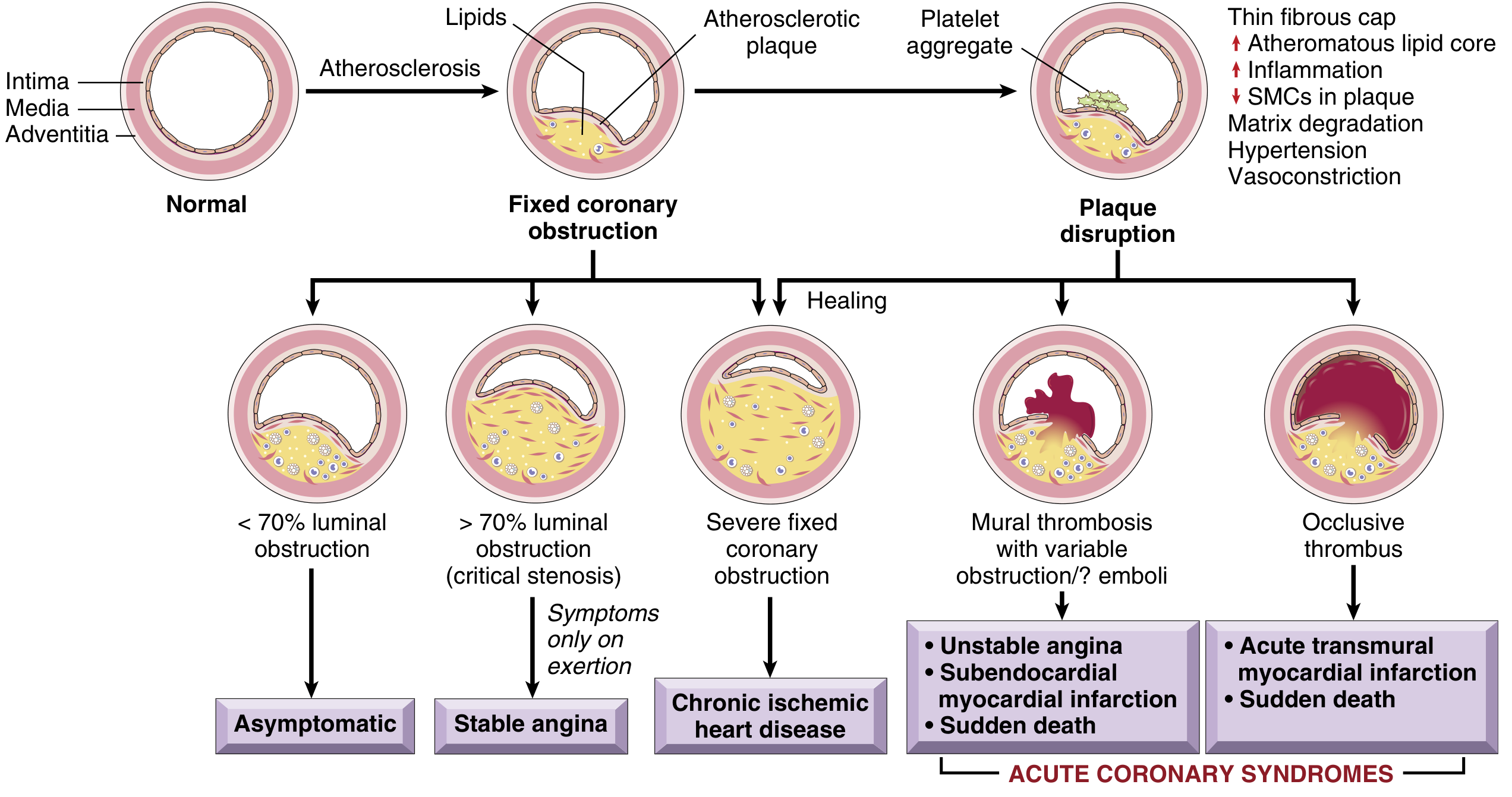
MI is commonly called a heart attack. Most cases result from acute coronary thrombosis following rupture or erosion of an atherosclerotic plaque. Robbins & Kumar Basic Pathology, p. 353.
Etiology and risk factors
Major cause
- Coronary atherosclerosis with plaque rupture or plaque erosion, followed by platelet aggregation and thrombus formation.
Other causes
- Coronary vasospasm, for example cocaine-induced or Prinzmetal angina
- Coronary embolism, such as in atrial fibrillation or infective endocarditis
- Coronary dissection
- Severe anemia, shock, sepsis, tachyarrhythmia, or severe hypertension causing oxygen supply-demand mismatch
- Small-vessel disease, vasculitis, or sickle-cell disease
Risk factors
- Non-modifiable: increasing age, male sex, family history of premature coronary artery disease
- Modifiable: smoking, hypertension, diabetes mellitus, dyslipidemia, obesity, sedentary lifestyle, unhealthy diet, and psychosocial stress.
Pathogenesis
The typical sequence is:
- An atherosclerotic plaque in a coronary artery ruptures, erodes, or hemorrhages.
- Subendothelial collagen and lipid-rich necrotic material are exposed.
- Platelets adhere, become activated, and aggregate. They release ADP, thromboxane A2, and serotonin, which amplify platelet aggregation and vasospasm.
- Tissue factor activates the coagulation cascade.
- A thrombus forms and may completely occlude the coronary artery.
- Ischemia causes loss of contractility within seconds to minutes. Persistent ischemia causes irreversible myocyte necrosis, beginning in the subendocardium and progressing outward as a wavefront.
- The extent of infarction depends on site of occlusion, duration of occlusion, collateral circulation, and myocardial oxygen demand.
Early restoration of coronary blood flow by primary percutaneous coronary intervention or fibrinolysis can limit infarct size. Robbins & Kumar Basic Pathology, p. 353.
Types of MI
1. STEMI
- Usually caused by complete and persistent coronary artery occlusion.
- ECG shows ST-segment elevation in contiguous leads or a new equivalent ischemic pattern.
- Requires immediate reperfusion therapy.
2. NSTEMI
- Usually due to subtotal or transient coronary occlusion.
- Troponin is elevated, but there is no persistent ST-segment elevation.
- ECG may show ST depression, T-wave inversion, or may be initially normal.
3. Type 1 MI
- Spontaneous MI due to atherosclerotic plaque rupture/erosion and acute coronary thrombosis.
4. Type 2 MI
- MI due to mismatch between myocardial oxygen supply and demand, without acute plaque rupture necessarily.
5. Procedure-related MI
- Occurs in association with PCI, CABG, or stent thrombosis.
Clinical features
Typical symptoms
- Severe, crushing, constricting retrosternal chest pain lasting more than 20 minutes
- Radiation to left arm, shoulder, neck, jaw, back, or epigastrium
- Pain is usually not fully relieved by rest or sublingual nitrates
- Sweating, pallor, anxiety, nausea, vomiting, dyspnea, and palpitations
Atypical presentation
More common in elderly people, women, and patients with diabetes:
- Dyspnea
- Syncope
- Unexplained fatigue
- Epigastric discomfort
- Silent MI, especially in diabetes
Examination findings
- Tachycardia or bradycardia
- Hypotension
- Cool clammy skin
- Fourth heart sound
- Basal crepitations and raised JVP in heart failure
- New systolic murmur in papillary muscle dysfunction or ventricular septal rupture
Diagnosis
1. ECG
A 12-lead ECG should be performed and repeated if initially non-diagnostic.
- STEMI: ST elevation in anatomically contiguous leads
- NSTEMI/unstable ischemia: ST depression and/or T-wave inversion may occur
- Development of pathological Q waves suggests myocardial necrosis
2. Cardiac biomarkers
- High-sensitivity cardiac troponin I or T is the preferred test.
- A dynamic rise and/or fall supports acute myocardial injury.
- CK-MB may assist in suspected reinfarction because it returns to baseline earlier than troponin.
3. Echocardiography
May demonstrate:
- Regional wall-motion abnormality
- Reduced left ventricular ejection fraction
- Mechanical complications, such as acute mitral regurgitation or ventricular septal rupture
4. Coronary angiography
Identifies the culprit coronary lesion and permits PCI.
5. Additional tests
- Complete blood count, blood glucose, renal function, electrolytes, lipid profile
- Chest radiograph if heart failure or another diagnosis is suspected
Management
MI is a medical emergency. Patients require admission to a coronary care unit, continuous ECG monitoring, IV access, and rapid cardiology assessment.
Initial management
- Rest and cardiac monitoring
- Oxygen only if hypoxemic, in respiratory distress, or shock
- Aspirin, unless contraindicated
- A P2Y12 inhibitor in appropriate patients
- Anticoagulation
- Nitrates for persistent ischemic pain if blood pressure permits
- Analgesia for severe pain
- Treat arrhythmias, pulmonary edema, hypotension, or cardiogenic shock promptly
Reperfusion therapy
STEMI
- Primary PCI is preferred when it can be performed rapidly.
- If timely PCI is unavailable and there are no contraindications, fibrinolytic therapy may be considered in eligible patients with recent symptom onset, followed by transfer for angiography/PCI.
NSTEMI
- Fibrinolysis is not indicated.
- Management is based on risk stratification, antithrombotic therapy, and an early invasive strategy for high-risk patients.
Current practice is guided by the 2025 ACC/AHA acute coronary syndrome guideline, which integrates treatment of STEMI and NSTEMI across the acute and post-discharge phases.
Drugs for secondary prevention
Unless contraindicated, long-term treatment includes:
- Dual antiplatelet therapy for the indicated duration after ACS/PCI
- High-intensity statin
- Beta-blocker, particularly with left ventricular dysfunction, arrhythmia, or ongoing angina
- ACE inhibitor or ARB, particularly in anterior MI, hypertension, diabetes, chronic kidney disease, or reduced LVEF
- Mineralocorticoid receptor antagonist when LVEF is reduced with heart failure or diabetes, provided renal function and potassium allow
Complications
Early complications
- Arrhythmias: ventricular tachycardia, ventricular fibrillation, atrial fibrillation, sinus bradycardia, and heart block
- Acute left ventricular failure and pulmonary edema
- Cardiogenic shock
- Pericarditis: usually within days after transmural MI
- Right ventricular infarction: can cause hypotension with raised JVP and clear lungs
Mechanical complications
Usually occur several days after a large transmural infarct:
- Papillary muscle rupture: acute severe mitral regurgitation and pulmonary edema
- Ventricular septal rupture: harsh pansystolic murmur with acute heart failure/shock
- Free-wall rupture: hemopericardium and cardiac tamponade, often rapidly fatal
Late complications
- Ventricular aneurysm
- Mural thrombus with systemic embolism
- Chronic heart failure
- Recurrent MI
- Post-MI pericarditis, also called Dressler syndrome
- Persistent ventricular arrhythmias and sudden cardiac death
Prevention and rehabilitation
Secondary prevention includes:
- Complete smoking cessation
- Control of hypertension, diabetes, and dyslipidemia
- Regular exercise and cardiac rehabilitation
- Weight management and a heart-healthy diet
- Adherence to antiplatelet, statin, and other prescribed therapy
- Psychological support and return-to-work counseling
Conclusion
Myocardial infarction is usually caused by acute thrombotic occlusion of an atherosclerotic coronary artery. Prompt recognition using symptoms, ECG, and troponin testing, followed by rapid reperfusion and evidence-based antithrombotic therapy, reduces mortality and preserves left ventricular function. Long-term secondary prevention is essential to prevent recurrent cardiovascular events.
Investigations in bactriology
Reading File
“Investigations in bacteriology” is a broad topic. It generally includes:
-
Specimen collection and transport
Blood, urine, sputum, stool, pus, CSF, throat swab, etc. Correct timing, aseptic collection, proper container, and rapid transport are essential. -
Direct examination of specimens
Microscopy using Gram stain, acid-fast stain, wet mount, and special stains to demonstrate organisms and inflammatory cells. -
Culture methods
Inoculation on appropriate media such as blood agar, MacConkey agar, chocolate agar, and selective/enrichment media, followed by incubation. -
Identification of bacteria
Colony morphology, haemolysis, pigment, motility, biochemical reactions, antigen detection, automated identification, MALDI-TOF, and molecular tests. -
Antimicrobial susceptibility testing
Kirby-Bauer disc diffusion, MIC determination, E-test, automated susceptibility systems, and detection of resistance mechanisms. -
Serology and antigen detection
Detection of bacterial antigen or antibody in selected infections. -
Molecular diagnosis
PCR, nucleic-acid amplification tests, sequencing, and rapid multiplex assays. -
Blood culture and culture-negative infection work-up
Especially important in septicemia, endocarditis, and enteric fever.
Would you like a 15-mark exam essay, a short note, or details on one section such as culture media, staining, or antibiotic sensitivity testing?
This is a shared conversation. Sign in to Orris to start your own chat.