Describe thyroid swelling differential diagnosis investigation and management of solitary thyroid nodule 14 marker general Surgery

Reading File
Finding Sources
Finding Sources
Searching the Web

solitary thyroid nodule differential diagnosis investigation management surgery 2024

Reading File
Finding Sources
Finding Sources
Reading File
Reading File
Reading File
Reading File
Reading File
Finding Sources
Reading File
Finding Sources
Reading File
Reading File
Searching Images

thyroid nodule workup algorithm flowchart FNAC management

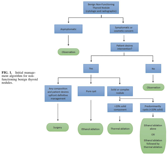
Summary : This flowchart presents the initial management algorithm for non-functioning benign thyroid nodules, guiding clinical decisions based on symptoms, patient preferences, and nodule characteristics.

flowchart:
# Nodes :
  • Start (rectangle): "Benign Non-Functioning Thyroid Nodule (cytologic and radiographic)"
  • Decision (rectangle): "Asymptomatic"
  • Action (ellipse): "Observation"
  • Decision (rectangle): "Symptomatic or cosmetic concern"
  • Decision (rectangle): "Patient desires intervention?"
  • Decision (rectangle): "Yes"
  • Decision (rectangle): "No"
  • Action (ellipse): "Observation"
  • Decision (rectangle): "Any composition and patient desires upfront definitive management"
  • Action (ellipse): "Surgery"
  • Decision (rectangle): "Pure cyst"
  • Action (ellipse): "Ethanol ablation"
  • Decision (rectangle): "Solid or complex nodule"
  • Decision (rectangle): ">20% solid component"
  • Action (ellipse): "Thermal ablation"
  • Decision (rectangle): "Predominantly cystic (<20% solid)"
  • Action (ellipse): "Ethanol ablation alone OR Ethanol ablation followed by thermal ablation"

# Connectors :
  • The flow starts at "Benign Non-Functioning Thyroid Nodule".
  • Splits into "Asymptomatic" (left) and "Symptomatic or cosmetic concern" (right).
  • "Asymptomatic" leads directly to "Observation".
  • "Symptomatic or cosmetic concern" leads to "Patient desires intervention?".
  • "Patient desires intervention?" splits into "Yes" and "No".
  • "No" leads to "Observation".
  • "Yes" branches into three paths:
      – "Any composition and patient desires upfront definitive management" → "Surgery".
      – "Pure cyst" → "Ethanol ablation".
      – "Solid or complex nodule" splits into:
          • ">20% solid component" → "Thermal ablation".
          • "Predominantly cystic (<20% solid)" → "Ethanol ablation alone OR Ethanol ablation followed by thermal ablation".

# Layout :
  • Top-down hierarchical structure.
  • Initial node at the top, branching into two main paths (asymptomatic vs. symptomatic).
  • Decision diamonds and action ellipses are used.
  • Multiple branches and merges, with final management options at the bottom.

# Analysis :
  • The algorithm prioritises observation for asymptomatic patients and those not desiring intervention.
  • For symptomatic patients desiring intervention, management is tailored by nodule composition: pure cysts are treated with ethanol ablation, solid/complex nodules with >20% solid component receive thermal ablation, and predominantly cystic nodules (<20% solid) may receive ethanol ablation alone or combined with thermal ablation.
  • Surgery is reserved for patients who desire upfront definitive management regardless of nodule composition.
  • The flowchart provides a clear, stepwise approach to decision-making in benign non-functioning thyroid nodules, balancing patient preference and nodule characteristics.

Summary : This flowchart presents the initial management algorithm for non-functioning benign thyroid nodules, guiding clinical decisions based on symptoms, patient preferences, and nodule characteristics. flowchart: # Nodes : • Start (rectangle): "Benign Non-Functioning Thyroid Nodule (cytologic and radiographic)" • Decision (rectangle): "Asymptomatic" • Action (ellipse): "Observation" • Decision (rectangle): "Symptomatic or cosmetic concern" • Decision (rectangle): "Patient desires intervention?" • Decision (rectangle): "Yes" • Decision (rectangle): "No" • Action (ellipse): "Observation" • Decision (rectangle): "Any composition and patient desires upfront definitive management" • Action (ellipse): "Surgery" • Decision (rectangle): "Pure cyst" • Action (ellipse): "Ethanol ablation" • Decision (rectangle): "Solid or complex nodule" • Decision (rectangle): ">20% solid component" • Action (ellipse): "Thermal ablation" • Decision (rectangle): "Predominantly cystic (<20% solid)" • Action (ellipse): "Ethanol ablation alone OR Ethanol ablation followed by thermal ablation" # Connectors : • The flow starts at "Benign Non-Functioning Thyroid Nodule". • Splits into "Asymptomatic" (left) and "Symptomatic or cosmetic concern" (right). • "Asymptomatic" leads directly to "Observation". • "Symptomatic or cosmetic concern" leads to "Patient desires intervention?". • "Patient desires intervention?" splits into "Yes" and "No". • "No" leads to "Observation". • "Yes" branches into three paths: – "Any composition and patient desires upfront definitive management" → "Surgery". – "Pure cyst" → "Ethanol ablation". – "Solid or complex nodule" splits into: • ">20% solid component" → "Thermal ablation". • "Predominantly cystic (<20% solid)" → "Ethanol ablation alone OR Ethanol ablation followed by thermal ablation". # Layout : • Top-down hierarchical structure. • Initial node at the top, branching into two main paths (asymptomatic vs. symptomatic). • Decision diamonds and action ellipses are used. • Multiple branches and merges, with final management options at the bottom. # Analysis : • The algorithm prioritises observation for asymptomatic patients and those not desiring intervention. • For symptomatic patients desiring intervention, management is tailored by nodule composition: pure cysts are treated with ethanol ablation, solid/complex nodules with >20% solid component receive thermal ablation, and predominantly cystic nodules (<20% solid) may receive ethanol ablation alone or combined with thermal ablation. • Surgery is reserved for patients who desire upfront definitive management regardless of nodule composition. • The flowchart provides a clear, stepwise approach to decision-making in benign non-functioning thyroid nodules, balancing patient preference and nodule characteristics.

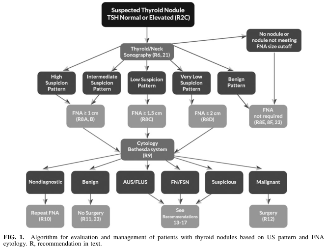
Summary : This figure presents an algorithmic flowchart for the evaluation and management of patients with suspected thyroid nodules, based on ultrasound (US) pattern and fine-needle aspiration (FNA) cytology, referencing the Bethesda system and specific recommendations.

flowchart:
# Nodes :
  • Start (rectangle): "Suspected Thyroid Nodule TSH Normal or Elevated (R2C)"
  • Decision (rectangle): "Thyroid/Neck Sonography (R6, 21)"
  • Branch (rectangle): "High Suspicion Pattern"
  • Branch (rectangle): "Intermediate Suspicion Pattern"
  • Branch (rectangle): "Low Suspicion Pattern"
  • Branch (rectangle): "Very Low Suspicion Pattern"
  • Branch (rectangle): "Benign Pattern"
  • Terminal (rectangle): "No nodule or nodule not meeting FNA size cutoff"
  • Action (rectangle): "FNA ≥ 1 cm (R8A, B)"
  • Action (rectangle): "FNA ≥ 1.5 cm (R8C)"
  • Action (rectangle): "FNA ≥ 2 cm (R8D)"
  • Terminal (rectangle): "FNA not required (R8E, 8F, 23)"
  • Action (rectangle): "Cytology Bethesda system (R9)"
  • Outcome (rectangle): "Nondiagnostic"
  • Outcome (rectangle): "Benign"
  • Outcome (rectangle): "AUS/FLUS"
  • Outcome (rectangle): "FN/FSN"
  • Outcome (rectangle): "Suspicious"
  • Outcome (rectangle): "Malignant"
  • Action (rectangle): "Repeat FNA (R10)"
  • Action (rectangle): "No Surgery (R11, 23)"
  • Action (rectangle): "See Recommendations 13–17"
  • Action (rectangle): "Surgery (R12)"

# Connectors :
  • Arrows direct flow from "Suspected Thyroid Nodule" to "Thyroid/Neck Sonography" and to "No nodule or nodule not meeting FNA size cutoff".
  • From "Thyroid/Neck Sonography", arrows branch to "High Suspicion Pattern", "Intermediate Suspicion Pattern", "Low Suspicion Pattern", "Very Low Suspicion Pattern", and "Benign Pattern".
  • Each suspicion pattern node leads to a corresponding FNA size threshold or "FNA not required".
  • All FNA nodes converge to "Cytology Bethesda system (R9)".
  • "Cytology Bethesda system (R9)" branches to six outcomes: "Nondiagnostic", "Benign", "AUS/FLUS", "FN/FSN", "Suspicious", "Malignant".
  • Each outcome leads to a specific management action (e.g., "Repeat FNA", "No Surgery", "See Recommendations", "Surgery").

# Layout :
  • Top-down hierarchical structure.
  • Initial decision splits into five main US pattern branches.
  • FNA thresholds filter which nodules proceed to cytology.
  • Cytology results further stratify management.
  • Terminal actions at the bottom row.

# Analysis :
  • The flowchart provides a stepwise, pattern-based approach to thyroid nodule management, integrating US risk stratification and cytology.
  • FNA is only performed if nodules meet specific size and suspicion criteria.
  • Cytology results (Bethesda system) dictate subsequent management, ranging from repeat FNA, observation, further recommendations, or surgery.
  • The algorithm emphasizes minimizing unnecessary procedures for benign or low-risk nodules and prioritizing intervention for suspicious or malignant findings.

Summary : This figure presents an algorithmic flowchart for the evaluation and management of patients with suspected thyroid nodules, based on ultrasound (US) pattern and fine-needle aspiration (FNA) cytology, referencing the Bethesda system and specific recommendations. flowchart: # Nodes : • Start (rectangle): "Suspected Thyroid Nodule TSH Normal or Elevated (R2C)" • Decision (rectangle): "Thyroid/Neck Sonography (R6, 21)" • Branch (rectangle): "High Suspicion Pattern" • Branch (rectangle): "Intermediate Suspicion Pattern" • Branch (rectangle): "Low Suspicion Pattern" • Branch (rectangle): "Very Low Suspicion Pattern" • Branch (rectangle): "Benign Pattern" • Terminal (rectangle): "No nodule or nodule not meeting FNA size cutoff" • Action (rectangle): "FNA ≥ 1 cm (R8A, B)" • Action (rectangle): "FNA ≥ 1.5 cm (R8C)" • Action (rectangle): "FNA ≥ 2 cm (R8D)" • Terminal (rectangle): "FNA not required (R8E, 8F, 23)" • Action (rectangle): "Cytology Bethesda system (R9)" • Outcome (rectangle): "Nondiagnostic" • Outcome (rectangle): "Benign" • Outcome (rectangle): "AUS/FLUS" • Outcome (rectangle): "FN/FSN" • Outcome (rectangle): "Suspicious" • Outcome (rectangle): "Malignant" • Action (rectangle): "Repeat FNA (R10)" • Action (rectangle): "No Surgery (R11, 23)" • Action (rectangle): "See Recommendations 13–17" • Action (rectangle): "Surgery (R12)" # Connectors : • Arrows direct flow from "Suspected Thyroid Nodule" to "Thyroid/Neck Sonography" and to "No nodule or nodule not meeting FNA size cutoff". • From "Thyroid/Neck Sonography", arrows branch to "High Suspicion Pattern", "Intermediate Suspicion Pattern", "Low Suspicion Pattern", "Very Low Suspicion Pattern", and "Benign Pattern". • Each suspicion pattern node leads to a corresponding FNA size threshold or "FNA not required". • All FNA nodes converge to "Cytology Bethesda system (R9)". • "Cytology Bethesda system (R9)" branches to six outcomes: "Nondiagnostic", "Benign", "AUS/FLUS", "FN/FSN", "Suspicious", "Malignant". • Each outcome leads to a specific management action (e.g., "Repeat FNA", "No Surgery", "See Recommendations", "Surgery"). # Layout : • Top-down hierarchical structure. • Initial decision splits into five main US pattern branches. • FNA thresholds filter which nodules proceed to cytology. • Cytology results further stratify management. • Terminal actions at the bottom row. # Analysis : • The flowchart provides a stepwise, pattern-based approach to thyroid nodule management, integrating US risk stratification and cytology. • FNA is only performed if nodules meet specific size and suspicion criteria. • Cytology results (Bethesda system) dictate subsequent management, ranging from repeat FNA, observation, further recommendations, or surgery. • The algorithm emphasizes minimizing unnecessary procedures for benign or low-risk nodules and prioritizing intervention for suspicious or malignant findings.

Summary : This flowchart presents the management algorithm for intrathyroid thymic carcinoma (ITC), detailing treatment pathways for resectable, locally advanced, and recurrent/metastatic disease, with node colours indicating treatment modalities and recommendations.

flowchart:
# Nodes :
  • ITC (purple rectangle, algorithm title)
  • Resectable (white rectangle)
  • Resect according to ESMO CPG on thyroid cancer [IV, A] (orange rectangle, surgery)
  • Adjuvant RTb [IV, B] (dark green rectangle, radiotherapy)
  • Observation [V, C] (white rectangle, non-treatment aspect)
  • Locally advanced (white rectangle)
  • RT or CRT [V, B] (dark green rectangle, radiotherapy/chemoradiotherapy)
  • Recurrent or metastatic (white rectangle)
  • Salvage surgerya [IV, A] (orange rectangle, surgery)
  • Platinum-based ChTc [V, B] (blue rectangle, systemic anticancer therapy)

# Connectors :
  • ITC splits into three branches: Resectable, Locally advanced, Recurrent or metastatic.
  • Resectable → Resect according to ESMO CPG on thyroid cancer [IV, A]
    – Resect according to ESMO CPG → Adjuvant RTb [IV, B] and Observation [V, C]
    – Adjuvant RTb [IV, B] → Observation [V, C]
  • Locally advanced → RT or CRT [V, B]
  • Recurrent or metastatic → Salvage surgerya [IV, A] → Platinum-based ChTc [V, B]

# Layout :
  • Three main vertical branches from the central ITC node.
  • Each branch represents a disease stage: resectable (left), locally advanced (middle), recurrent/metastatic (right).
  • Nodes are colour-coded: purple (title), orange (surgery), dark green (RT), blue (systemic therapy), white (other management/non-treatment).

# Analysis :
  • The flowchart visually separates management strategies by disease stage, with clear progression from initial assessment to specific treatments.
  • Surgery is recommended for resectable and salvageable cases, followed by possible adjuvant radiotherapy or observation.
  • Locally advanced cases are managed with radiotherapy or chemoradiotherapy.
  • Recurrent/metastatic cases may undergo salvage surgery and then platinum-based chemotherapy.
  • Colour coding aids in distinguishing treatment modalities, and footnotes clarify specific recommendations and regimen options.

Summary : This flowchart presents the management algorithm for intrathyroid thymic carcinoma (ITC), detailing treatment pathways for resectable, locally advanced, and recurrent/metastatic disease, with node colours indicating treatment modalities and recommendations. flowchart: # Nodes : • ITC (purple rectangle, algorithm title) • Resectable (white rectangle) • Resect according to ESMO CPG on thyroid cancer [IV, A] (orange rectangle, surgery) • Adjuvant RTb [IV, B] (dark green rectangle, radiotherapy) • Observation [V, C] (white rectangle, non-treatment aspect) • Locally advanced (white rectangle) • RT or CRT [V, B] (dark green rectangle, radiotherapy/chemoradiotherapy) • Recurrent or metastatic (white rectangle) • Salvage surgerya [IV, A] (orange rectangle, surgery) • Platinum-based ChTc [V, B] (blue rectangle, systemic anticancer therapy) # Connectors : • ITC splits into three branches: Resectable, Locally advanced, Recurrent or metastatic. • Resectable → Resect according to ESMO CPG on thyroid cancer [IV, A] – Resect according to ESMO CPG → Adjuvant RTb [IV, B] and Observation [V, C] – Adjuvant RTb [IV, B] → Observation [V, C] • Locally advanced → RT or CRT [V, B] • Recurrent or metastatic → Salvage surgerya [IV, A] → Platinum-based ChTc [V, B] # Layout : • Three main vertical branches from the central ITC node. • Each branch represents a disease stage: resectable (left), locally advanced (middle), recurrent/metastatic (right). • Nodes are colour-coded: purple (title), orange (surgery), dark green (RT), blue (systemic therapy), white (other management/non-treatment). # Analysis : • The flowchart visually separates management strategies by disease stage, with clear progression from initial assessment to specific treatments. • Surgery is recommended for resectable and salvageable cases, followed by possible adjuvant radiotherapy or observation. • Locally advanced cases are managed with radiotherapy or chemoradiotherapy. • Recurrent/metastatic cases may undergo salvage surgery and then platinum-based chemotherapy. • Colour coding aids in distinguishing treatment modalities, and footnotes clarify specific recommendations and regimen options.

I now have all the comprehensive content needed. Let me compile the complete answer.

Thyroid Swelling: Differential Diagnosis, Investigation, and Management of Solitary Thyroid Nodule

(General Surgery - 14 Marker)

PART 1: DIFFERENTIAL DIAGNOSIS OF THYROID SWELLING

Any swelling in the anterior triangle of the neck that moves with deglutition (swallowing) is considered thyroid until proven otherwise. The swelling may also move with tongue protrusion if it is a thyroglossal cyst.

A. Classification of Thyroid Swellings

Based on size/pattern:
TypeDescription
Diffuse goiterEntire gland enlarged
Uninodular goiterSingle discrete nodule
Multinodular goiterMultiple nodules in an enlarged gland
Based on function:
TypeTSHClinical
EuthyroidNormalNon-toxic goiter
HyperthyroidLow/suppressedToxic goiter (Graves', toxic adenoma, toxic MNG)
HypothyroidElevatedHashimoto's, iodine deficiency

B. Differential Diagnosis of a Neck/Thyroid Swelling

Thyroid causes (moves with swallowing):
  1. Simple/colloid goiter - iodine deficiency, dietary goitrogens
  2. Multinodular goiter (MNG) - most common thyroid enlargement worldwide
  3. Graves' disease - diffuse toxic goiter, autoimmune (TSH-R antibody)
  4. Hashimoto's thyroiditis - autoimmune; most common cause of hypothyroidism
  5. Subacute (de Quervain's) thyroiditis - viral, painful, tender; elevated ESR >100 mm/hr
  6. Follicular adenoma - benign solitary nodule, well encapsulated
  7. Thyroid cyst - colloid/degenerative/hemorrhagic
  8. Thyroid carcinoma - papillary (most common), follicular, medullary, anaplastic
  9. Thyroglossal duct cyst - midline, moves with tongue protrusion AND swallowing
Non-thyroid causes in anterior neck (does NOT move with swallowing):
  • Cervical lymphadenopathy (reactive, lymphoma, metastatic)
  • Branchial cyst (lateral, at angle of jaw/SCM)
  • Lipoma
  • Dermoid cyst
  • Carotid body tumor (pulsatile)
  • Submandibular salivary gland pathology

PART 2: SOLITARY THYROID NODULE (STN)

Definition

A single, discrete nodule in an otherwise normal thyroid gland. Present in ~4% of the US population. The primary concern is ruling out malignancy - thyroid cancer occurs in approximately 5-15% of solitary nodules.

Features Suggesting MALIGNANCY in STN

History (High-risk features):
  • Age <20 years or >70 years
  • Male sex
  • Rapid growth of nodule
  • Hoarseness / dysphagia / stridor (local invasion)
  • Cervical lymphadenopathy
  • History of neck/head radiation (linear risk increase from 6.5 to 2000 cGy; ~40% chance of cancer if prior radiation)
  • Family history of thyroid cancer (PTC, MTC/MEN2)
  • Previous thyroid cancer
Examination (Features favouring malignancy):
  • Firm or hard, stony consistency
  • Fixed/immobile nodule
  • Irregular margins
  • Size >3-4 cm
  • Ipsilateral cervical lymphadenopathy
  • Hoarseness suggesting RLN involvement
Features favouring BENIGN nodule:
  • Soft, smooth, mobile
  • Painful/tender (suggests thyroiditis or hemorrhagic cyst)
  • Transient/fluctuating size
  • Autonomously functioning ("hot") nodule on scintigraphy (risk of malignancy <1%)
- Sabiston Textbook of Surgery, Biological Basis of Modern Surgical Practice

PART 3: INVESTIGATIONS OF SOLITARY THYROID NODULE

Step 1 - Thyroid Function Tests

Serum TSH - First investigation:
  • Suppressed TSH → suggests hyperfunctioning nodule → proceed to radionuclide scintigraphy
  • Normal/elevated TSH → proceed to ultrasound + FNAC
  • Serum T3, T4 (if TSH abnormal)
  • Anti-TPO antibodies, anti-Tg antibodies (if Hashimoto's suspected)
  • Serum calcitonin - if medullary thyroid cancer (MTC) suspected (family history, MEN2)

Step 2 - Neck Ultrasound (Most Important Imaging)

Neck ultrasound is the most important radiographic study for evaluation of thyroid nodules. It provides:
  • Highest resolution imaging of thyroid parenchyma
  • Characterizes nodule composition, echogenicity, shape, margins, calcifications
  • Assesses cervical lymphadenopathy (levels II-VI)
  • Guides FNA biopsy
ATA Sonographic Risk Stratification (2015/2016 Guidelines):
Sonographic PatternFeaturesEst. Risk of MalignancyFNA Size Cutoff
High suspicionSolid hypoechoic + microcalcifications OR irregular margins OR taller-than-wide OR extrathyroidal extension>70-90%FNA at ≥1 cm
Intermediate suspicionHypoechoic solid with smooth margins, no microcalcifications or ETE10-20%FNA at ≥1 cm
Low suspicionIso/hyperechoic solid or partially cystic with eccentric solid areas5-10%FNA at ≥1.5 cm
Very low suspicionSpongiform or partially cystic without any high-risk features<3%Consider FNA ≥2 cm; observation reasonable
BenignPurely cystic (no solid component)<1%No biopsy; aspiration for symptoms
(Source: Haugen et al. ATA 2016 Guidelines, cited in Sabiston)
ACR TI-RADS (alternative scoring): Points-based system scoring composition, echogenicity, shape, margin, and echogenic foci → TR1-TR5.
Here is the ATA nodule management algorithm:
Thyroid nodule workup algorithm - ATA based, Bethesda system

Step 3 - Fine Needle Aspiration Cytology (FNAC)

Most accurate and cost-effective invasive investigation. Sensitivity >80%, Specificity >90%.
  • Performed with 23-27 gauge needle, ideally ultrasound-guided
  • On-site cytopathological evaluation confirms adequacy
Bethesda System for Reporting Thyroid Cytopathology (Updated 2023 - 3rd edition):
Bethesda CategoryDiagnosisRisk of MalignancyUsual Management
INondiagnostic / Unsatisfactory5-10%Repeat FNA
IIBenign~2-4%Observation / clinical follow-up
IIIAtypia of Undetermined Significance (AUS) / FLUS~10-30%Repeat FNA or molecular testing
IVFollicular Neoplasm (FN) / Suspicious for FN25-40%Lobectomy (diagnostic)
VSuspicious for Malignancy50-75%Near-total / total thyroidectomy
VIMalignant97-99%Total thyroidectomy
Note: FNA cannot distinguish between benign follicular adenoma and follicular thyroid carcinoma (capsular/vascular invasion only seen on histology) - hence Bethesda IV nodules require surgical excision.

Step 4 - Radionuclide Scintigraphy (Thyroid Scan)

Using Technetium-99m (pertechnetate) or I-131:
  • Hot nodule (hyperfunctioning) - increased uptake; malignancy risk <1% - rarely biopsied
  • Cold nodule (non-functioning) - decreased uptake; malignancy risk ~10-15%
  • Warm nodule - same uptake as surrounding tissue
Indicated especially when TSH is suppressed (to identify toxic adenoma).

Step 5 - Additional Investigations

  • CT scan/MRI neck and chest: Assess substernal extension, tracheal compression/deviation, vascular encasement, mediastinal lymphadenopathy. Non-contrast preferred (iodinated contrast delays RAI therapy).
  • Chest X-ray: Tracheal deviation, retrosternal shadow, lung metastases (cannon-ball deposits in follicular cancer)
  • Laryngoscopy: Pre-operative vocal cord assessment; mandatory if hoarseness present
  • Molecular testing of FNA specimen: BRAF V600E mutation (papillary CA), RAS, RET/PTC fusions, PAX8-PPARγ (follicular CA) - used for indeterminate (Bethesda III/IV) nodules to guide surgery vs surveillance

PART 4: MANAGEMENT OF SOLITARY THYROID NODULE

Management algorithm depends on FNAC result, clinical features, and nodule characteristics.

A. BENIGN STN (Bethesda II)

Observation:
  • Majority of patients with small, asymptomatic benign nodules do not require treatment
  • Regular follow-up with clinical examination + USS every 6-12 months
  • Repeat FNA in 12 months if initially non-diagnostic
Indications for surgery in benign STN:
  1. Nodule continues to enlarge despite observation
  2. Compressive symptoms (dysphagia, dyspnoea, stridor)
  3. Cosmetically unacceptable goiter
  4. Substernal extension
  5. Concern for malignancy not resolved by FNAC
  6. Patient preference for definitive management
Minimally invasive options (for non-surgical candidates):
  • Thermal ablation (radiofrequency ablation, microwave, laser): For solid/complex nodules >20% solid component
  • Ethanol ablation: For pure or predominantly cystic nodules
  • Radioactive iodine (RAI) I-131: For hyperfunctioning (hot) autonomous nodule; also second-line to surgery per 2023 ETA guidelines

B. INDETERMINATE STN (Bethesda III/IV)

  • Bethesda III: Repeat FNA; if repeat indeterminate → molecular testing → surgery if positive
  • Bethesda IV: Diagnostic thyroid lobectomy (ipsilateral lobe + isthmus)
    • If final histology benign → no further surgery
    • If malignant → completion total thyroidectomy

C. MALIGNANT STN (Bethesda V/VI) - Surgical Management

Papillary Thyroid Carcinoma (PTC) - Most common (80%):
  • Total thyroidectomy is standard for:
    • Tumors >1 cm
    • Bilateral disease
    • Extrathyroidal extension
    • Cervical lymph node metastasis
    • Prior radiation history
  • Lobectomy alone acceptable for: low-risk PTC <1 cm (papillary microcarcinoma), no extrathyroidal extension, no node involvement, no distant metastasis
  • Post-op: RAI ablation + TSH suppression with levothyroxine
Follicular Thyroid Carcinoma (FTC):
  • Hematogenous spread (lung, bone) > lymphatic
  • Minimally invasive: lobectomy may suffice
  • Widely invasive: total thyroidectomy + RAI
Medullary Thyroid Carcinoma (MTC):
  • Arises from parafollicular C cells (calcitonin-secreting)
  • May be sporadic or familial (MEN2A/2B - RET proto-oncogene mutation)
  • Total thyroidectomy + bilateral central neck dissection (prophylactic)
  • RAI not effective (C cells don't take up iodine)
  • Monitor with serum calcitonin + CEA post-op
Anaplastic Thyroid Carcinoma (ATC):
  • Most aggressive, rapidly fatal
  • Multimodality: external beam radiotherapy + chemotherapy (doxorubicin); surgery if resectable
  • Targeted therapy: dabrafenib + trametinib if BRAF V600E mutation positive
Lymphoma:
  • Often arises in background of Hashimoto's thyroiditis
  • Chemotherapy/radiotherapy; surgery not primary treatment

D. Surgical Procedures

ProcedureExtentIndication
Lobectomy + isthmusectomyOne lobe + isthmusBenign solitary nodule, diagnostic (Bethesda IV), low-risk PTC <1 cm
Near-total thyroidectomy>95% of glandLarge benign goiter
Total thyroidectomyEntire glandMalignancy, bilateral disease, MNG with cancer, recurrent goiter
Modified radical neck dissectionCentral (level VI) and lateral (II-V) nodesClinically positive nodes
Complications of thyroidectomy:
  1. Recurrent laryngeal nerve (RLN) injury: Hoarseness (unilateral), respiratory distress (bilateral) - risk ~1%
  2. Hypoparathyroidism: Hypocalcaemia (tetany, Chvostek's sign, Trousseau's sign) - most common after total thyroidectomy
  3. Hypothyroidism: Requires lifelong levothyroxine after total thyroidectomy
  4. Thyroid storm: Rare post-operative; treated with beta-blockers, PTU, Lugol's iodine, steroids
  5. Hemorrhage/haematoma: Can cause airway compromise; reopening at bedside if acute
  6. Wound infection, seroma
  7. Superior laryngeal nerve injury: Loss of high-pitched phonation (opera singer sign)

Summary Algorithm for STN Management

STN detected
    │
    ├─ TSH ─────── Suppressed → Scintigraphy → Hot nodule → RAI/observation
    │                                        → Cold nodule → USS + FNAC
    │              Normal/High ──────────────────────────→ USS + FNAC
    │
    ├─ FNAC (Bethesda)
    │       │
    │       ├─ I (Non-diagnostic) → Repeat FNAC
    │       ├─ II (Benign) → Observation / surgery if large/compressive
    │       ├─ III (AUS) → Repeat FNAC / molecular testing
    │       ├─ IV (FN) → Diagnostic lobectomy
    │       ├─ V (Suspicious) → Total thyroidectomy
    │       └─ VI (Malignant) → Total thyroidectomy ± neck dissection

Key teaching points for 14-marker exam:
  1. A solitary cold nodule on scintigraphy has ~10-15% malignancy risk
  2. A hot nodule rarely (<1%) is malignant
  3. FNAC is the gold standard investigation - sensitivity >80%, specificity >90%
  4. FNA cannot distinguish follicular adenoma from follicular carcinoma (needs histology)
  5. The Bethesda system has 6 categories - memorize the malignancy risks
  6. Prior radiation exposure increases thyroid cancer risk by ~40%
  7. Always do pre-operative laryngoscopy
  8. Most common post-thyroidectomy complication = hypoparathyroidism (hypocalcaemia)
Sources: Sabiston Textbook of Surgery (Biological Basis of Modern Surgical Practice); Schwartz's Principles of Surgery, 11th Ed.

Give me to the point for 15 marks so I can score full marks

Solitary Thyroid Nodule - 15 Marks (Exam Answer)


DEFINITION (1 mark)

A single discrete palpable lesion in an otherwise normal thyroid gland. Present in ~4% of population. Key concern = rule out malignancy (10-15% risk).

DIFFERENTIAL DIAGNOSIS OF THYROID SWELLING (3 marks)

Benign:
  • Colloid/simple goiter
  • Follicular adenoma ← most common STN
  • Thyroid cyst (colloid, hemorrhagic)
  • Hashimoto's thyroiditis
  • Subacute thyroiditis
Malignant:
  • Papillary carcinoma (80%) ← most common thyroid cancer
  • Follicular carcinoma
  • Medullary carcinoma
  • Anaplastic carcinoma
  • Lymphoma
Non-thyroid mimics (don't move with swallowing):
  • Cervical lymphadenopathy, branchial cyst, lipoma

FEATURES SUGGESTING MALIGNANCY (2 marks)

BenignMalignant
Soft, smooth, mobileHard, irregular, fixed
Painful/tenderPainless
Hot on scanCold on scan
Female, young adultMale, age <20 or >70
High-risk history: neck irradiation, family history MTC/PTC, rapid growth, hoarseness, cervical nodes

INVESTIGATIONS (4 marks)

1. Blood Tests

  • Serum TSH → first test always
  • T3, T4 (if TSH abnormal)
  • Anti-TPO antibodies (Hashimoto's)
  • Serum calcitonin (if MTC/MEN2 suspected)

2. Ultrasound Neck ← Most important imaging

Features of malignancy on USS:
  • Hypoechoic, solid nodule
  • Microcalcifications
  • Irregular margins
  • Taller-than-wide shape
  • Extrathyroidal extension
ATA Risk (size for FNA):
  • High suspicion → FNA ≥1 cm
  • Intermediate → FNA ≥1 cm
  • Low suspicion → FNA ≥1.5 cm
  • Very low → FNA ≥2 cm
  • Purely cystic → No FNA needed

3. FNAC ← Gold standard investigation

  • Sensitivity >80%, Specificity >90%
  • 23-27 gauge needle, USS-guided preferred
Bethesda Classification:
CategoryDiagnosisMalignancy RiskManagement
INon-diagnostic5-10%Repeat FNA
IIBenign~2-4%Observe
IIIAUS / FLUS10-30%Repeat / molecular test
IVFollicular Neoplasm25-40%Lobectomy (diagnostic)
VSuspicious for malignancy50-75%Total thyroidectomy
VIMalignant97-99%Total thyroidectomy
Key: FNA cannot distinguish follicular adenoma from follicular carcinoma - needs surgical histology

4. Radionuclide Scan (Tc-99m / I-131)

  • Hot nodule = hyperfunctioning; malignancy risk <1%
  • Cold nodule = non-functioning; malignancy risk 10-15%
  • Do when TSH is suppressed

5. Other

  • CXR: tracheal deviation, retrosternal shadow, lung mets
  • CT neck/chest: substernal extension, airway compression
  • Pre-op laryngoscopy: mandatory to assess vocal cords
  • Molecular testing (BRAF V600E, RAS, RET/PTC): for indeterminate Bethesda III/IV

MANAGEMENT (5 marks)

Algorithm

TSH suppressed → Scan → Hot nodule → RAI / observe
TSH normal     → USS + FNAC → Bethesda category → Management below

Bethesda-based Management

Bethesda II (Benign):
  • Observe with USS every 12 months
  • Surgery if: enlarging, compressive symptoms, retrosternal, cosmesis
Bethesda III:
  • Repeat FNA; if still indeterminate → molecular testing / lobectomy
Bethesda IV:
  • Diagnostic thyroid lobectomy + isthmusectomy
  • If histology malignant → completion total thyroidectomy
Bethesda V/VI (Malignant):
  • Total thyroidectomy ← standard for most thyroid cancers
  • Lobectomy only acceptable for: low-risk PTC <1 cm, no ETE, no nodes
  • Neck dissection if palpable nodes

Surgery Type Summary

OperationIndication
Lobectomy + isthmusectomyBenign STN, Bethesda IV, low-risk microcarcinoma
Total thyroidectomyMalignancy, bilateral disease, >1 cm cancer
Central neck dissection (level VI)MTC, clinically +ve central nodes
Modified radical neck dissectionLateral node metastasis

Post-op for Thyroid Cancer

  • RAI (I-131) ablation: differentiated thyroid cancer (PTC/FTC) after total thyroidectomy
  • TSH suppression with levothyroxine (reduces stimulation of residual/recurrent cancer)
  • RAI not effective for MTC or anaplastic cancer
  • Monitor: serum thyroglobulin (PTC/FTC), calcitonin (MTC)

COMPLICATIONS OF THYROIDECTOMY (1 mark)

  1. Hypoparathyroidism → hypocalcaemia → tetany (most common after total thyroidectomy)
  2. RLN injury → hoarseness (unilateral), respiratory distress (bilateral)
  3. Hypothyroidism → lifelong levothyroxine
  4. Hemorrhage/hematoma → airway emergency
  5. Thyroid storm → rare; treat with beta-blockers, PTU, Lugol's iodine, steroids, cooling

EXAM QUICK-RECALL POINTS

  • STN incidence 4%; malignancy risk 10-15%
  • Purely cystic = <1% malignancy (no FNA)
  • Hot nodule = <1% malignancy
  • Cold nodule = 10-15% malignancy
  • History of radiation → 40% risk of cancer in the nodule
  • FNA is gold standard but cannot differentiate benign vs malignant follicular lesions
  • Bethesda 6 categories - malignancy risk increases I → VI
  • Most common complication after total thyroidectomy = hypocalcaemia
  • Pre-op laryngoscopy is mandatory
This is a shared conversation. Sign in to Orris to start your own chat.