
Immune complex mediated membranous proliferative glomerulonephritis (mpgn) presenting as adult onset nephrotic syndrome complicated by aki- ckd rf with hypertension explain this
Reading File
Now let me run parallel searches across the library for all components of this complex presentation.
Finding Sources
Finding Sources
Finding Sources
Searching PubMed
membranoproliferative glomerulonephritis immune complex nephrotic syndrome
Good results. Now let me read the most detailed sources in parallel - Comprehensive Clinical Nephrology, Goldman-Cecil, Robbins Pathology, and the NKF Primer.
Reading File
Reading File
Reading File
Finding Sources
Searching PubMed
membranoproliferative glomerulonephritis treatment outcomes
Excellent data. Let me now read the Goldman-Cecil continuation and get NKF Primer details on nephrotic syndrome + AKI-CKD complications.
Reading File
Finding Sources
Finding Sources
Reading File
Finding Sources
Finding Sources
I now have comprehensive data from multiple authoritative sources. Let me fetch the MPGN histology image and compile the full answer.
Loading Image
I now have all the information needed for a thorough, well-structured answer. Let me compile it.
Immune Complex-Mediated MPGN: Adult-Onset Nephrotic Syndrome with AKI-CKD and Hypertension
1. Understanding the Full Clinical Label
The diagnosis you describe breaks down into:
| Component | Meaning |
|---|---|
| IC-MPGN | Immune complex-mediated membranoproliferative (mesangiocapillary) glomerulonephritis |
| Adult-onset nephrotic syndrome | Heavy proteinuria (>3.5 g/day), hypoalbuminemia, edema, hyperlipidemia presenting in adulthood |
| AKI-CKD | Superimposed acute kidney injury on a background of chronic kidney disease |
| RF | Renal failure (loss of GFR) |
| Hypertension | Secondary to fluid retention + RAAS activation + reduced renal autoregulation |
2. What is IC-MPGN?
MPGN is not a single disease - it is a pattern of glomerular injury defined histologically. The modern classification is based on immunofluorescence:
- Immunoglobulin/Immune complex-mediated (IC-MPGN) - the type in this patient
- Complement-mediated (C3 glomerulopathy: C3GN and Dense Deposit Disease)
- Neither (thrombotic microangiopathy pattern)
In IC-MPGN, antigen-antibody complexes deposit in the glomerulus, activating both the classical and alternative complement pathways, leading to glomerular inflammation and structural remodeling. - Robbins, Cotran & Kumar Pathologic Basis of Disease
3. Causes of IC-MPGN (Secondary Causes to Rule Out)
As per Goldman-Cecil Medicine, all patients need a full work-up:
Infections:
- Hepatitis C virus (most common, often with cryoglobulinemia)
- Hepatitis B virus
- Bacterial: infective endocarditis, infected ventriculo-atrial shunts, visceral abscesses, leprosy, meningococcal meningitis
- Protozoal: malaria, schistosomiasis, leishmaniasis, filariasis
Autoimmune:
- Systemic lupus erythematosus (lupus nephritis class III/IV can mimic MPGN)
- Sjogren syndrome
- Rheumatoid arthritis
- Mixed connective tissue disease
Dysproteinemia:
- Monoclonal gammopathy (plasma cell or B-cell dyscrasia)
If none found: evaluate for complement dysregulation (complement factor H, I, or C3 nephritic factor)
- Goldman-Cecil Medicine, Table 107-8
4. Pathogenesis of the MPGN Pattern
- Circulating immune complexes (antigen + IgG antibody) deposit in subendothelial locations and the mesangium
- Complement activation (C1q, C4, C3 all deposited - hallmark of classical pathway activation)
- Mesangial cells proliferate and extend outward along the capillary wall
- New basement membrane is synthesized in response to subendothelial deposits
- This creates the characteristic "tram-track" or double-contour appearance (GBM duplication/splitting) on PAS/silver stain
- Leukocytes infiltrate capillary lumens, and the glomerulus takes on a lobular, hypercellular appearance
- Crescents may form in severe cases, progressing to RPGN
- Robbins, Cotran & Kumar Pathologic Basis of Disease, p. 850
5. Histopathology (Renal Biopsy Findings)
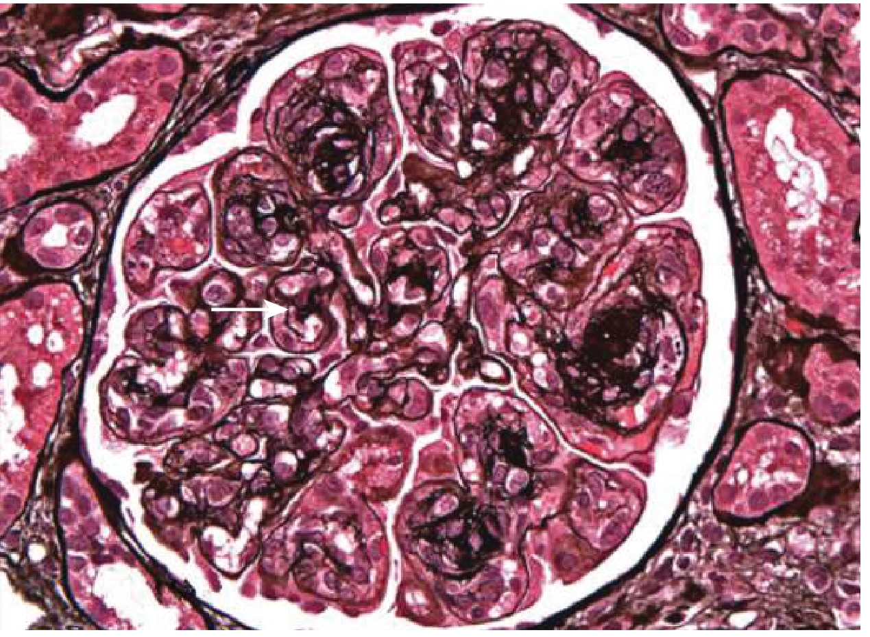
MPGN: Note the lobulation of the glomerular tuft and duplication (tram-tracking) of the basement membrane on silver stain. - Goldman-Cecil Medicine, Figure 107-13
| Technique | Findings in IC-MPGN |
|---|---|
| Light Microscopy | Glomerular hypercellularity, lobular architecture, tram-track GBM, mesangial expansion, possible crescents |
| Immunofluorescence | Granular deposits of IgG + C3 along capillary walls and mesangium; C1q and C4 also positive (classical pathway activation) |
| Electron Microscopy | Subendothelial electron-dense deposits (primary finding); mesangial and occasional subepithelial deposits also present |
The presence of IgG + C3 + C1q + C4 on IF is what distinguishes IC-MPGN from complement-mediated C3 glomerulopathy (which shows isolated C3 without immunoglobulin). - Robbins, Cotran & Kumar Pathologic Basis of Disease
6. Why Does This Present as Adult-Onset Nephrotic Syndrome?
Classic teaching says MPGN most commonly presents in adolescence and young adulthood, but adult-onset (especially in the 4th-6th decade) is increasingly recognized, often linked to a secondary cause (HCV, monoclonal gammopathy) that becomes prevalent with age.
Nephrotic Syndrome occurs because:
- Massive subendothelial and mesangial immune deposits damage the glomerular filtration barrier (podocytes + GBM)
- Complement-mediated inflammation increases GBM permeability
- Result: heavy proteinuria (>3.5 g/day) → hypoalbuminemia → reduced oncotic pressure → edema + ascites
- Compensatory hepatic lipoprotein synthesis → hyperlipidemia and lipiduria (oval fat bodies, fatty casts)
The MPGN pattern presents as pure nephrotic syndrome in 10% of cases, but more commonly as a mixed nephrotic-nephritic picture - the nephritic features (hematuria, RBC casts, hypertension, azotemia) often co-exist with the nephrotic features. - Robbins, Cotran & Kumar Pathologic Basis of Disease
7. Complement Levels as a Diagnostic Clue
This is one of the most clinically important features of IC-MPGN:
- Both C3 and C4 are reduced (intermittently) in IC-MPGN - because classical pathway activation consumes both
- In C3 glomerulopathy (complement-mediated MPGN): only C3 is low (alternative pathway only)
- C3 nephritic factor (C3NeF) stabilizes C3 convertase and is found in complement-mediated subtypes
Low C4 + low C3 in the context of MPGN pattern on biopsy strongly supports an IC-mediated mechanism. - Goldman-Cecil Medicine
8. Complications Explained
A. AKI-on-CKD (Acute Kidney Injury on Chronic Kidney Disease)
MPGN follows a slowly progressive, unrelenting course. The mechanism of CKD development:
- Ongoing immune complex deposition → persistent glomerular inflammation → glomerulosclerosis
- Tubulointerstitial damage from proteinuria (filtered proteins are toxic to tubular cells)
- ~50% of untreated patients develop end-stage kidney disease within 10-15 years
- Only 25-40% maintain normal renal function without treatment
- Spontaneous remission occurs in <10% of cases
AKI superimposed on this CKD can occur from:
-
Severe nephrotic syndrome causing renal hypoperfusion (reduced effective circulating volume)
-
Crescentic transformation (RPGN) - rapid GFR loss from >50% crescent formation
-
Thrombotic events (renal vein thrombosis - nephrotic syndrome increases coagulation risk)
-
Sepsis (infections that triggered MPGN can also precipitate AKI)
-
Contrast nephropathy or nephrotoxic drugs in a susceptible CKD patient
-
Goldman-Cecil Medicine, Prognosis section
B. Hypertension
Hypertension in IC-MPGN + CKD results from multiple converging mechanisms:
- Fluid retention: proteinuria → hypoalbuminemia → secondary hyperaldosteronism → Na+ and water retention → volume overload
- RAAS activation: reduced renal perfusion (from volume redistribution in nephrotic syndrome) → renin release → angiotensin II → vasoconstriction + aldosterone
- Reduced renal mass (CKD): loss of pressure-natriuresis mechanism
- Endothelin and sympathetic activation: inflammatory cytokines from glomerular injury activate sympathetic outflow
Hypertension in turn accelerates CKD progression by causing glomerular hypertension (when autoregulation is lost), creating a vicious cycle.
9. Clinical Presentation Summary
A typical adult patient with this diagnosis will have:
- Edema (periorbital in the morning, pedal/ankle, ascites in severe cases)
- Frothy urine (proteinuria)
- Hematuria (often microscopic - RBCs and RBC casts from nephritic component)
- Hypertension (moderate to severe)
- Oliguria or reduced urine output if AKI is present
- Fatigue and anorexia from uremia (if CKD advanced)
- Features of the underlying cause (jaundice/right upper quadrant pain in HCV; joint pain/rash in SLE; pallor/bone pain in monoclonal gammopathy)
10. Investigations
| Investigation | Finding/Purpose |
|---|---|
| Urine dipstick + microscopy | Proteinuria 3+ to 4+, hematuria, RBC casts, granular casts, lipid oval bodies |
| 24-hour urine protein / urine PCR | >3.5 g/day (nephrotic range) |
| Serum albumin | Low (<3 g/dL) |
| Serum lipids | Elevated total cholesterol and LDL |
| Creatinine, BUN, eGFR | Elevated (AKI-CKD) |
| Complement C3, C4 | Both low in IC-MPGN |
| ANA, anti-dsDNA, ANCA | Rule out lupus/vasculitis |
| HCV antibody + RNA, HBsAg | Rule out viral causes |
| Serum protein electrophoresis + immunofixation | Rule out monoclonal gammopathy |
| Cryoglobulins | Rule out mixed cryoglobulinemia (HCV-associated) |
| Renal biopsy | Definitive diagnosis (LM + IF + EM required) |
11. Treatment
Treatment is cause-driven first, then disease-directed:
Step 1 - Treat the underlying cause:
- HCV-associated IC-MPGN: Direct-acting antivirals (DAAs) - highly effective, protect against recurrence
- SLE: hydroxychloroquine + appropriate immunosuppression for LN class
- Monoclonal gammopathy: clone-directed therapy (rituximab, bortezomib)
- Bacterial infections: antibiotics + source control
Step 2 - Supportive / Renoprotective care (all patients):
- RAAS blockade: ACE inhibitor or ARB - first-line for blood pressure control AND antiproteinuric effect (target BP <130/80 mmHg)
- Salt restriction and loop diuretics for edema/fluid overload
- Statins for hyperlipidemia
- Anticoagulation: consider prophylactic anticoagulation if serum albumin <2.5 g/dL (high VTE risk)
Step 3 - Immunosuppression (for patients with impaired kidney function or nephrotic-range proteinuria):
From [Goldman-Cecil Medicine]:
- Prednisone 1 mg/kg/day (max 60-80 mg/day) for 12-16 weeks
- Mycophenolate mofetil 2-3 g/day may be added for 6-12 months if suboptimal response
- Refractory: Cyclophosphamide 2 mg/kg/day + prednisone 10 mg/day (3-6 months), OR Rituximab 1 g x2 doses 2 weeks apart + third dose at 6 months
Step 4 - Crescentic/RPGN presentation (rapidly declining GFR):
- IV methylprednisolone pulse (1-3 g) followed by oral corticosteroids + oral cyclophosphamide (ANCA-vasculitis regimen)
Emerging therapy: A 2025 Phase III RCT (PMID: 41337715) published in NEJM evaluated pegcetacoplan (complement C3 inhibitor) in both C3 glomerulopathy and IC-MPGN, signaling that targeted complement inhibition may become part of treatment in select patients.
12. Prognosis
-
50-60% of untreated patients progress to ESKD within 10-15 years
-
With treatment: remission in 30-50% of observational studies
-
Worse prognosis with: crescents, nephrotic-range proteinuria, elevated creatinine at presentation, persistent hypertension, and failure to achieve remission
-
Post-transplant: recurrence rate 20-45%; graft loss in up to 50% of those with recurrence
-
Goldman-Cecil Medicine, Prognosis section
Summary Diagram (Pathophysiologic Flow)
Antigenic trigger (HCV / bacteria / SLE / monoclonal protein)
        ↓
Circulating immune complexes (Ag-Ab-C3)
        ↓
Subendothelial + mesangial deposition in glomerulus
        ↓
Classical complement activation (C1q → C4 → C3 → C5-9 MAC)
        ↓
Mesangial proliferation + GBM duplication (tram-track)
        ↓
Glomerular filtration barrier disruption
        ↓
Heavy proteinuria (>3.5 g/day)
        ↓
┌─────────────────────┬────────────────────────┐
│ Nephrotic Syndrome  │   Nephritic Component  │
│ - Hypoalbuminemia   │   - Hematuria          │
│ - Edema             │   - RBC casts          │
│ - Hyperlipidemia    │   - Hypertension       │
│ - Lipiduria         │   - Oliguria/AKI       │
└─────────────────────┴────────────────────────┘
        ↓ (progressive glomerulosclerosis)
    CKD → ESKD (50% at 10-15 years)
Sources:
- Robbins, Cotran & Kumar Pathologic Basis of Disease - MPGN Morphology and Pathogenesis (p. 850)
- Goldman-Cecil Medicine, International Edition - MPGN Classification, Treatment and Prognosis (Ch. 107)
- Comprehensive Clinical Nephrology, 7th Edition - IC-MPGN in Transplantation
- NEJM 2025 - Pegcetacoplan in C3G and IC-MPGN (PMID: 41337715)
This is a shared conversation. Sign in to Orris to start your own chat.