
Create a clean hand-drawn histopathology illustration in oral pathology textbook style, showing the microscopic features of muco-epidermpid carcinoma in a circular histological slide view on a white background. Use realistic H&E stain colors (pink, purple, lilac, light blue). The drawing should look like a neat medical student practical record diagram made with colored pencils and fine ink pen. Include clearly labeled arrows pointing to the important histopathological features. Labels should be handwritten in neat uppercase medical-note style. The illustration should have: - Soft pastel histology shading - Detailed cellular architecture - Clear stromal and epithelial components - Educational and exam-oriented appearance - Minimalist white background - Circular microscope field in center - Accurate histopathological arrangement - High-resolution anatomy illustration style - No watermark - No extra decorative elements Label all characteristic microscopic features of [NAME OF LESION/TUMOR]. Style reference: Traditional oral pathology practical notebook diagram, hand-drawn histology art, H&E stained microscopic illustration, pathology atlas style, educational medical sketch. Make the diagram scientifically accurate and suitable for BDS oral pathology record submission.
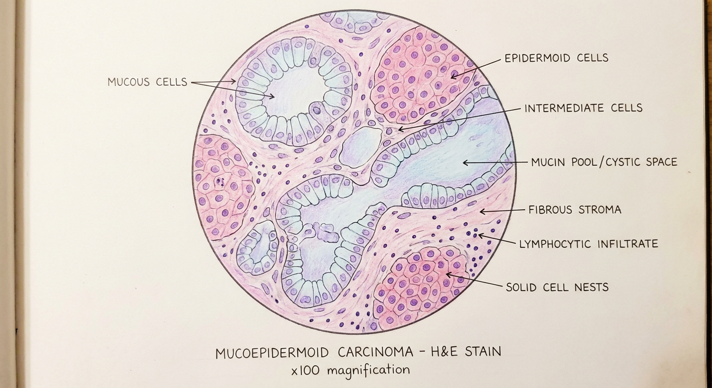
Hand-drawn oral pathology practical record diagram of mucoepidermoid carcinoma histopathology, drawn with colored pencils and fine ink pen, medical student notebook style, BDS oral pathology practical submission quality. Circular microscope field view centered on white background. H&E stain colors: soft pink eosin stroma, lavender/lilac nuclei, light purple mucous cells, pale blue mucin pools, deep purple epidermoid cell nuclei. The circular field shows: - Multiple irregular cystic spaces lined by tall columnar mucous cells with pale cytoplasm filled with mucin (goblet-like, light blue tinted) - Islands and nests of epidermoid (squamous-like) cells with pink cytoplasm and round purple nuclei, arranged in solid sheets - Intermediate cells: small basaloid cells with scant cytoplasm between mucous and epidermoid areas - Large mucin-filled cystic spaces (pale blue/light lavender pools) occupying central areas - Fibrous connective tissue stroma in pink with scattered lymphocytes (small dark purple dots) - Some areas of solid cell nesting without cysts - Mild nuclear pleomorphism and occasional mitotic figures in epidermoid cells Neat handwritten uppercase labels with fine ink arrows pointing to: - MUCOUS CELLS - EPIDERMOID CELLS - INTERMEDIATE CELLS - MUCIN POOL / CYSTIC SPACE - FIBROUS STROMA - LYMPHOCYTIC INFILTRATE - SOLID CELL NESTS Bottom text in neat handwriting: "MUCOEPIDERMOID CARCINOMA - H&E STAIN" with "x100 magnification" underneath Style: Traditional oral pathology practical notebook, hand-drawn histology sketch with colored pencils, soft pastel shading, thin precise ink outlines, educational atlas quality, neat and clean, minimalist white background outside the circle, no watermark, scientifically accurate cellular arrangement
| Label | Histopathological Significance |
|---|---|
| MUCOUS CELLS | Tall columnar goblet-like cells with pale mucin-filled cytoplasm; diagnostic hallmark |
| EPIDERMOID CELLS | Squamous-like cells with pink cytoplasm and round nuclei; form solid nests |
| INTERMEDIATE CELLS | Small basaloid cells between mucous and epidermoid zones; most numerous in high-grade |
| MUCIN POOL / CYSTIC SPACE | Large pale blue lumina filled with mucin; dominant in low-grade tumors |
| FIBROUS STROMA | Pink collagenous connective tissue separating tumor islands |
| LYMPHOCYTIC INFILTRATE | Small dark purple nuclei scattered in stroma; inflammatory response |
| SOLID CELL NESTS | Compact islands without cyst formation; more common in intermediate/high-grade |
- Low-grade: Predominantly cystic, >50% mucous cells, minimal atypia
- Intermediate-grade: Mix of cysts and solid nests, moderate atypia
- High-grade: Predominantly solid, nuclear atypia, mitoses, necrosis; mucin focal and must be demonstrated for diagnosis (Robbins Basic Pathology)