
Gestational hypertension: pathophysiology and management
"gestational hypertension" AND management
gestational hypertension pathophysiology management 2025 guidelines
gestational hypertension preeclampsia placenta pathophysiology diagram
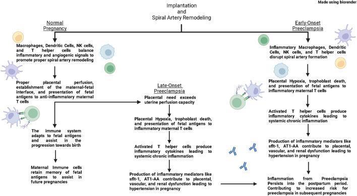
This pathophysiology diagram illustrates the immunological mechanisms underlying three distinct maternal states: Normal Pregnancy, Early-Onset Preeclampsia, and Late-Onset Preeclampsia. The flowchart begins with the process of implantation and spiral artery remodeling. In a normal pregnancy, macrophages, dendritic cells, NK cells, and T helper cells balance inflammatory and angiogenic signals to promote proper vascular remodeling and anti-inflammatory maternal T cell responses. Early-onset preeclampsia is characterized by the disruption of spiral artery formation by inflammatory immune cells, leading to placental hypoxia, trophoblast death, and polarization toward inflammatory T cells. Late-onset preeclampsia follows a normal initial vascular remodeling phase, but subsequently enters a pathological state when placental needs exceed uterine perfusion capacity. Both preeclampsia pathways converge on systemic chronic inflammation, driven by inflammatory cytokines and mediators like sflt-1 and AT1-AA. These factors result in placental, vascular, and renal dysfunction, culminating in gestational hypertension. The diagram highlights that the inflammatory environment persists into the postpartum period, increasing future pregnancy risks.
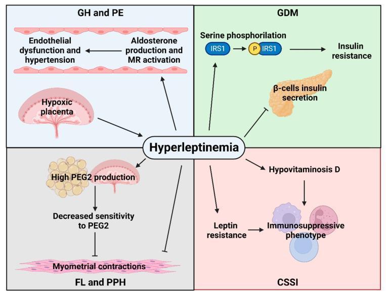
A pathophysiology diagram illustrating the central role of hyperleptinemia in the development of maternal obstetric complications, divided into four quadrants by condition. 1) GH and PE (Gestational Hypertension and Pre-eclampsia): Hyperleptinemia is linked to placental hypoxia and increased aldosterone production/MR activation, leading to endothelial dysfunction and hypertension. 2) GDM (Gestational Diabetes Mellitus): The diagram shows hyperleptinemia inducing serine phosphorylation of IRS1, leading to insulin resistance, while simultaneously inhibiting beta-cell insulin secretion. 3) FL and PPH (Failed Labor and Postpartum Hemorrhage): Hyperleptinemia stimulates high PGE2 production from adipose tissue and the placenta, which correlates with decreased sensitivity to PGE2 and subsequent inhibition of myometrial contractions. 4) CSSI (C-section Surgical Site Infection): Hyperleptinemia contributes to hypovitaminosis D and leptin resistance, both resulting in an immunosuppressive phenotype. Arrows indicate stimulatory pathways, while T-bars represent inhibitory actions, providing a comprehensive overview of hormonal and molecular signaling in obese pregnancies.
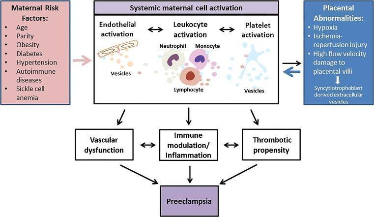
A pathophysiology diagram illustrating the integrated role of extracellular vesicles (EVs) in the pathogenesis of preeclampsia. The flowchart identifies two primary triggers: 'Maternal Risk Factors' (including age, obesity, diabetes, and hypertension) and 'Placental Abnormalities' (such as hypoxia, ischemia-reperfusion injury, and syncytiotrophoblast-derived EV release). These triggers lead to 'Systemic maternal cell activation,' which is depicted as a central mechanism involving three main pathways: 1) Endothelial activation, 2) Leukocyte activation (neutrophils, monocytes, and lymphocytes), and 3) Platelet activation. Each cell type is shown releasing vesicles that mediate cross-talk between systems. The diagram shows how these cellular activations result in three downstream clinical consequences: vascular dysfunction, immune modulation/inflammation, and increased thrombotic propensity. These pathological processes collectively culminate in the clinical manifestation of Preeclampsia. This diagram serves as an educational summary of how maternal-placental interactions through EVs drive systemic inflammation and vascular damage in pregnancy-induced hypertension disorders.
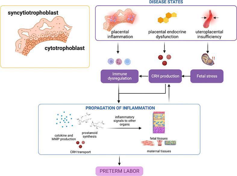
A pathophysiology diagram illustrating the disease states in the placenta that contribute to spontaneous preterm birth (sPTB). The top section identifies three primary placental 'Disease States': placental inflammation (represented by inflamed trophoblast tissue), placental endocrine dysfunction (depicted by hexagonal cellular units), and uteroplacental insufficiency (illustrated by a constricted blood vessel). These states lead through a central mediation layer involving immune dysregulation, CRH (Corticotropin-Releasing Hormone) production, and fetal stress. These pathways converge into a 'Propagation of Inflammation' stage, which details the biochemical cascade including cytokine and MMP production, prostanoid synthesis, and CRH transport. This cascade generates inflammatory signals that target both fetal and maternal tissues (including the decidua and myometrium). The flowchart concludes with the clinical outcome: Preterm Labor. An inset on the left provides anatomical context of the syncytiotrophoblast and cytotrophoblast layers. The diagram highlights the predominant roles of the NF-κB pathway and CRH-mediated feed-forward mechanisms in gestational tissue inflammatory activation.
Gestational Hypertension: Pathophysiology and Management
Definition and Diagnostic Criteria
- Two separate BP measurements ≥140/90 mmHg are required
- Absence of proteinuria distinguishes it from preeclampsia
- Gestational hypertension is a provisional diagnosis - it includes women who will eventually develop preeclampsia but have not yet shown proteinuria
Pathophysiology
1. Relationship to Preeclampsia
2. Impaired Trophoblast Invasion and Spiral Artery Remodeling
- Spiral arteries remain narrow and high-resistance
- Uteroplacental perfusion is reduced
- Downstream placental ischemia follows
3. Angiogenic Imbalance (sFlt-1 / PlGF)
- sFlt-1 (soluble fms-like tyrosine kinase 1) - an anti-angiogenic factor - is overproduced by the ischemic placenta
- PlGF (placental growth factor) - a pro-angiogenic factor - is reduced
- sFlt-1 sequesters free VEGF and PlGF in the circulation, depriving the maternal endothelium of these survival signals
- A ratio of circulating sFlt-1/PlGF ≥40 is associated with an increased risk of developing preeclampsia with severe features within 2 weeks
4. Endothelial Dysfunction
- Endothelial cells produce less prostacyclin (vasodilator) and more thromboxane A2 (vasoconstrictor), shifting the balance toward vasoconstriction
- Increased vascular permeability leads to edema
- Endothelial dysfunction in the kidney causes the characteristic glomerular lesion (glomerular endotheliosis)
- Systemic vasoconstriction raises total peripheral resistance and thus blood pressure
5. Subgroups Within Gestational Hypertension
- Unmasked chronic hypertension - women with underlying essential hypertension whose BP dipped in the second trimester (the physiologic nadir at ~20 weeks), leading to incorrect labeling as normotensive; BP rises in the third trimester. These women have a strong family history, low uric acid, no proteinuria, and are at risk for later chronic hypertension.
- True GH / attenuated preeclampsia - shares the same endovascular pathology as preeclampsia but the clinical expression is milder (no proteinuria), perhaps reflecting host differences in compensatory capacity.
6. Metabolic Contributions
7. Cerebrovascular Autoregulation
Risk Factors
| Category | Examples |
|---|---|
| Primigravida | First pregnancy doubles risk |
| Obesity | ~3× increased risk; linear relationship with BMI |
| Multiple gestation | Twin: 6.7%, triplet: 12.7% |
| Chronic hypertension | Superimposed preeclampsia in 21-25% |
| Diabetes mellitus | Up to 20% risk overall; up to 70% in class F/R |
| Antiphospholipid syndrome | Strong independent risk factor |
| Renal disease | Elevated creatinine, glomerulonephritis |
| Family history | Genetic predisposition to endothelial dysfunction |
| Previous preeclampsia | High recurrence risk |
Management
1. Monitoring and Surveillance
- Regular BP checks (outpatient or remote monitoring)
- Urinalysis/urine protein:creatinine ratio at each visit
- Fetal growth surveillance with serial ultrasound
- Assessment for symptoms of severe disease (headache, visual disturbances, epigastric pain)
- Laboratory workup: CBC, liver enzymes, creatinine, urate
2. Blood Pressure Targets
- BP 140-159 / 90-109 mmHg: antihypertensive therapy should be initiated to achieve <140/90 mmHg to prevent maternal and perinatal morbidity
- BP ≥160/110 mmHg: confirmed on repeat measurement within 15 minutes - treat urgently to bring BP to <160/<110 mmHg within 30-60 minutes to prevent stroke
3. Antihypertensive Pharmacotherapy
First-Line Oral Agents
| Drug | Mechanism | Notes |
|---|---|---|
| Labetalol | α + β blocker | Preferred; α-blockade preserves uteroplacental flow; avoid in asthma |
| Long-acting nifedipine | CCB (dihydropyridine) | Once-daily dosing; may cause edema |
| Methyldopa | Central α2 agonist | Longest safety record; multiple daily dosing required |
IV Agents for Severe/Urgent Hypertension
| Drug | Notes |
|---|---|
| IV Labetalol | First-line; safe and effective; short duration of action |
| IV Nicardipine | Established safety record as tocolytic; effective for hypertensive emergencies |
| IV/oral Hydralazine | Historically first-line; now second-line due to meta-analysis showing increased maternal hypotension, oliguria, placental abruption, and low Apgar scores vs labetalol/nifedipine |
| Oral short-acting nifedipine | Acceptable where IV access unavailable |
Contraindicated Agents
- ACE inhibitors (ACEIs) and angiotensin receptor blockers (ARBs): contraindicated in 2nd and 3rd trimesters - cause renal dysgenesis, oligohydramnios, pulmonary hypoplasia, hypocalvaria, IUGR, and increased stillbirth risk. The 2025 AHA/ACC guidelines also now add ARBs to the explicit contraindications list.
- Nitroprusside: risk of fetal cyanide poisoning if used >4 hours; generally avoided
- Mineralocorticoid receptor antagonists (e.g. spironolactone): added to avoid list in 2025 guidelines
Diuretics
4. Magnesium Sulfate
- Seizure prophylaxis in preeclampsia with severe features
- Acute management of eclamptic seizures
- Dose: 4-6 g IV loading dose, then 1-2 g/hour maintenance
5. Aspirin for Prevention
6. Timing of Delivery
- Without severe features: expectant management is appropriate with close monitoring; delivery at 37 weeks is the general recommendation
- With severe features (BP ≥160/110): treat as preeclampsia with severe features; delivery recommended unless gestational age is <34 weeks and patient is eligible for expectant management in a tertiary center
7. Intrapartum and Postpartum Management
- BP should be monitored closely intrapartum
- Hypertension often persists or worsens in the first days postpartum; close surveillance for 72 hours and at the 1-2 week postpartum visit is essential
- Antihypertensives used in pregnancy are generally safe in breastfeeding; labetalol and nifedipine are commonly continued
Long-Term Consequences
- Increased risk of chronic hypertension, cardiovascular disease, chronic kidney disease, and diabetes mellitus later in life
- Both conditions share metabolic and vascular risk factors; the hypertensive pregnancy may itself accelerate subclinical vascular disease
- Cardiovascular risk reduction strategies should be initiated at postnatal health encounters
Summary Flowchart
New BP ≥140/90 after 20 weeks, no prior hypertension
            ↓
  Gestational Hypertension (provisional)
            ↓
  Monitor closely for proteinuria / end-organ signs
       ↙                    ↘
No progression            Progresses to Preeclampsia
(true GH)                 (10-25% of cases)
       ↓                         ↓
Antihypertensives if ≥140/90   Severe features? → Delivery vs. expectant
Deliver at 37 weeks             MgSO4 for seizure prophylaxis
Postpartum follow-up            IV labetalol / nicardipine for BP ≥160/110