Detailed description of gestational trophoblastic disease both malignant and benign from barek and Novak and eecwnt updates
Reading File
Finding Sources
Finding Sources
Searching PubMed
"gestational trophoblastic disease" management
Finding Sources
Reading File
Reading File
Reading File
Reading File
Reading File
Reading File
Reading File
Reading File
Reading File
Reading File
Reading File
Reading File
Reading File
Reading File
Reading File
Reading File
Reading File
Searching PubMed
"gestational trophoblastic neoplasia" immunotherapy checkpoint inhibitor
I now have all the content I need for a comprehensive answer. Let me compile everything into a detailed, well-structured response.
Gestational Trophoblastic Disease (GTD)
From Berek & Novak's Gynecology, Chapter 41 - Berkowitz, Horowitz & Goldstein
Overview and Classification
Gestational trophoblastic disease (GTD) describes a heterogeneous group of interrelated lesions arising from abnormal proliferation of placental trophoblasts. These lesions are histologically distinct and can be benign or malignant:
Benign GTD:
- Complete hydatidiform mole (CHM)
- Partial hydatidiform mole (PHM)
Malignant GTD (= Gestational Trophoblastic Neoplasia, GTN):
- Invasive mole
- Choriocarcinoma
- Placental-site trophoblastic tumor (PSTT)
- Epithelioid trophoblastic tumor (ETT)
GTNs are among the rare human tumors curable even with widespread dissemination. They can follow any gestational event - molar pregnancy, induced or spontaneous abortion, ectopic pregnancy, or term pregnancy.
PART 1: BENIGN GTD - HYDATIDIFORM MOLES
Epidemiology
Incidence varies dramatically by geography:
- Japan: 2:1,000 pregnancies (threefold higher than the West)
- Europe/North America: 0.6-1.1 per 1,000 pregnancies
- Taiwan: 1 in 125 pregnancies
- United States: 1 in 1,500 live births
- Complete moles: 1:1,945 pregnancies
- Partial moles: 1:695 pregnancies (Ireland data from thorough pathologic review)
Age extremes (adolescence and perimenopausal) carry increased risk. Prior molar pregnancy also elevates risk.
Pathology and Cytogenetics
Table: Features Distinguishing Complete vs. Partial Hydatidiform Moles
| Feature | Complete Mole | Partial Mole |
|---|---|---|
| Fetal/embryonic tissue | Absent | Present |
| Hydatidiform swelling of villi | Diffuse | Focal |
| Trophoblastic hyperplasia | Diffuse | Focal |
| Scalloping of chorionic villi | Absent | Present |
| Trophoblastic stromal inclusions | Absent | Present |
| Karyotype | 46,XX (90%); 46,XY (10%) | Triploid (69 chromosomes) |
| p57 immunostaining | Negative (complete mole only) | Positive |
Complete Hydatidiform Mole (CHM)
- Exhibits characteristic swelling and diffuse trophoblastic hyperplasia
- 46,XX karyotype in ~90%; 46,XY in ~10%
- Chromosomes are entirely of paternal (androgenetic) origin - molar chromosomes arise from an ovum fertilized by a haploid sperm that duplicates its chromosomes; the ovum nucleus is absent or inactivated
- Mitochondrial DNA is of maternal origin
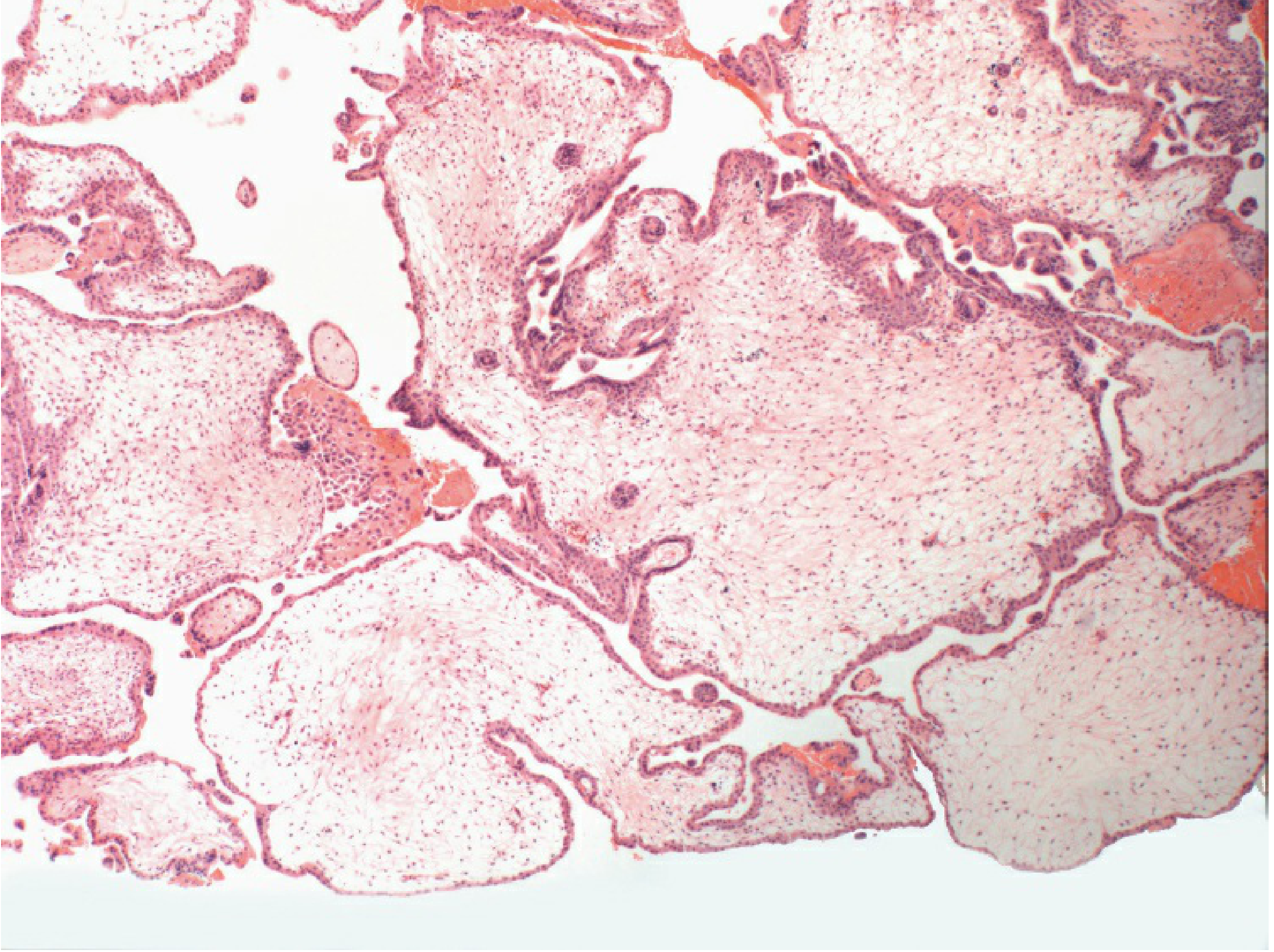
Partial Hydatidiform Mole (PHM)
Pathologic features include:
- Chorionic villi of varying size with focal hydatidiform swelling, cavitation, and trophoblastic hyperplasia
- Marked villous scalloping
- Prominent stromal trophoblastic inclusions
- Identifiable embryonic or fetal tissues
- Triploid karyotype (69 chromosomes) - the extra haploid set is paternally derived
- Associated fetus exhibits stigmata of triploidy: growth retardation, syndactyly, hydrocephaly
Advances in Pathologic Diagnosis
When molar pregnancy is diagnosed early in the first trimester, distinguishing CHM from PHM or hydropic abortion can be difficult due to: smaller villi, less trophoblastic hyperplasia, more primitive villous stroma, and less global necrosis.
Diagnostic adjuncts:
- Flow cytometry - determines ploidy (diploid vs. triploid)
- p57 immunostaining - p57 is a paternally imprinted, maternally expressed gene product:
- Complete mole: diploid + p57 NEGATIVE (no maternal chromosomes)
- Hydropic abortion: diploid (sometimes triploid) + p57 POSITIVE
- Partial mole: triploid + p57 POSITIVE
Familial Recurrent Molar Pregnancy
Rare condition characterized by recurrent complete hydatidiform moles of biparental origin (rather than the usual androgenetic origin). Caused by mutations in the NLRP7 gene located on chromosome 19q13.4, resulting in dysregulation of imprinting in the female germline with abnormal development of embryonic and extraembryonic tissue.
Clinical Features of Complete Hydatidiform Mole
With widespread use of early hCG measurement and transvaginal ultrasonography, patients are now diagnosed much earlier - before classic signs develop.
| Clinical Feature | Classic Frequency | Current Frequency |
|---|---|---|
| Vaginal bleeding | 97% | 46% |
| Excessive uterine size | >50% | 28% |
| Anemia (Hb <10 g/dL) | 50% | 5% |
| Preeclampsia | Common in 2nd trimester | Less common |
| Hyperemesis gravidarum | Common | Less common |
| Hyperthyroidism | Common | Rare |
| Theca lutein cysts >6 cm | ~50% | ~50% |
Key features:
- Vaginal bleeding - most common symptom; retained blood distends endometrial cavity; large volumes may be passed
- Excessive uterine size - endometrial cavity expanded by chorionic tissue and retained blood; associated with markedly elevated hCG
- Preeclampsia - may develop before 24 weeks (unusual in normal pregnancy); patients with very large uteri may also have hyperemesis
- Hyperthyroidism - develops almost exclusively with very high hCG levels; hCG appears to be the thyroid stimulator (positive correlations between hCG and T4/T3). Anesthesia or surgery may precipitate thyroid storm - beta-adrenergic blocking agents should be administered prophylactically
- Theca lutein ovarian cysts - develop in about half of complete mole patients from hCG-mediated ovarian hyperstimulation; cysts reach >6 cm; regress spontaneously within 2-4 months after evacuation; may be complicated by torsion or rupture
- Trophoblastic embolization - rare respiratory distress from trophoblastic emboli to lungs; usually in patients with excessive uterine size and markedly elevated hCG; manifests as chest pain, dyspnea, tachypnea, bilateral pulmonary infiltrates; resolves within 72 hours with cardiopulmonary support
Clinical Features of Partial Hydatidiform Mole
Patients usually do not have the dramatic clinical features of complete mole. In a survey of 81 patients:
- Main sign: vaginal bleeding (72.8%)
- Excessive uterine enlargement: 3.7%
- Preeclampsia: 2.5%
- Initial clinical diagnosis: incomplete or missed abortion in 91.3%, hydatidiform mole in only 6.2%
- Pre-evacuation hCG >100,000 mIU/mL in only 6.6%
- No patient had theca lutein cysts, hyperemesis, or hyperthyroidism
Natural History
Complete mole:
- After evacuation, hCG levels spontaneously regress to normal in most patients
- GTN (persistent trophoblastic neoplasia) develops in approximately 20% of patients after CHM evacuation
- Locally invasive mole: 15%
- Metastatic GTN: 4%
Partial mole:
- GTN develops in only about 2-4% of patients
- Metastatic GTN is very rare
Diagnosis
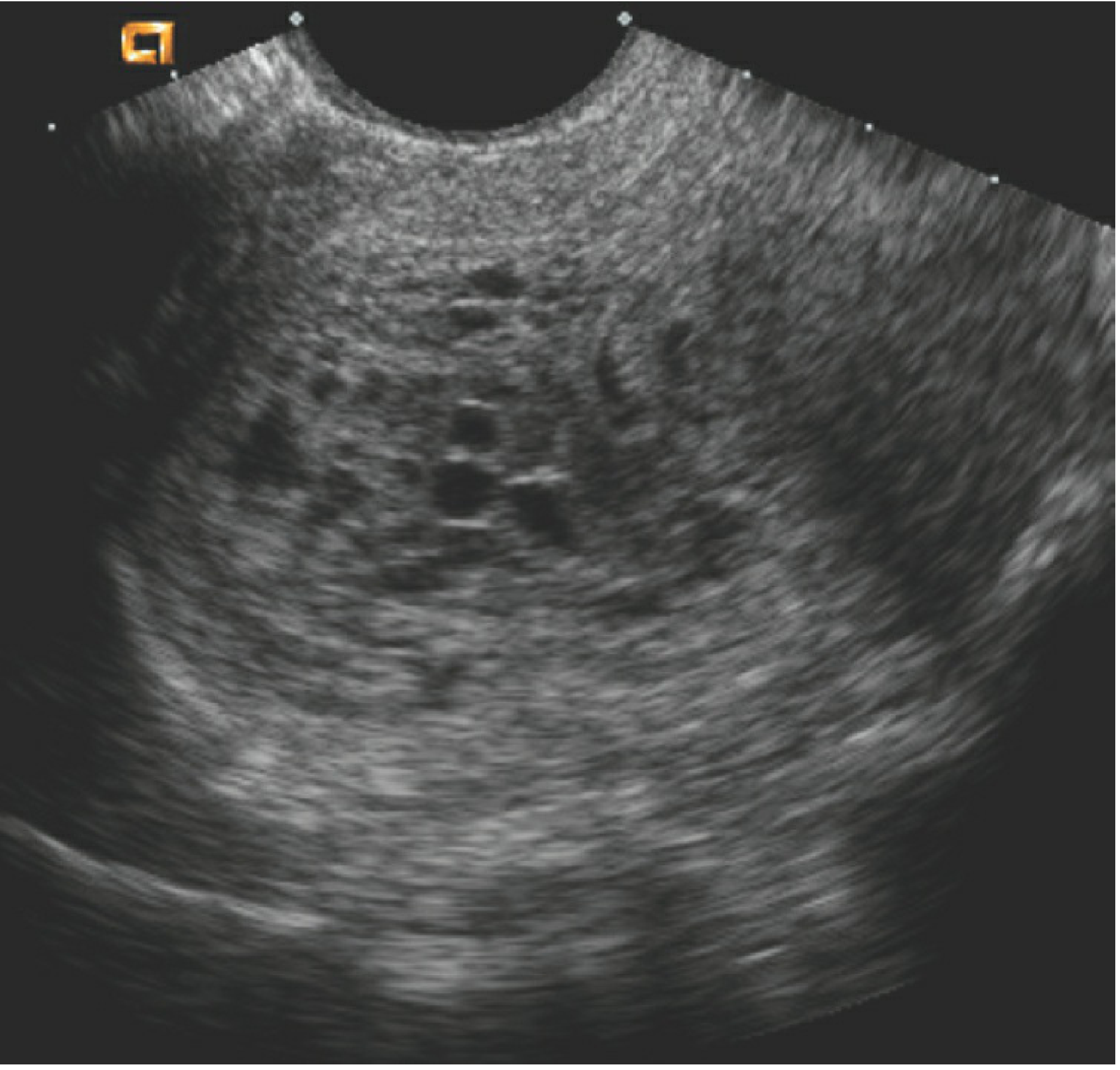
- Ultrasonography is the primary diagnostic tool
- Complete mole: characteristic vesicular pattern (diffuse hydropic swelling) detectable as early as first trimester - described as "snowstorm" appearance
- Partial mole: focal cystic spaces in placenta + increased transverse diameter of gestational sac (positive predictive value 90% when both criteria present)
- hCG levels - markedly elevated; very high levels (>100,000 mIU/mL) associated with more complications
- Histopathology of evacuated tissue - essential for definitive diagnosis
Treatment of Hydatidiform Mole
Pre-evacuation workup: Evaluate for preeclampsia, hyperthyroidism, electrolyte imbalance, anemia.
Suction Curettage (Preferred method)
Preferred regardless of uterine size when fertility is desired. Steps:
- Oxytocin infusion - begun before anesthesia induction
- Cervical dilation - active uterine bleeding should not delay completion
- Suction curettage - uterine size decreases dramatically within minutes; 12-mm cannula strongly advised; for uterus >14 weeks, manual fundal massage to stimulate contraction
- Sharp curettage - gentle curettage to remove residual molar tissue
- Rh-negative patients should receive Rh immune globulin (trophoblast cells express RhD factor)
Hysterectomy
- Preferred if patient desires sterilization (mole in situ)
- Ovaries may be preserved even with prominent theca lutein cysts
- Does not prevent metastasis - hCG follow-up is still required
Prophylactic Chemotherapy
Controversial - only ~20% are at risk of GTN, exposing all patients to potentially toxic treatment. In a study of 247 patients with high-risk complete mole features (hCG >100,000 mIU/mL, uterine size larger than dates, or theca lutein cysts >6 cm), prophylactic chemotherapy reduced the rate of postmolar GTN. May be considered in high-risk patients where follow-up is unreliable.
Post-Molar Follow-Up
After molar evacuation:
- Weekly hCG measurements until normal for 3 consecutive weeks
- Monthly hCG measurements until normal for 6 consecutive months (complete mole)
- Effective contraception throughout the entire follow-up interval
Average time to first normal hCG after evacuation: approximately 9 weeks. After achieving nondetectable serum hCG levels, the risk of developing GTN approaches zero.
Contraception: Oral contraceptives may be used safely after molar evacuation. IUDs should not be inserted until hCG has normalized (risk of uterine perforation, bleeding, infection).
PART 2: MALIGNANT GTD - GESTATIONAL TROPHOBLASTIC NEOPLASIA (GTN)
Overview
GTN is diagnosed when hCG levels fail to normalize or rise during post-molar follow-up, or when metastases are identified. Key features:
- GTN can follow any gestational event (molar or non-molar)
- Tendency for early vascular invasion and widespread dissemination
- Trophoblastic tumors are often perfused by fragile vessels and are frequently hemorrhagic
Criteria for diagnosing post-molar GTN (FIGO):
- Plateau of hCG for 4 measurements over 3 weeks
- Rise of hCG ≥10% over 3 weekly measurements
- Persistence of hCG for ≥6 months
- Histologic diagnosis of choriocarcinoma
Nonmetastatic (Locally Invasive) GTN
Locally invasive GTN develops in ~15% of patients after complete mole evacuation. Symptoms:
- Irregular vaginal bleeding
- Theca lutein cysts
- Uterine subinvolution or asymmetric enlargement
- Persistently elevated serum hCG levels
- Trophoblastic tumor may perforate the myometrium (intraperitoneal bleeding) or erode into uterine vessels (vaginal hemorrhage)
- Bulky, necrotic tumor may cause uterine sepsis (purulent vaginal discharge, acute pelvic pain)
- After molar evacuation, persistent GTN may have histology of either hydatidiform mole or choriocarcinoma
- After nonmolar pregnancy, persistent GTN always has histology of choriocarcinoma
Choriocarcinoma
- Histologic hallmark: sheets of anaplastic syncytiotrophoblast and cytotrophoblast without chorionic villi
- Highly malignant; tendency for early vascular invasion with widespread hematogenous dissemination
- May follow molar or non-molar gestations; GTN after nonmolar pregnancies always exhibits choriocarcinoma histology
- Contains both cytotrophoblast and syncytiotrophoblast but no villi
Placental-Site Trophoblastic Tumor (PSTT) and Epithelioid Trophoblastic Tumor (ETT)
- Uncommon but important variants consisting predominantly of intermediate trophoblast
- Produce small amounts of hCG and human placental lactogen (hPL) relative to their mass
- Tend to remain confined to the uterus, metastasizing late
- Relatively insensitive to chemotherapy (in contrast to other GTN)
- Hysterectomy is the primary curative treatment for nonmetastatic disease
- Patients with metastatic PSTT may still achieve remission but tumors are less responsive to chemotherapy
Metastatic GTN
Metastatic GTN occurs in ~4% of patients after complete mole evacuation and more commonly after nonmolar pregnancies.
Most common sites of metastases:
- Lungs: 80%
- Vagina: 30%
- Pelvis: 20%
- Liver: 10%
- Brain: 10%
Pulmonary Metastases
- 80% have lung involvement visible on chest radiography at diagnosis
- Symptoms: chest pain, cough, hemoptysis, dyspnea, or asymptomatic
- Four radiographic patterns:
- Alveolar or "snowstorm" pattern
- Discrete rounded densities
- Pleural effusion
- Embolic pattern (pulmonary arterial occlusion)
- Some patients have minimal gynecologic symptoms with extensive pulmonary disease
Vaginal Metastases
- Vaginal metastases are highly vascular; biopsy is associated with significant hemorrhage
- They may appear as bluish-purple submucosal lesions
Hepatic Metastases
- Usually indicate advanced disease
- Hepatic arterial infusion or hepatic resection may be required for resistant hepatic metastasis
Cerebral Metastases
- Plasma-to-CSF hCG ratio tends to be <60 in the presence of cerebral metastases
- Managed with whole-brain radiation (3,000 cGy in 10 fractions) or stereotactic radiosurgery + combination chemotherapy
FIGO Staging
| Stage | Description |
|---|---|
| I | Disease confined to uterus |
| II | GTN extending outside uterus but limited to genital structures (adnexa, vagina, broad ligament) |
| III | GTN extending to lungs with or without known genital tract involvement (based on plain chest X-ray, not CT) |
| IV | All other metastatic sites (brain, liver, kidney, GI tract) - highest risk |
Stage IV is the highest risk category - most likely to be resistant to chemotherapy; choriocarcinoma usually present; commonly follows nonmolar pregnancy.
WHO Prognostic Scoring System
In addition to staging, the WHO prognostic score guides chemotherapy selection:
| Variable | Score 0 | Score 1 | Score 2 | Score 4 |
|---|---|---|---|---|
| Age (years) | ≤39 | >39 | - | - |
| Antecedent pregnancy | Hydatidiform mole | Abortion | Term | - |
| Interval (end of pregnancy to chemo) | <4 months | 4-6 months | 7-12 months | >12 months |
| hCG at GTN diagnosis (IU/L) | <10³ | 10³-10⁴ | 10⁴-10⁵ | >10⁵ |
| ABO blood group | - | O or A | B or AB | - |
| Largest tumor (including uterine) | - | 3-5 cm | ≥5 cm | - |
| Site of metastases | Lung/vagina | Spleen/kidney | GI | Brain/liver |
| Number of metastases | - | 1-4 | 5-8 | >8 |
| Prior chemotherapy | - | - | Single drug | ≥2 drugs |
- Score ≤6 = Low risk → single-agent chemotherapy
- Score >6 = High risk → multimodal therapy with intensive combination chemotherapy ± surgery ± radiation
Diagnostic Evaluation
Initial workup for GTN:
- History and physical examination
- hCG level (serum)
- Complete blood count, renal and liver function tests
- Chest X-ray (plain - used for staging, not CT)
- CT or MRI scan of head as indicated
For patients with pulmonary or other metastases:
- Liver ultrasonography and CT/MRI for hepatic metastases (in patients with abnormal liver function)
- CT/MRI head for early diagnosis of asymptomatic cerebral lesions
- Chest CT detects micrometastases not visible on plain X-ray in ~40% of presumed nonmetastatic patients, but presence of micrometastases has not been shown to substantially change outcome
- CSF hCG measurement if brain CT/MRI is negative in patients with choriocarcinoma
- Pelvic ultrasonography to detect extensive uterine trophoblastic involvement
Management of GTN
Protocol overview (Table 41-4 from Berek & Novak):
| Stage | Initial Treatment | Resistant Disease |
|---|---|---|
| Stage I | Single-agent chemotherapy OR hysterectomy + adjuvant chemo | Combination chemo; hysterectomy; local resection |
| Stages II-III (low risk) | Single-agent chemotherapy | Combination chemotherapy |
| Stages II-III (high risk) | Combination chemotherapy | Second-line combination chemo |
| Stage IV | Combination chemotherapy ± brain RT ± hepatic resection/embolization | Second-line combination chemo; hepatic arterial infusion |
Low-Risk Disease (Stage I and Low-Risk Stages II-III, Score <7)
Hysterectomy + adjuvant single-agent chemotherapy - for patients not wishing to preserve fertility
- Adjuvant chemo rationale: reduces dissemination of viable tumor cells at surgery; treats occult micrometastases
- In a series of 31 patients: 100% achieved complete remission with no additional therapy
- Mandatory for PSTT and ETT stage I (relatively chemoresistant)
Single-agent chemotherapy - preferred when fertility preservation is desired
At the NETDC (July 1965 - December 2016):
- 592 patients with stage-I GTN treated with primary single-agent chemotherapy
- 82.3% (487 patients) achieved complete remission
- Remaining 105 patients achieved remission after combination chemotherapy or surgery
For low-risk stages II-III:
- 168 patients treated with single-agent chemotherapy
- 77.3% (130 patients) achieved complete remission
- Remaining 38 patients required combination chemotherapy
Single-Agent Chemotherapy Regimens
Both methotrexate (MTX) and actinomycin-D (ActD) achieve comparable excellent remission rates:
MTX regimens:
- 5-day regimen
- 8-day regimen
- Pulsatile weekly (30 mg/m² IM)
ActD regimens:
- 5-day regimen
- Pulsatile (biweekly 1.25 mg/m² IV bolus)
GOG Phase III RCT findings:
- 216 patients randomized to biweekly ActD (1.25 mg/m²) vs. weekly MTX (30 mg/m² IM)
- Remission rates: MTX 58%, ActD 73%
- ActD appeared superior to this weekly MTX regimen, but all patients ultimately achieved remission
- Comparison with 5-day or 8-day MTX regimens remains needed
MTX with folinic acid (MTX-FA):
- 8-day MTX-FA: complete remission in 87.6% of 185 patients
- Stage-I GTN: 90.2% remission; low-risk stages II-III: 68.2% remission
- Resistance more common with: choriocarcinoma, metastasis, pre-treatment hCG >50,000 mIU/mL
- Side effects: thrombocytopenia, granulocytopenia, hepatotoxicity (generally well tolerated)
High-Risk Metastatic GTN (Stages II-IV, Score >6)
EMA-CO (Etoposide, MTX, ActD, Cyclophosphamide, Vincristine) - preferred primary treatment:
- Etoposide alone: complete remission in 95% of 60 patients with nonmetastatic/low-risk metastatic GTN
- EMA-CO in high-risk GTN: 83% remission rate (initial studies); 90.6% complete sustained remission in 96 high-risk patients (score >6) in another series
- Remission with EMA-CO in 86% of patients with brain metastasis
- Well tolerated; treatment seldom suspended due to toxicity
At NETDC (July 1965 - December 2016):
- 144 patients with high-risk GTN stages II-IV
- 84% (121 patients) achieved complete remission
- Before 1975 (single-agent primary therapy): only 30% achieved remission for stage IV
- After 1975 (multimodal intensive therapy): 75.7% achieved remission for stage IV
Triple therapy (MTX + ActD + cyclophosphamide): Pre-EMA-CO era treatment; still useful in selected low-risk patients resistant to single agents.
Management of Refractory GTN
Agents and regimens for patients resistant to all standard protocols:
- EMA-EP (EMA-CO with substitution of etoposide + cisplatin on day 8): remission in 76% of 21 patients resistant to EMA-CO
- PVB (cisplatin, vinblastine, bleomycin)
- Ifosfamide and paclitaxel
- TE/TP (paclitaxel, etoposide, cisplatin): 3 complete + 4 partial responses in 7 EMA-CO resistant patients
- FUDR-containing regimens: complete remission in all 21 patients with drug-resistant GTN (Wan et al.)
- 5-FU + ActD: remission in 9 of 11 (82%) patients with drug resistance
- Autologous bone marrow transplantation / stem cell rescue: complete remissions reported; role not yet fully defined
Special Situations
Thoracotomy: For persistent viable pulmonary metastasis following intensive chemotherapy; thorough workup to exclude other disease sites is required first; fibrotic nodules may persist indefinitely after complete remission.
Hysterectomy in metastatic disease: For uterine hemorrhage or sepsis control; may substantially reduce tumor burden and limit need for multiple chemotherapy courses.
Cerebral metastases: Whole-brain radiation 3,000 cGy in 10 fractions OR stereotactic radiosurgery + combination chemotherapy. Irradiation may be hemostatic and tumoricidal, reducing risk of spontaneous cerebral hemorrhage. Excellent remission rates (86%) with intensive IV combination chemotherapy + intrathecal MTX.
Craniotomy: For complications (bleeding) - not routine treatment.
Hepatic metastases: Hepatic arterial infusion for resistant disease; hepatic resection or arterial embolization for acute bleeding or resistant tumor focus.
Duration of Therapy and Follow-Up
- Combination chemotherapy given as frequently as toxicity permits until 3 consecutive normal hCG levels
- After normal hCG attained: at least 3 additional courses to reduce relapse risk
- All GTN patients: weekly hCG until normal for 3 consecutive weeks, then monthly until normal for 12 consecutive months
False-Positive hCG Tests
Heterophile antibodies can cause falsely positive hCG results in some commercial assay systems. This concept is critical when following patients after molar gestation or GTN - a false positive can lead to unnecessary treatment. Verification with a different assay or urine hCG (heterophile antibodies are not excreted in urine) should be performed when there is clinical doubt.
Persistent low-level "real" hCG: Some patients may have a persistent low-level hCG from a physiologic source (e.g., pituitary hCG) rather than active GTN. Further diagnostic evaluation is warranted before initiating or continuing treatment.
Subsequent Pregnancies
After uncomplicated hydatidiform mole:
- Patients can anticipate normal future reproduction
- No increased risk of spontaneous abortion, stillbirth, or congenital malformations compared to the general population
- Pregnancy should be deferred until completion of the follow-up period
After GTN:
- After achieving remission with chemotherapy, patients can anticipate normal future reproduction
- No increased teratogenicity or adverse obstetric outcomes demonstrated in post-chemotherapy pregnancies
Key Points Summary (from Berek & Novak)
- GTD is a group of interrelated tumors including complete/partial moles, invasive moles, choriocarcinoma, PSTT, and ETT.
- Complete and partial moles differ in chromosomal origin, histopathology, clinical presentation, and risk of GTN.
- hCG is a critical marker for diagnosing GTN, monitoring chemotherapy response, and detecting relapse.
- Metastases can occur after any type of GTD but are more common after nonmolar pregnancies; most common sites are lung and vagina.
- Single-agent chemotherapy achieves high remission rates in nonmetastatic and low-risk metastatic GTN.
- High-risk GTN requires primary combination chemotherapy, surgery for complications, and occasionally radiation.
- After achieving remission with chemotherapy, patients can anticipate normal future reproduction.
Recent Updates (2023-2026)
1. Immunotherapy/Checkpoint Inhibitors - Emerging Paradigm
Multiple recent reviews signal a new era for refractory GTN:
- PD-L1 is highly expressed on trophoblastic tissue, providing biological rationale for checkpoint blockade
- Pembrolizumab (anti-PD-1) has shown responses in refractory GTN, with complete responses documented
- The systematic review by Wang et al. [PMID 37594534] and the 2024 review by Baas et al. [PMID 37703867] describe "Immunotherapy for GTN: A New Paradigm" - cases of chemotherapy-refractory disease responding to immune checkpoint inhibitors
- A 2026 narrative review (Barcellos et al., PMID 41627371) specifically reviews checkpoint inhibition in GTN with a focus on anti-tumor immune reawakening
2. Twin Pregnancy with Complete Mole and Coexisting Normal Fetus
- A 2026 meta-analysis by Salmeri et al. [PMID 41066778] analyzed clinical outcomes in multiple pregnancy with complete hydatidiform mole and coexisting normal fetus - this rare condition carries significant maternal and fetal risks, and the evidence base for management is now more systematically characterized.
3. Epidemiology
- Systematic reviews (2025) on magnitude of GTD in Africa and East Africa confirm higher incidence in low-resource settings and highlight histopathologic patterns (PMID 41120163, 41029567).
4. Psychological Impact
- A 2025 meta-analysis (PMID 40555372) characterized the prevalence of depression and anxiety in GTD, highlighting the psychological burden that should be addressed alongside medical management.
Primary source: Berek & Novak's Gynecology (ISBN 9781496380357), Chapter 41, pp. 2493-2521 - Berkowitz RS, Horowitz NS, Goldstein DP
Recent literature: PubMed PMIDs 37594534, 37703867, 40292281, 41627371, 41066778
This is a shared conversation. Sign in to Orris to start your own chat.