
Explain me Great Saphenous Vein in simple way
great saphenous vein anatomy diagram leg
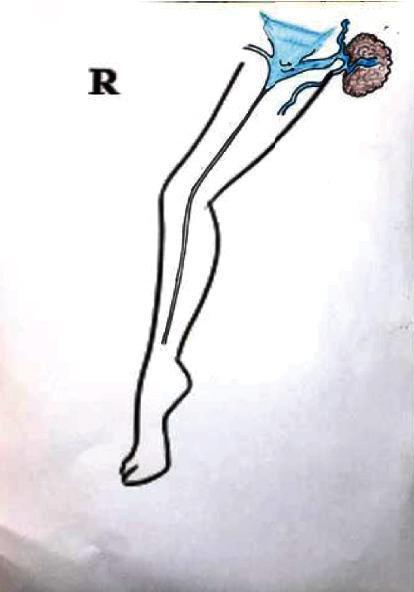
This medical schematic illustrates the vascular anatomy of the right lower extremity, focusing on mechanisms for the recurrence of varicose veins in the groin region. The diagram depicts the outline of a leg with a primary vertical vessel, representing the great saphenous vein (GSV), extending from the foot to the thigh. In the inguinal (groin) area, specifically within Scarpa's triangle, the illustration highlights a complex vascular network in blue. Key components include a triangular-shaped structure representing the saphenofemoral junction or a venous confluence, and a brown, textured cluster representing a lymph node. Small blue vessels are shown interconnecting the lymph node with the main venous system, demonstrating lymphatic-venous communication. The diagram serves an educational purpose by visualizing how accessory lymph node vessels can serve as pathways for venous reflux, leading to recurrent varicosities after surgical or endovenous treatments like radiofrequency ablation (RFA). This clinical illustration is designed for vascular surgery and phlebology education.
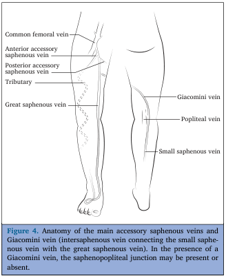
Summary : This anatomical illustration shows the main superficial veins of the lower limb, focusing on the accessory saphenous veins and the Giacomini vein, which connects the small saphenous vein with the great saphenous vein. illustration: # Scene Overview : • Diagram of the posterior and medial aspects of the lower limb, showing the course and connections of major superficial veins. • Black-and-white line drawing with labeled anatomical structures. • The left leg is shown from the medial side, and the right leg from the posterior side. # Technical Details : • No scale bar or magnification indicated. • Anatomical labels include: Common femoral vein, Anterior accessory saphenous vein, Posterior accessory saphenous vein, Tributary, Great saphenous vein, Giacomini vein, Popliteal vein, Small saphenous vein. # Spatial Relationships : • The great saphenous vein runs along the medial aspect of the leg and thigh, draining into the common femoral vein. • The small saphenous vein runs along the posterior aspect of the calf, draining into the popliteal vein. • The Giacomini vein connects the small saphenous vein to the great saphenous vein, running obliquely across the posterior thigh. • Anterior and posterior accessory saphenous veins branch from the great saphenous vein in the thigh. # Analysis : • The illustration highlights the anatomical variability of superficial venous connections in the lower limb, particularly the presence of the Giacomini vein, which may alter the typical drainage pattern at the saphenopopliteal junction. • The diagram is useful for understanding venous anatomy relevant to clinical procedures and venous insufficiency.
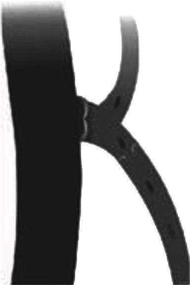
This medical anatomical diagram illustrates the venous architecture of the Great Saphenous Vein (GSV) at the saphenofemoral junction. The illustration features a primary vertical trunk (representing the deep femoral system or proximal GSV) with two prominent tributary veins branching to the right in a Y-shaped configuration. A critical educational focus is placed on the valvular anatomy: terminal and subterminal valves are depicted at the junction where the tributaries enter the main vessel. Small perforations are indicated along the length of the tributary vessels, which represent the superficial circumflex iliac, superficial epigastric, or external pudendal veins. The diagram highlights the pathophysiological mechanism of venous incompetence, where despite competent terminal valves, reflux through these arch tributaries can lead to incompetent GSV and the recurrence of varicose veins following radiofrequency ablation (RFA). This content is designed for advanced medical education in vascular surgery and phlebology, specifically for understanding the causes of post-surgical recurrence in the groin region.
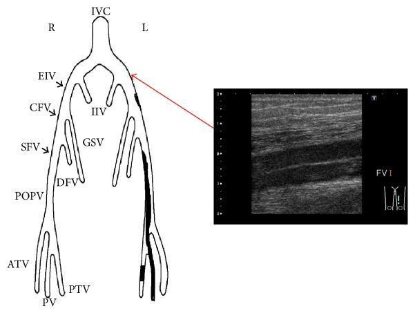
This composite image consists of an anatomical diagram and a corresponding diagnostic ultrasound, illustrating the deep venous system of the lower extremities. The left side features a schematic representation of the venous anatomy starting from the inferior vena cava (IVC), bifurcating into the internal (IIV) and external iliac veins (EIV). It follows the progression down to the common femoral vein (CFV), superficial femoral vein (SFV), deep femoral vein (DFV), great saphenous vein (GSV), popliteal vein (POPV), and distal leg veins (ATV, PTV, PV). The left leg in the diagram contains dark shaded regions representing deep vein thrombosis (DVT) extending from the iliac to the popliteal segments. A red arrow connects the left external iliac region to a B-mode ultrasonography image on the right. The ultrasound shows a longitudinal view of a leg vein with a hypoechoic lumen and hyperechoic walls. The absence of significant echogenic material within the lumen indicates a reduction in thrombus burden following anticoagulant therapy and endovascular treatment (EVT). This figure is intended for educational use in vascular medicine and radiology to demonstrate DVT localization and therapeutic monitoring.
The Great Saphenous Vein (GSV) - Simply Explained
What Is It?
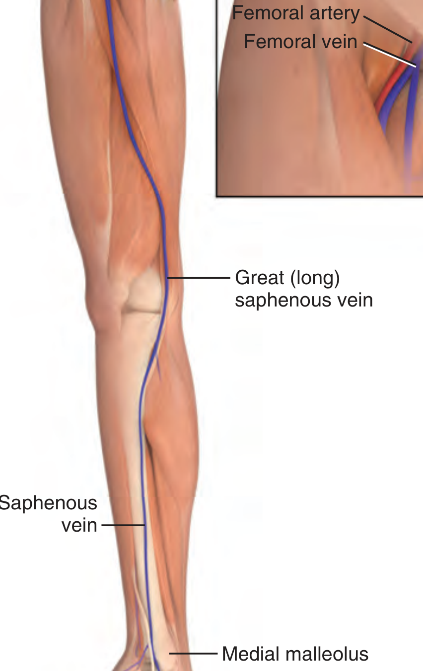
Where Does It Start and End?
| Location | What Happens |
|---|---|
| Foot | Starts from the dorsal venous arch on the top/back of the foot |
| Ankle | Runs just in front of the medial malleolus (the bony bump on the inner ankle) - easily felt here |
| Lower leg | Travels up the anteromedial (inner-front) side of the leg, accompanied by the saphenous nerve |
| Knee | Passes superficially (just under skin) on the medial side |
| Thigh | Crosses slightly anterolaterally (front-outer) as it ascends |
| Groin | Passes through a hole in the deep fascia called the saphenous ring (fossa ovalis) and drains into the femoral vein at the femoral triangle |
Think of It This Way
Key Features (What Makes It Special)
Why Is It Clinically Important?
1. IV Access / Venous Cutdown
2. Bypass Surgery (CABG)
3. Varicose Veins
4. Treatment: High Ligation and Stripping
- Ligating (tying off) the vein at its junction with the femoral vein in the groin (the saphenofemoral junction, SFJ)
- Stripping (physically pulling out) the vein from inside the leg using a stripper tool
Anatomical Diagram - Superficial Venous Connections
Quick Summary
- What: Longest vein in the body
- Where: Medial (inner) side of the leg, from foot to groin
- Job: Drain superficial blood from leg back to femoral vein
- Runs with: Saphenous nerve
- Drains into: Femoral vein (at the saphenofemoral junction in the groin)
- Clinical uses: IV cutdown, bypass graft harvest
- When it fails: Varicose veins