I now have comprehensive content from Berek & Novak's Gynecology. Let me compile the full, detailed answer.
Amenorrhea - As per OBG (Berek & Novak's Gynecology, Chapter 34)
Definition
Normal menstruation requires an intact hypothalamic-pituitary-ovarian-uterine axis. If any component fails, bleeding cannot occur.
- Primary amenorrhea: Absence of menses by age 13 when there is no visible development of secondary sexual characteristics, OR by age 15 in the presence of normal secondary sexual characteristics.
- Secondary amenorrhea: Absence of menses for 3 months in a woman who previously had regular cycles, or for 6 months in a woman who had irregular cycles.
- Oligomenorrhea: Cycles occurring at intervals > 35 days (but not complete absence).
Pregnancy must always be excluded first regardless of the type of amenorrhea.
Physiology of Normal Menstruation
For menstruation to occur, a precise sequence must be intact:
- Hypothalamus secretes GnRH in a pulsatile fashion (modulated by neurotransmitters and hormones)
- GnRH stimulates pituitary to release FSH and LH
- FSH/LH promote ovarian follicular development and ovulation
- Pre-ovulatory follicle secretes estrogen; post-ovulation corpus luteum secretes progesterone + estrogen
- These hormones stimulate endometrial development
- If no pregnancy, estrogen and progesterone fall → withdrawal bleeding
Any disruption at the hypothalamus, pituitary, ovary, uterus, or outflow tract prevents menstruation.
Classification
Amenorrhea is best classified anatomically and functionally:
| Category | Level |
|---|
| Outflow tract / uterine | Cervix, vagina, uterus |
| Ovarian | Primary ovarian insufficiency, gonadal dysgenesis |
| Pituitary | Prolactinoma, Sheehan syndrome, empty sella |
| Hypothalamic | Functional hypothalamic amenorrhea, Kallmann syndrome |
A clinically useful subdivision is based on the presence or absence of secondary sexual characteristics.
PRIMARY AMENORRHEA
A. Amenorrhea WITHOUT Secondary Sexual Characteristics
This implies gonadal failure or hypothalamic/pituitary dysfunction occurring before puberty.
1. Hypergonadotropic Hypogonadism (High FSH)
The ovaries have failed - no estrogen is produced to suppress pituitary, so FSH rises.
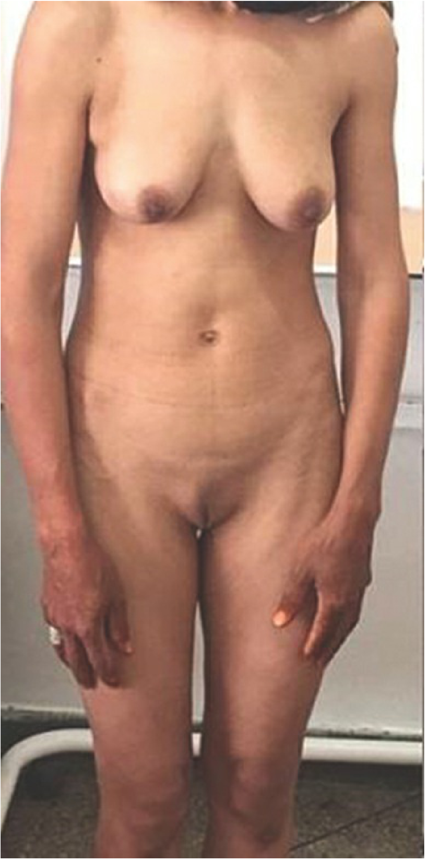
- Gonadal dysgenesis (Turner syndrome - 45,X): Most common cause. Short stature, webbed neck, shield chest, streak gonads, horseshoe kidney, cardiac defects (coarctation of aorta). Mosaic forms (45X/46XX) may have some development.
- Pure gonadal dysgenesis (Swyer syndrome - 46,XY): Normal female phenotype with streak gonads. Y chromosome material warrants gonadectomy due to ~25-30% risk of gonadoblastoma/dysgerminoma.
- Other causes of primary ovarian failure: Autoimmune, galactosemia, FSH receptor mutations.
When gonadal failure occurs with primary amenorrhea, there is a high incidence of abnormal karyotype.
2. Hypogonadotropic Hypogonadism (Low FSH/LH)
The brain/pituitary is not signaling properly.
- Kallmann syndrome: GnRH deficiency + anosmia/hyposmia (failure of GnRH neurons to migrate from olfactory placode). X-linked (KAL1 gene) or autosomal. Characterized by absent/diminished sense of smell.
- Idiopathic hypogonadotropic hypogonadism: Low GnRH without anosmia. Various gene mutations (GNRH1, KISS1, etc.)
- GnRH receptor mutations: G-protein coupled receptor mutations → decreased GnRH binding → no FSH/LH stimulation. Patients are normosomic.
- FSH β-subunit deficiency: Low FSH, elevated LH (paradoxically), low androgens. Rare autosomal recessive.
3. Androgen Insensitivity Syndrome (AIS) - 46,XY
- Complete AIS: XY karyotype with female phenotype. Testes present (undescended), functioning AMH (no mullerian structures), absent pubic/axillary hair (hallmark), blind vaginal pouch, breast development at puberty (due to testosterone aromatization to estrogen).
- Testes must be removed after puberty due to malignancy risk.
4. 5α-Reductase Deficiency - 46,XY
- Testosterone cannot be converted to dihydrotestosterone (DHT) → ambiguous external genitalia at birth. Virilization occurs at puberty. No breast development (testosterone intact, suppresses breast development). Distinguishes from AIS. Normal AMH → no mullerian structures.
B. Amenorrhea WITH Secondary Sexual Characteristics (Normal Pubertal Development but No Menses)
This indicates normal estrogen production but a structural or outflow problem.
Anatomic / Outflow Tract Causes
| Condition | Key Feature |
|---|
| Imperforate hymen | Cyclic pelvic pain, blue bulging hymen, hematocolpos |
| Transverse vaginal septum | Cyclic pain; vagina partially or fully obstructed |
| Cervical stenosis | Post-procedural (cone biopsy, LEEP) |
| Müllerian agenesis (MRKH syndrome) | Absent uterus and upper vagina, normal ovaries and secondary sexual characteristics, normal 46,XX karyotype, normal female hormones |
Anatomic causes are relatively few and can be diagnosed by history, physical examination, and imaging.
SECONDARY AMENORRHEA
Causes are classified by the level of the HPO axis:
1. Hypothalamic Causes - Functional Hypothalamic Amenorrhea (FHA)
Most common cause of secondary amenorrhea after pregnancy. Caused by disruption of pulsatile GnRH secretion.
- Weight loss / anorexia nervosa / malnutrition: Energy deficit suppresses GnRH. Low BMI is a red flag.
- Excessive exercise: Common in female athletes (part of the "female athlete triad": amenorrhea + low energy availability + low bone density).
- Psychosocial stress: Corticotropin-releasing hormone (CRH) and opioids suppress GnRH.
- Obesity: Adipose tissue produces leptin and estrone; can disrupt HPO axis.
Lab findings: Low/normal FSH, low estradiol, low LH (functional, not structural failure).
2. Pituitary Causes
- Hyperprolactinemia: Elevated prolactin → abnormal GnRH secretion → anovulation and amenorrhea.
- Causes: Prolactinoma (most common pituitary tumor), CNS lesions disrupting dopamine transport down the pituitary stalk, drugs (antidepressants, antipsychotics - especially risperidone, metoclopramide, antihypertensives, opiates, H2 blockers).
- Galactorrhea may or may not be present.
- Hypothyroidism elevates TRH, which stimulates prolactin → treat hypothyroidism first.
- Sheehan syndrome: Post-partum pituitary necrosis due to obstetric hemorrhage and hypovolemia. Presents with failure to lactate, inability to regrow shaved pubic hair, and secondary amenorrhea.
- Empty sella syndrome: CSF herniation into the sella; pituitary function usually preserved.
3. Ovarian Causes
- Primary Ovarian Insufficiency (POI) (formerly premature ovarian failure/premature menopause):
- Defined as amenorrhea for ≥4 months + two FSH levels in the menopausal range (>40 IU/L), measured ≥1 month apart, before age 40.
- Causes: Autoimmune (most common identifiable cause - associated with thyroid disease, Addison's disease), chromosomal (Turner mosaic, fragile X premutation), iatrogenic (chemotherapy, radiation), idiopathic (~90%).
- Unlike menopause, POI is not always permanent - spontaneous ovulation and even pregnancy can occasionally occur.
- Health consequences: Osteoporosis, cardiovascular disease, sexual dysfunction, psychological impact.
- Management: Hormone therapy (estrogen + progestogen) until age ~51.
4. Uterine / Outflow Tract Causes
- Asherman syndrome (intrauterine adhesions / synechiae):
- Most common cause: Vigorous curettage (especially post-abortion or post-partum with infection).
- Presents as hypomenorrhea or amenorrhea after a D&C.
- Diagnosis: Hysteroscopy (gold standard), sonohysterography.
- Treatment: Hysteroscopic adhesiolysis + post-operative estrogen therapy to promote endometrial regeneration.
5. Other Endocrine Causes
- Hypothyroidism: Elevated TRH → elevated prolactin → amenorrhea. Also direct effect on menstrual cycle.
- Hyperthyroidism: Can also disrupt cycle; usually oligomenorrhea.
- PCOS (Polycystic Ovary Syndrome):
- Prevalence 6-10%; most common endocrine disorder in women of reproductive age.
- Rotterdam criteria (2003): Diagnosis requires 2 of 3: (1) hyperandrogenism (clinical or biochemical), (2) oligomenorrhea or amenorrhea, (3) polycystic ovaries on ultrasound.
- Although PCOS more commonly causes irregular bleeding, it is one of the most common causes of amenorrhea.
- Associated with insulin resistance, obesity, endometrial hyperplasia/cancer risk, diabetes, and cardiovascular disease.
- Not all hirsute amenorrheic women have PCOS - consider androgen-secreting tumors (rapid-onset hirsutism), congenital adrenal hyperplasia (non-classic).
- Cushing syndrome: Hypercortisolism suppresses GnRH.
- Congenital Adrenal Hyperplasia (non-classical): Elevated androgens disrupt HPO axis.
EVALUATION OF AMENORRHEA
History
- Age of onset, duration
- Weight changes, exercise habits, stress
- Galactorrhea, hot flushes (suggest hypoestrogenism)
- Hirsutism, acne (suggest hyperandrogenism)
- Headaches, visual field defects (suggest pituitary mass)
- Cyclic pelvic pain (suggest outflow obstruction)
- Prior uterine procedures, obstetric history
- Medications
Physical Examination
- BMI, height, weight
- Secondary sexual characteristics (Tanner staging)
- Signs of Turner syndrome, androgen excess, Cushing features
- Pelvic exam: vaginal patency, uterine presence, cervical abnormalities
Investigations (Step-wise)
Step 1 - Exclude pregnancy: Serum or urine β-hCG (always first).
Step 2 - Basic hormonal panel:
- Prolactin - if elevated, MRI pituitary
- TSH - if elevated, treat hypothyroidism
- FSH + Estradiol:
- High FSH + low estradiol → hypergonadotropic hypogonadism (ovarian failure)
- Low/normal FSH + low estradiol → hypogonadotropic hypogonadism (hypothalamic/pituitary)
- Normal FSH + normal/slightly low estradiol with anatomic uterus → consider outflow/uterine cause
- AMH (anti-Müllerian hormone): Can be helpful to assess ovarian reserve
Step 3 - Targeted investigations:
- Karyotype: If primary amenorrhea with absent secondary sexual characteristics, or POI <30 years
- Pelvic ultrasound: Ovarian morphology (PCOS, POI), uterine anatomy, hematocolpos
- MRI brain/pituitary: If elevated prolactin, visual symptoms, hypogonadotropic hypogonadism
- Hysteroscopy/sonohysterography: If Asherman syndrome suspected
- Testosterone, DHEA-S: If hyperandrogenism present
- 17-OHP: To rule out CAH
Progestogen Challenge Test (historical)
Administer progestogen for 5-10 days; withdrawal bleed = adequate estrogen + patent outflow tract. No bleed = either insufficient estrogen or outflow obstruction (low sensitivity; used less now).
TREATMENT
Treatment is directed at the underlying cause:
| Cause | Treatment |
|---|
| Hypothalamic (FHA) | Weight restoration, reduce exercise, stress management; GnRH pulsatile therapy or gonadotropins for fertility |
| Hyperprolactinemia | Dopamine agonists (cabergoline first-line, bromocriptine); surgery if large macroadenoma |
| Hypothyroidism | Levothyroxine |
| PCOS | Lifestyle modification; OCP for cycle regulation; metformin; clomiphene/letrozole for ovulation induction |
| POI | HRT (estrogen + progestogen) until age ~51; calcium + vitamin D; psychological support; donor oocytes for fertility |
| Asherman syndrome | Hysteroscopic adhesiolysis + post-op estrogen |
| Gonadal dysgenesis (XY) | Gonadectomy (malignancy risk) + HRT |
| Anatomic outflow obstruction | Surgical correction (hymenotomy, septum excision) |
Bone Health
Women with hypoestrogenic amenorrhea (FHA, POI) are at risk for osteoporosis. Management includes:
- Hormone therapy (estrogen)
- Adequate calcium (1000-1300 mg/day) and Vitamin D
- Weight-bearing exercise (once energy balance is restored)
Ovulation Induction (for women desiring pregnancy)
- Hypothalamic cause: Pulsatile GnRH therapy or FSH injections
- PCOS: Letrozole (first-line) or clomiphene citrate; +/- metformin
- POI: Oocyte donation (spontaneous pregnancy is rare but possible)
KEY POINTS SUMMARY (Berek & Novak)
- Primary amenorrhea = no menses by age 13 (no secondary sex characteristics) or age 15 (with secondary sex characteristics).
- Absent/irregular menses may indicate an underlying medical condition affecting overall health - always pursue an etiology.
- Gonadal failure with primary amenorrhea has a high incidence of abnormal karyotype.
- Most important diagnostic steps: β-hCG, prolactin, TSH, FSH + estradiol, physical exam for secondary sexual characteristics and anatomy. AMH may also be helpful.
- Management includes: treating the primary cause, hormone therapy (for hypoestrogenism, bone protection, symptom relief), and ovulation induction for women desiring pregnancy.
- Anatomic causes are relatively few and can be diagnosed by history, physical exam, and imaging.
Source: Berek & Novak's Gynecology, Chapter 34 - Amenorrhea (Baker VL, Beall SA), pp. 1856-1885.