
Answer for 7-10marks answer for Hallmarks of cancer based on Robins pathology testbook don't miss any important points
Reading File
Now let me search the medical library for Hallmarks of Cancer from Robbins Pathology.
Finding Sources
Finding Sources
Excellent! Found both Robbins Basic Pathology and Robbins Pathologic Basis of Disease. Let me read all the relevant sections in parallel.
Reading File
Reading File
These are table of contents pages. Let me read the actual content sections from Robbins Pathologic Basis of Disease (the more detailed one).
Reading File
Reading File
Good content is coming through. Let me read the detailed content for each hallmark from Robbins Basic Pathology (more concise, exam-oriented) and also from the full Pathologic Basis of Disease.
Reading File
Reading File
Excellent content! Let me now read more of both texts to get full detail on each hallmark - especially the remaining ones like RB, TP53, metabolism, apoptosis, telomeres, angiogenesis, invasion, and immune evasion.
Reading File
Reading File
Let me read the remaining hallmarks content from Robbins Pathologic Basis of Disease:
Reading File
Reading File
Reading File
I now have comprehensive content. Let me also fetch the hallmarks diagram image to include it for the user.
Loading Image
I now have all the content I need to write a comprehensive 7-10 mark answer. Let me compile this into a detailed, well-structured response.
Hallmarks of Cancer
Based on Robbins & Kumar - Robbins Pathologic Basis of Disease
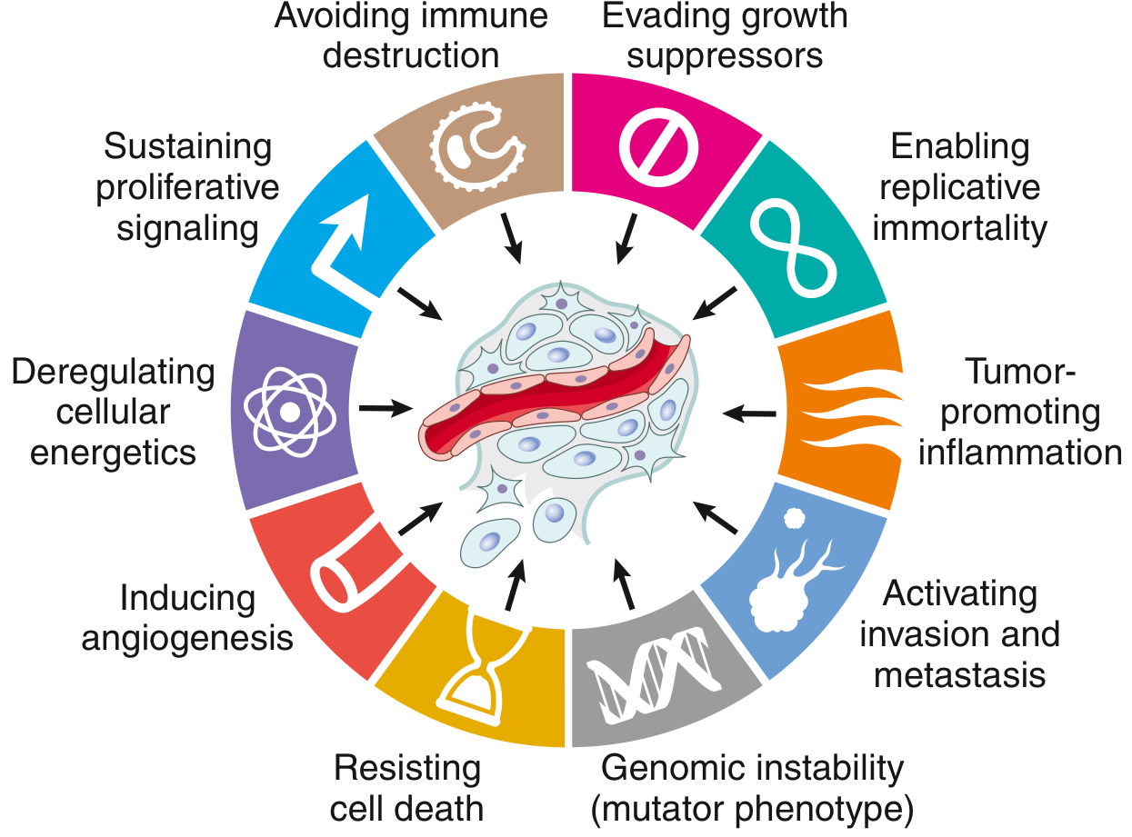
Fig. 7.20 / Fig. 6.15 - Eight hallmarks of cancer and two enabling characteristics. (Hanahan D, Weinberg RA: Cell 2011)
All cancers display eight fundamental changes in cell physiology known as the hallmarks of cancer. Their acquisition is accelerated by two enabling characteristics: genomic instability and tumor-promoting inflammation. (Robbins Pathologic Basis of Disease, p. 251-286)
1. Self-Sufficiency in Growth Signals (Oncogenes)
Normal cell proliferation requires binding of external growth factors. Cancer cells bypass this by acquiring gain-of-function mutations converting proto-oncogenes to oncogenes, encoding constitutively active oncoproteins.
Mechanisms:
- Growth factors (autocrine loops): Tumor cells secrete their own growth factors and co-express receptors (e.g., glioblastomas secrete PDGF and overexpress PDGF receptors; sarcomas secrete TGF-α and express EGF receptors).
- Growth factor receptors: Mutations cause constitutive signaling without ligand binding. Examples:
- EGF receptor - point mutations in lung cancer
- HER2/ERBB2 - gene amplification in breast cancer
- JAK2 - point mutations in myeloproliferative neoplasms
- Downstream signal transducers:
- RAS - most commonly mutated oncogene in human cancers; point mutations (especially codon 12) impair GTPase activity, causing constitutive RAS-GTP signaling through MAPK and PI3K/AKT pathways
- BCR-ABL fusion in CML - constitutive ABL tyrosine kinase activity
- BRAF and PI3K mutations in many cancers
- Transcription factors: MYC is a master transcription factor upregulated via chromosomal translocation (Burkitt lymphoma), gene amplification (neuroblastoma), or upstream signaling. MYC drives expression of cyclins, CDKs, and metabolic genes supporting rapid growth.
- Cyclins and CDKs: CDK4/cyclin D complex mutations accelerate G1-S progression.
2. Insensitivity to Growth-Inhibitory Signals (Tumor Suppressor Genes)
Tumor suppressors are negative regulators of growth; their loss-of-function leads to uncontrolled proliferation. They obey the "two-hit hypothesis" (Knudson) - both alleles must be inactivated.
RB Gene - "Governor of the Cell Cycle"
- RB protein (pRB) normally keeps cells in G1 by binding and inhibiting the E2F family of transcription factors.
- In response to mitogenic signals, CDK4-6/cyclin D complexes phosphorylate RB, releasing E2F and permitting S-phase entry.
- In cancer: loss of RB function (by mutation, deletion, or hyperphosphorylation) keeps E2F constitutively active.
- Inactivation by CDK4 amplification, cyclin D overexpression, or loss of CDK inhibitor p16/INK4a all effectively disable the RB pathway.
- Germline loss-of-function mutation → familial retinoblastoma.
TP53 - "Guardian of the Genome"
- p53 is activated by DNA damage, hypoxia, oncogene activation, telomere shortening - essentially any cellular stress.
- Activated p53 acts as a transcription factor inducing:
- p21 - CDK inhibitor causing G1 arrest (for DNA repair)
- GADD45 - DNA repair gene
- BAX - pro-apoptotic BCL2 family member (triggers apoptosis if damage irreparable)
- p53 is the most commonly mutated gene in human cancers (~50% of all cancers). In Li-Fraumeni syndrome, germline TP53 mutation confers increased risk of multiple tumors.
- MDM2 is the chief negative regulator of p53; MDM2 amplification (seen in many sarcomas) inactivates p53 without mutation.
Other Tumor Suppressors:
- CDKN2A (p16/INK4a): Inhibits CDK4/cyclin D; also encodes p14/ARF which stabilizes p53 by inhibiting MDM2. Mutated in melanoma, bladder cancer.
- TGF-β pathway: In normal cells, TGF-β activates SMAD proteins to arrest growth. In cancer, SMAD4 is frequently mutated (pancreatic cancer); TGF-β receptors are mutated in colon/endometrial cancers.
- E-cadherin (CDH1): Loss allows β-catenin release and activation of Wnt target genes driving proliferation; germline CDH1 mutations cause familial gastric carcinoma.
- NF2 (Merlin), VHL, APC, PTEN are other important tumor suppressor genes.
3. Altered Cellular Metabolism (Warburg Effect)
Even when oxygen is plentiful, cancer cells preferentially use aerobic glycolysis (glucose → lactate) rather than oxidative phosphorylation. This is the Warburg effect.
- Aerobic glycolysis is less efficient in ATP production but generates biosynthetic precursors (nucleotides, lipids, amino acids) needed for rapid cell growth.
- This is driven by oncogenic activation of HIF-1α (hypoxia-inducible factor), RAS, and MYC, which upregulate glucose transporters (GLUT1) and glycolytic enzymes.
- Autophagy - cancer cells under nutrient stress can activate autophagy to degrade organelles and macromolecules for energy. Autophagy may also help cancer cells survive initial growth in an avascular environment.
- Oncometabolism: Mutations in IDH1/IDH2 (in gliomas and AML) produce a neomorphic product, 2-hydroxyglutarate, which inhibits α-ketoglutarate-dependent enzymes, causing widespread epigenetic dysregulation.
4. Evasion of Apoptosis (Resisting Cell Death)
Tumor cells develop resistance to programmed cell death through:
Mechanisms of apoptosis evasion:
- Loss of TP53 function: p53 normally triggers apoptosis via BAX and PUMA; its loss in cancer prevents this. TP53 mutation frequency is even higher in relapsed tumors (after chemotherapy).
- BCL2 overexpression: BCL2 is an anti-apoptotic protein. Translocation t(14;18) in follicular lymphoma fuses BCL2 to the immunoglobulin heavy chain locus → BCL2 overexpression → cells resist apoptosis. These tumors are often indolent (low proliferation but low cell death). In CLL, loss of specific microRNAs leads to BCL2 upregulation.
- Overexpression of IAP (inhibitor of apoptosis) proteins prevents caspase activation.
- Apoptosis via the intrinsic (mitochondrial) pathway requires pro-apoptotic BH3-only proteins (PUMA, BIM, BAD) to overcome anti-apoptotic BCL2, BCLXL, and MCL1.
- Therapeutic targeting: Venetoclax (BCL2 inhibitor) is used in CLL; MDM2 inhibitors are in clinical trials.
5. Limitless Replicative Potential (Immortality)
Normal cells can divide only a finite number of times (Hayflick limit) due to progressive telomere shortening with each cell division. When telomeres become critically short, cells enter senescence or undergo crisis/apoptosis.
- Telomeres are TTAGGG repeats at chromosome ends that protect chromosomal integrity.
- Cancer cells bypass this limit by upregulating telomerase, the ribonucleoprotein enzyme that adds telomeric repeats to chromosome ends.
- Telomerase is expressed at high levels in ~90% of human cancers but is silent in most somatic cells.
- Without telomerase reactivation, cancer cells in crisis show chromosomal end-to-end fusions, breakage-fusion-bridge cycles, and massive genomic instability.
- In addition, cancer cells may have stem cell-like properties allowing indefinite self-renewal.
6. Sustained Angiogenesis
Like normal tissues, tumors require blood supply. Tumors >1-2 mm cannot grow without new blood vessel formation (neovascularization).
- The balance between pro-angiogenic and anti-angiogenic factors is called the angiogenic switch.
- VEGF (Vascular Endothelial Growth Factor) is the chief pro-angiogenic factor; its expression is upregulated by hypoxia (via HIF-1α), RAS, and TP53 loss.
- VEGF-A, FGF-2 (basic FGF): Released from tumor cells and from the ECM by proteolytic enzymes.
- Anti-angiogenic factors (thrombospondin-1, endostatin, angiostatin) are downregulated in cancers. TP53 normally upregulates thrombospondin-1; loss of p53 shifts the balance toward angiogenesis.
- Tumor vasculature is often abnormal - leaky, tortuous, and inefficient, contributing to hypoxia.
- Anti-VEGF therapy (bevacizumab) is used clinically in many solid tumors.
7. Ability to Invade and Metastasize
Invasion and metastasis are the most clinically dangerous properties of cancer and cause the majority of cancer deaths. The process involves several steps:
Invasion of the Extracellular Matrix (ECM)
Occurs in 4 steps:
- Loosening of cell-cell contacts: Loss of E-cadherin (by mutation, epigenetic silencing, or SNAIL/TWIST-mediated repression). E-cadherin maintains normal epithelial integrity; its loss promotes Epithelial-Mesenchymal Transition (EMT).
- Degradation of ECM: By matrix metalloproteinases (MMPs) and cathepsins secreted by tumor and stromal cells. Proteolysis also releases growth factors (IGF, TGF-β) and angiogenic fragments from ECM.
- Attachment to novel ECM components: Altered integrins allow cancer cells to bind type IV collagen and laminin.
- Migration: Directed by chemotactic factors, proteolytic fragments, and actin cytoskeleton reorganization.
Vascular Dissemination and Metastasis
- Tumor cells enter lymphatics or blood vessels (intravasation), survive as circulating tumor cells (CTCs), and extravasate at distant sites.
- Metastatic patterns are often predictable: lung, liver, brain, and bone are common sites.
- Organ tropism: Some tumors preferentially metastasize to specific organs due to matching chemokine receptors on tumor cells and their ligands on target endothelium (e.g., CXCR4 on tumor cells/CXCL12 on bone marrow endothelium).
- Epithelial-Mesenchymal Transition (EMT): Transcription factors TWIST and SNAIL drive EMT, enabling cells to acquire mesenchymal, migratory properties. At the metastatic site, reverse MET (mesenchymal-epithelial transition) occurs for colonization.
- Tumor dormancy: Metastatic cells may fail to grow at distant sites for years due to immune suppression or unfavorable microenvironment, then suddenly reactivate.
- Breast cancer metastatic to bone: cancer cells secrete PTHRP → osteoblasts make RANKL → osteoclast activation → bone resorption → release of IGF and TGF-β → cancer cell growth.
8. Evasion of Immune Surveillance
The immune system normally recognizes and eliminates cancer cells bearing abnormal antigens. Cancer cells develop multiple strategies to escape:
Tumor Antigens (targets of immune recognition)
- Tumor-specific antigens (TSAs): Arise from mutated proteins (neoantigens) unique to tumor cells - driver mutations or passenger mutations.
- Tumor-associated antigens (TAAs): Overexpressed normal proteins (e.g., HER2), cancer-testis antigens (MAGE family), and viral antigens (HPV E6/E7).
Antitumor Immune Effectors
- CD8+ cytotoxic T lymphocytes (CTLs): Most important; recognize tumor peptide-MHC I complexes and kill cancer cells.
- NK cells: Kill cells with low MHC I expression.
- Macrophages, antibody-dependent cellular cytotoxicity (ADCC).
Mechanisms of Immune Evasion
- Loss of MHC class I expression: Cancer cells downregulate MHC I or TAP (antigen processing), preventing CTL recognition.
- Lack of co-stimulatory molecules: Without CD80/86 (B7-1/B7-2), T cells become anergic rather than activated.
- Expression of immune checkpoint ligands: PD-L1 (programmed death ligand 1) on cancer cells binds PD-1 on T cells, inhibiting their cytotoxic activity. This is the basis of anti-PD-1/PD-L1 checkpoint immunotherapy (pembrolizumab, nivolumab, atezolizumab).
- CTLA-4 pathway: CTLA-4 on T cells competes with CD28 for B7 binding, suppressing T cell activation. Anti-CTLA-4 (ipilimumab) blocks this.
- Immunosuppressive tumor microenvironment: Tumor cells recruit regulatory T cells (Tregs) and M2 macrophages that suppress immune responses; secrete immunosuppressive cytokines (TGF-β, IL-10).
- FAS/FAS-L: Tumor cells may express FAS-L, inducing apoptosis in infiltrating T cells.
Enabling Characteristics
These two features accelerate the acquisition of hallmarks:
A. Genomic Instability
- Cancer cells acquire mutations at a faster rate than normal cells due to impaired DNA repair.
- Mismatch repair deficiency: Inactivation of MLH1, MSH2, MSH6, PMS2 → microsatellite instability (MSI-high); associated with Lynch syndrome (hereditary non-polyposis colorectal cancer - HNPCC) and sporadic colorectal, endometrial cancers. MSI-high tumors are highly responsive to PD-1 checkpoint inhibitors.
- Nucleotide excision repair: Deficiency causes xeroderma pigmentosum; UV-induced skin cancers.
- BRCA1/BRCA2 mutations: Impair homologous recombination repair; associated with familial breast and ovarian cancers. BRCA-deficient tumors are susceptible to PARP inhibitors.
- Chromosomal instability (CIN): Defects in mitotic spindle checkpoint → aneuploidy and chromosomal rearrangements.
- Epigenetic changes: DNA methylation (gene silencing), histone modifications, and chromatin remodeling also drive cancer without mutating DNA sequences.
- Noncoding RNAs: MicroRNAs can act as oncogenes (miR-17-92 cluster) or tumor suppressors (miR-15a, miR-16-1 in CLL); long noncoding RNAs (lncRNAs) also play roles.
B. Tumor-Promoting Inflammation
- Chronic inflammation promotes carcinogenesis by:
- Supplying bioactive molecules: growth factors, survival signals (EGF, TNF), pro-angiogenic factors (VEGF), ECM-remodeling enzymes (MMPs).
- Generating reactive oxygen species (ROS) that damage DNA.
- Providing chemokines and cytokines that attract immunosuppressive cells.
- Examples: H. pylori → gastric cancer; HCV → hepatocellular carcinoma; IBD → colorectal cancer.
Molecular Basis of Multistep Carcinogenesis
The acquisition of multiple hallmarks requires accumulation of multiple mutations over time - a process called multistep carcinogenesis. The classic example is colorectal carcinogenesis (Vogelstein model):
- Normal epithelium → adenoma (APC mutation) → intermediate adenoma (KRAS mutation) → late adenoma (SMAD4/TGF-β loss) → carcinoma (TP53 mutation) → metastasis.
Each step confers a selective growth advantage. Driver mutations confer hallmark properties; passenger mutations accumulate randomly but do not contribute to growth advantage.
Summary Table
| Hallmark | Key Genes/Mechanisms | Example |
|---|---|---|
| Self-sufficiency in growth | RAS, EGFR, HER2, MYC, BCR-ABL | CML (BCR-ABL), Lung ca (EGFR mut) |
| Insensitivity to growth inhibition | RB, TP53, CDKN2A, SMAD4, TGF-β | Retinoblastoma (RB), Li-Fraumeni (p53) |
| Altered metabolism | Warburg effect, HIF-1α, IDH1/2 | Aerobic glycolysis in all cancers |
| Evasion of apoptosis | BCL2, TP53, MDM2 | Follicular lymphoma (BCL2) |
| Limitless replication | Telomerase reactivation | ~90% of human cancers |
| Sustained angiogenesis | VEGF, FGF2, HIF-1α | Anti-VEGF (bevacizumab) |
| Invasion and metastasis | E-cadherin loss, MMPs, EMT (TWIST/SNAIL) | Most cancer deaths |
| Immune evasion | PD-L1, MHC I loss, Tregs | Anti-PD-1/CTLA-4 therapy |
| Enabling: Genomic instability | DNA repair (MLH1, BRCA1, TP53) | Lynch syndrome, HNPCC |
| Enabling: Tumor-promoting inflammation | ROS, VEGF, MMPs from immune cells | H. pylori → gastric cancer |
Source: Robbins & Kumar Basic Pathology (Robbins Pathology), Chapter 6; Robbins, Cotran & Kumar Pathologic Basis of Disease, Chapter 7, pp. 251-286
Answer for 7-10marks answer for Hallmarks of cancer based on Robins pathology testbook don't miss any important points
I already have all the content from the previous turn from Robbins Pathology. Here is the complete, exam-ready answer:
Hallmarks of Cancer
(Robbins Pathologic Basis of Disease / Robbins & Kumar Basic Pathology)
All cancers share eight fundamental changes in cell physiology - the hallmarks of cancer - plus two enabling characteristics that accelerate their acquisition.
INTRODUCTION
Cancer is not one disease but many, all sharing a profound dysregulation of growth. While hundreds of genes are mutated across different cancers, a conceptually satisfying framework is to consider the common biologic properties imparted by these diverse mutations. These are the hallmarks of cancer.
"It appears that all cancers display eight fundamental changes in cell physiology, which are considered the hallmarks of cancer." - Robbins Pathologic Basis of Disease, p. 268
HALLMARK 1 - Self-Sufficiency in Growth Signals
Normal cell proliferation requires external growth factor binding to receptors. Cancer cells bypass this by gain-of-function mutations converting proto-oncogenes to oncogenes, which encode constitutively active oncoproteins that promote growth even without external signals.
Steps of normal growth signaling (subverted in cancer):
- Growth factor binds its specific receptor
- Transient activation of receptor tyrosine kinase
- Signal transduction via second messengers (RAS, PI3K/AKT, MAPK)
- Activation of transcription factors (MYC)
- Cell cycle entry and division
Mechanisms of oncogene activation:
A. Growth Factors (Autocrine loops)
- Tumor cells secrete growth factors AND express their own receptors
- Glioblastomas secrete PDGF + overexpress PDGF receptors
- Sarcomas secrete TGF-α + express EGF receptors
B. Growth Factor Receptors
- EGFR - point mutations in lung cancer (targeted by gefitinib/erlotinib)
- HER2/ERBB2 - gene amplification in breast cancer (targeted by trastuzumab)
- JAK2 - point mutations in myeloproliferative neoplasms
- Mutant receptors signal constitutively without ligand
C. Downstream Signal Transducers
- RAS - most commonly mutated oncogene in human cancers (point mutations at codon 12 impair GTPase activity → constitutive RAS-GTP → MAPK + PI3K/AKT signaling)
- BCR-ABL fusion (t(9;22), Philadelphia chromosome) in CML - constitutive ABL tyrosine kinase activity (targeted by imatinib)
- BRAF V600E - melanoma, colorectal cancer
- PI3K mutations - many cancers
D. Transcription Factors
- MYC - master transcription factor; drives expression of cyclins, CDKs, metabolic genes
- Upregulated by: chromosomal translocation t(8;14) in Burkitt lymphoma, gene amplification in neuroblastoma, or upstream signaling (many cancers)
E. Cyclins and CDKs
- CDK4/cyclin D complex phosphorylates RB → cell cycle progression
- CDK4 amplification or cyclin D overexpression bypasses normal G1 checkpoint
HALLMARK 2 - Insensitivity to Growth-Inhibitory Signals (Tumor Suppressor Genes)
Tumor suppressors are negative regulators of cell proliferation. Their inactivation (loss-of-function) leads to unchecked growth. They follow the "two-hit hypothesis" (Knudson) - both alleles must be inactivated.
RB Gene - "Governor of the Cell Cycle"
- pRB is the key brake on cell cycle progression at the G1/S checkpoint
- Normally, unphosphorylated pRB binds and inhibits E2F transcription factors, blocking S-phase entry
- Mitogenic signals → CDK4-6/cyclin D complexes → phosphorylate (inactivate) pRB → E2F released → S-phase genes transcribed
- In cancer: loss of RB function (mutation, deletion) keeps E2F constitutively active
- Same effect achieved by: CDK4 amplification, cyclin D overexpression, loss of p16/INK4a
- Germline RB mutation → familial retinoblastoma (autosomal dominant)
- Sporadic retinoblastoma: both alleles lost by somatic mutation (two hits)
- RB pathway disruption found in virtually all human cancers
TP53 - "Guardian of the Genome"
- p53 is activated by: DNA damage, hypoxia, oncogene activation, telomere shortening - any cellular stress
- Activated p53 (transcription factor) induces:
- p21 (CDKN1A) - CDK inhibitor → G1/S arrest for DNA repair
- GADD45 - DNA repair protein
- BAX, PUMA - pro-apoptotic BCL2 family members → apoptosis if damage irreparable
- p53 is the most commonly mutated gene in human cancers (~50%)
- MDM2 is the chief negative regulator of p53 (ubiquitin E3 ligase that degrades p53); MDM2 amplification inactivates p53 without TP53 mutation (seen in many sarcomas)
- Germline TP53 mutation → Li-Fraumeni syndrome (sarcomas, breast cancer, brain tumors, adrenocortical carcinoma)
- TP53 mutations are even more common in relapsed/chemoresistant tumors (p53 also mediates drug-induced apoptosis)
Other Tumor Suppressors
-
CDKN2A (p16/INK4a + p14/ARF):
- p16 inhibits CDK4/cyclin D → keeps pRB active (growth arrest)
- p14/ARF stabilizes p53 by inhibiting MDM2
- Mutated in melanoma, bladder cancer, head and neck tumors, ALL
- Often silenced by promoter hypermethylation in cervical cancer
-
TGF-β / SMAD pathway:
- In normal cells, TGF-β → SMAD2/3 phosphorylation → SMAD4 complex → transcription of CDK inhibitors; repression of MYC, cyclins
- SMAD4 mutation common in pancreatic cancer
- TGF-β receptor mutations in colon, stomach, endometrial cancers
-
E-cadherin (CDH1): Loss allows β-catenin release, activating Wnt target genes; germline loss → familial gastric carcinoma
-
APC, PTEN, NF2 (Merlin), VHL - other important tumor suppressors
HALLMARK 3 - Altered Cellular Metabolism (Warburg Effect)
Even when oxygen is sufficient, cancer cells preferentially use aerobic glycolysis (glucose → lactate) rather than oxidative phosphorylation. This is the Warburg effect.
Why aerobic glycolysis?
- Less efficient in ATP production but generates biosynthetic intermediates (nucleotides, lipids, amino acids) needed for rapid cell growth
- Rapidly dividing cells need building blocks more than they need ATP
Drivers:
- HIF-1α (hypoxia inducible factor) upregulates GLUT1 glucose transporter and glycolytic enzymes
- RAS and MYC oncoproteins upregulate metabolic genes
- PET scanning exploits the Warburg effect (18F-FDG uptake)
Autophagy:
- Under nutrient stress, cancer cells can degrade organelles and macromolecules for energy and biosynthetic building blocks
- Autophagy may be protective early (nutrient stress in avascular tumor) but may also paradoxically support cancer cell survival
Oncometabolism:
- Mutations in IDH1/IDH2 genes (gliomas, AML) produce the neomorphic metabolite 2-hydroxyglutarate (2-HG)
- 2-HG inhibits α-ketoglutarate-dependent dioxygenases → widespread epigenetic dysregulation (DNA/histone hypermethylation)
- IDH inhibitors (ivosidenib, enasidenib) are FDA-approved for IDH-mutant AML
HALLMARK 4 - Evasion of Apoptosis (Resisting Cell Death)
Tumor cells develop resistance to programmed cell death via the intrinsic (mitochondrial) pathway of apoptosis.
Normal apoptosis pathway:
- Cellular stress → pro-apoptotic BH3-only proteins (PUMA, BIM, BAD) activated
- These inhibit anti-apoptotic BCL2/BCLXL/MCL1
- BAX/BAK oligomerize on mitochondria → cytochrome c release → APAF-1 activation → caspase cascade → cell death
Cancer mechanisms of evasion:
-
Loss of TP53 function - prevents upregulation of PUMA and BAX in response to DNA damage; most cancer cells with intact TP53 would undergo apoptosis when stressed
-
BCL2 overexpression:
- Translocation t(14;18) in follicular lymphoma fuses BCL2 to immunoglobulin heavy chain locus → BCL2 overexpression
- These tumors are indolent (low proliferation rate AND low cell death rate)
- In CLL, loss of miR-15a and miR-16-1 allows BCL2 upregulation
-
Overexpression of IAP (inhibitor of apoptosis) proteins - directly inhibit caspases
-
MDM2 amplification - degrades p53, preventing apoptosis induction
Therapeutic applications:
- Venetoclax (BCL2 inhibitor) - approved for CLL, AML
- MDM2 inhibitors (nutlins) - clinical trials for MDM2-amplified sarcomas
HALLMARK 5 - Limitless Replicative Potential (Immortality)
Normal cells can divide a finite number of times - the Hayflick limit (~50-70 divisions) due to progressive telomere shortening.
Telomere biology:
- Telomeres are repetitive DNA sequences (TTAGGG)n at chromosome ends, maintained by telomerase (a ribonucleoprotein reverse transcriptase)
- With each cell division, telomeres shorten (lagging strand problem)
- Critically short telomeres trigger senescence (p53/p21 pathway) or crisis (mitotic catastrophe, apoptosis)
Cancer mechanism:
- Cancer cells reactivate telomerase (TERT gene) - expressed at high levels in ~90% of human cancers but silent in most somatic cells
- This maintains telomere length and enables unlimited replication
- Without telomerase, cells entering crisis show chromosome end-to-end fusions → breakage-fusion-bridge cycles → massive genomic instability - which paradoxically generates further mutations
- Some cancers use ALT (Alternative Lengthening of Telomeres) - a recombination-based mechanism
Stem cell-like properties:
- Cancer stem cells have self-renewal capacity; they express stem cell transcription factors (OCT4, SOX2, NANOG) and are often therapy-resistant
HALLMARK 6 - Sustained Angiogenesis
Solid tumors cannot grow beyond 1-2 mm without a blood supply (limits of oxygen diffusion). Tumors induce new vessel formation (neovascularization).
The angiogenic switch:
- Normally, pro- and anti-angiogenic factors are in balance
- Tumors tip this balance toward angiogenesis
Pro-angiogenic factors (upregulated in cancer):
- VEGF-A - chief driver; binds VEGFR on endothelial cells → proliferation, migration, vessel formation
- FGF-2 (basic FGF) - released from ECM by MMP proteolysis
- PDGF - stabilizes new vessels
Anti-angiogenic factors (downregulated in cancer):
- Thrombospondin-1 - normally upregulated by p53; loss of p53 removes this brake
- Endostatin, angiostatin - fragments of larger ECM proteins
Drivers of VEGF upregulation:
- HIF-1α (activated by hypoxia, RAS, VHL loss) is the master inducer of VEGF
- VHL tumor suppressor loss (von Hippel-Lindau syndrome, clear cell renal carcinoma) prevents HIF-1α degradation
Tumor vasculature is structurally abnormal - leaky, tortuous, poorly organized.
Therapeutic targeting:
- Bevacizumab (anti-VEGF monoclonal antibody) - colorectal, lung, ovarian, glioblastoma
- Sorafenib, sunitinib (VEGFR tyrosine kinase inhibitors) - renal cell carcinoma, HCC
HALLMARK 7 - Ability to Invade and Metastasize
Metastases cause ~90% of cancer deaths and are the most clinically important hallmark.
Invasion of Extracellular Matrix (ECM) - 4 Steps:
Step 1 - Loosening of cell-cell contacts:
- Loss of E-cadherin by mutation, promoter methylation, or SNAIL/TWIST-mediated transcriptional repression
- Epithelial-Mesenchymal Transition (EMT): cancer cells lose epithelial markers (E-cadherin) and gain mesenchymal markers (vimentin, N-cadherin), acquiring invasive, migratory properties
Step 2 - Degradation of ECM:
- Matrix metalloproteinases (MMPs) and cathepsins secreted by tumor cells and stromal cells degrade basement membrane and interstitial matrix
- Proteolysis also releases ECM-sequestered growth factors (VEGF, TGF-β, IGF)
Step 3 - Attachment to novel ECM components:
- Changed integrin expression allows attachment to laminin and type IV collagen in degraded basement membrane
Step 4 - Migration:
- Actin cytoskeleton reorganization, directed by chemotactic signals and growth factors
Vascular Dissemination and Metastasis:
- Intravasation: Tumor cells penetrate blood/lymphatic vessels
- Survival in circulation: As circulating tumor cells (CTCs), often in clusters (circulating tumor microemboli) resistant to anoikis
- Extravasation: At distant site, CTCs adhere to endothelium and penetrate vessel wall
- Colonization: Most cells die; few survive and proliferate to form metastasis
Organ tropism:
- Colon → liver (portal drainage)
- Lung → liver, brain, adrenals, bone
- Prostate → bone (osteoblastic metastases)
- Breast → bone (PTHRP → RANKL → osteoclast activation → bone resorption → IGF/TGF-β release → cancer cell growth), liver, lung, brain
Chemokine receptor-ligand matching determines organ tropism (e.g., CXCR4 on tumor cells / CXCL12 on bone marrow endothelium)
Tumor dormancy: Metastatic cells can remain quiescent for years then reactivate - explains late relapses in breast cancer.
EMT transcription factors: TWIST, SNAIL, ZEB1 drive EMT; at metastatic site, MET (reverse EMT) occurs for colonization.
HALLMARK 8 - Evasion of Immune Surveillance
The immune system normally detects and destroys cells expressing abnormal antigens. Cancer cells develop multiple strategies to escape.
Tumor Antigens:
Tumor-Specific Antigens (TSAs / Neoantigens):
- Arise from mutated proteins (driver or passenger mutations)
- Unique to tumor cells; strongest immunogenic targets
- Basis of neoantigen-based vaccines and adoptive T cell therapy
Tumor-Associated Antigens (TAAs):
- Overexpressed normal proteins (HER2, EpCAM)
- Cancer-testis antigens (MAGE, SSX) - normally expressed only in testis/placenta, aberrantly expressed in cancer
- Viral antigens (HPV E6/E7 in cervical cancer; EBV proteins in Burkitt lymphoma)
- Oncofetal antigens (CEA, AFP)
Antitumor Immune Effectors:
- CD8+ CTLs - most important; recognize peptide-MHC I complexes and kill tumor cells via perforin/granzyme
- NK cells - kill cells with downregulated MHC I ("missing self")
- CD4+ Th1 cells - help CTL activation
- Macrophages (M1 phenotype) - phagocytosis and ADCC
Mechanisms of Immune Evasion:
-
Loss of MHC class I expression - downregulation of MHC I or TAP (transporter associated with antigen processing) prevents CTL recognition; also downregulates beta-2 microglobulin
-
Lack of co-stimulation - absence of CD80/CD86 (B7-1/B7-2) on tumor cells causes T cell anergy
-
PD-L1/PD-1 checkpoint:
- Tumor cells express PD-L1 which binds PD-1 on CTLs → T cell exhaustion/apoptosis
- Anti-PD-1 (pembrolizumab, nivolumab) and anti-PD-L1 (atezolizumab) antibodies restore anti-tumor immunity
- Effective in melanoma, lung cancer, renal cell carcinoma, MSI-high tumors
-
CTLA-4 checkpoint:
- CTLA-4 on T cells competes with CD28 for B7 ligands → inhibits T cell activation
- Ipilimumab (anti-CTLA-4) - used in melanoma
-
Immunosuppressive tumor microenvironment:
- Recruitment of Regulatory T cells (Tregs) - suppress CTL activity via IL-10, TGF-β
- M2-polarized macrophages (tumor-associated macrophages) - promote angiogenesis and immune suppression
- Secretion of immunosuppressive cytokines: TGF-β, IL-10, VEGF
-
FAS/FAS-L counterattack - tumor cells express FAS ligand, inducing apoptosis in infiltrating T cells ("tumor counterattack")
ENABLING CHARACTERISTICS
These do not qualify as hallmarks themselves but accelerate acquisition of all hallmarks:
A. Genomic Instability ("Mutator Phenotype")
Cancer cells acquire mutations faster than normal due to impaired DNA repair systems:
| Repair Pathway | Gene(s) | Cancer Association |
|---|---|---|
| Mismatch repair | MLH1, MSH2, MSH6, PMS2 | Lynch syndrome (HNPCC) - colon, endometrial; MSI-high tumors respond to anti-PD-1 |
| Nucleotide excision repair | XPA-XPG | Xeroderma pigmentosum → UV-induced skin cancers |
| Homologous recombination | BRCA1, BRCA2 | Familial breast and ovarian cancer; respond to PARP inhibitors (olaparib) |
| Base excision repair | MUTYH | Familial colorectal polyposis |
| Spindle checkpoint | Various | Chromosomal instability (CIN) → aneuploidy |
Chromosomal changes in cancer:
- Translocations (BCR-ABL, MYC fusions), deletions, amplifications, inversions
- Gene amplification manifests as double minutes or homogeneously staining regions (HSRs) on karyotype
Epigenetic changes:
- DNA methylation of CpG islands in promoters silences tumor suppressor genes (e.g., MLH1, CDKN2A, E-cadherin)
- Histone modifications (acetylation, methylation) by mutated HATs, HDACs, and KMTs alter gene expression
- IDH1/2 mutations produce 2-HG → inhibit TET demethylases and KDM histone demethylases → global hypermethylation (CpG island methylator phenotype, CIMP)
Noncoding RNAs:
- MicroRNAs (miRNAs) as oncogenes: miR-17-92 cluster (B-cell lymphomas)
- MiRNAs as tumor suppressors: miR-15a/miR-16-1 (inhibit BCL2 in CLL; deleted in CLL)
- Long noncoding RNAs (lncRNAs): HOTAIR, MALAT1 - regulate chromatin and gene expression in cancer
B. Tumor-Promoting Inflammation
Chronic inflammation creates a pro-tumorigenic microenvironment by supplying:
- Growth factors (EGF, FGF, HGF) promoting cell survival and proliferation
- Pro-angiogenic factors (VEGF) from inflammatory cells
- ECM-remodeling enzymes (MMPs, cathepsins) enabling invasion
- Reactive oxygen and nitrogen species causing mutagenic DNA damage
- Immunosuppressive mediators (IL-10, TGF-β, prostaglandins)
Examples:
- H. pylori chronic gastritis → gastric adenocarcinoma and MALT lymphoma
- Hepatitis C virus → chronic hepatitis → hepatocellular carcinoma
- Ulcerative colitis → colorectal cancer
- Asbestosis → mesothelioma
MOLECULAR BASIS OF MULTISTEP CARCINOGENESIS
Acquiring all hallmarks requires multiple mutations accumulating over years - multistep carcinogenesis. The Vogelstein model of colorectal carcinogenesis is the paradigm:
Normal epithelium
    → APC mutation (adenoma initiation)
    → KRAS mutation (early adenoma growth)
    → SMAD4/TGF-β loss (intermediate adenoma)
    → TP53 mutation (late adenoma/carcinoma)
    → Additional mutations (metastasis)
Each step provides a selective growth advantage (clonal expansion). This explains why most solid cancers arise in middle-to-old age.
SUMMARY TABLE
| Hallmark | Key Genes/Proteins | Important Examples |
|---|---|---|
| 1. Self-sufficiency in growth | RAS, EGFR, HER2, BCR-ABL, MYC, cyclin D | CML (BCR-ABL), lung ca (EGFR), Burkitt lymphoma (MYC) |
| 2. Insensitivity to growth inhibition | RB, TP53, p16/INK4a, SMAD4, CDH1 | Retinoblastoma (RB), Li-Fraumeni (p53), pancreatic ca (SMAD4) |
| 3. Altered metabolism | Warburg effect, HIF-1α, IDH1/2, MYC | Aerobic glycolysis; IDH mutations in glioma, AML |
| 4. Evasion of apoptosis | BCL2, TP53, MDM2, BAX | Follicular lymphoma t(14;18), CLL |
| 5. Limitless replication | Telomerase (TERT), ALT | ~90% of all cancers |
| 6. Sustained angiogenesis | VEGF, FGF2, HIF-1α, TSP-1 loss | Anti-VEGF (bevacizumab); VHL loss in renal ca |
| 7. Invasion/Metastasis | E-cadherin, MMPs, EMT (TWIST/SNAIL) | Most cancer deaths; bone met via PTHRP |
| 8. Immune evasion | PD-L1, MHC I, Tregs, TGF-β | Anti-PD-1 (pembrolizumab), anti-CTLA-4 (ipilimumab) |
| Enabling: Genomic instability | MLH1, BRCA1/2, TP53, TET, DNMT | Lynch syndrome, HNPCC, xeroderma pigmentosum |
| Enabling: Tumor-promoting inflammation | ROS, VEGF, MMPs, IL-10, TGF-β | H. pylori → gastric ca; HCV → HCC |
Source: Robbins & Kumar Basic Pathology (10th ed.), Chapter 6, pp. 198-220; Robbins, Cotran & Kumar Pathologic Basis of Disease (10th ed.), Chapter 7, pp. 251-290
That's way too much I need you to make the answer for 7marks
Here is a focused, exam-ready 7-mark answer:
Hallmarks of Cancer
(Robbins Pathology)
All cancers share 8 fundamental changes in cell physiology called the hallmarks of cancer, plus 2 enabling characteristics.
1. Self-Sufficiency in Growth Signals
Cancer cells acquire gain-of-function mutations converting proto-oncogenes to oncogenes, producing constitutively active oncoproteins that drive growth without external signals.
- Autocrine loops: Tumor cells secrete their own growth factors and co-express receptors (e.g., glioblastoma - PDGF)
- Mutant receptors: EGFR mutations (lung cancer), HER2 amplification (breast cancer)
- RAS mutations: Most common oncogene mutation; impairs GTPase activity → constitutive MAPK/PI3K signaling
- MYC: Master transcription factor; upregulated in Burkitt lymphoma (t(8;14)), neuroblastoma
- BCR-ABL fusion (Philadelphia chromosome, t(9;22)) in CML - targeted by imatinib
2. Insensitivity to Growth-Inhibitory Signals
Tumor suppressor genes are loss-of-function mutations; both alleles must be lost (Knudson's two-hit hypothesis).
RB - "Governor of the Cell Cycle"
- Unphosphorylated pRB binds E2F, blocking G1→S transition
- Growth signals → CDK4/cyclin D phosphorylates RB → E2F released → cell cycle entry
- Loss of RB (retinoblastoma), or equivalent loss via p16 deletion or CDK4 amplification, drives unchecked proliferation
TP53 - "Guardian of the Genome"
- Activated by DNA damage, hypoxia, oncogene stress
- Induces p21 (G1 arrest), GADD45 (repair), BAX/PUMA (apoptosis)
- Most commonly mutated gene in all cancers (~50%)
- MDM2 amplification inactivates p53 without mutation
- Germline loss → Li-Fraumeni syndrome
Other suppressors: SMAD4 (pancreatic ca), TGF-β receptors (colon ca), E-cadherin/CDH1 (gastric ca), p16/INK4a (melanoma)
3. Altered Cellular Metabolism (Warburg Effect)
Even in the presence of oxygen, cancer cells preferentially use aerobic glycolysis (glucose → lactate). This produces biosynthetic precursors (nucleotides, lipids) needed for rapid growth. Driven by HIF-1α, MYC, and RAS.
- IDH1/2 mutations (glioma, AML) produce 2-hydroxyglutarate → epigenetic dysregulation
- PET scanning exploits this (18F-FDG uptake)
4. Evasion of Apoptosis
Cancer cells resist apoptosis by:
- Loss of TP53 → no BAX/PUMA upregulation after DNA damage
- BCL2 overexpression: t(14;18) in follicular lymphoma → BCL2 fused to Ig heavy chain locus → anti-apoptotic protection → indolent tumor
- MDM2 amplification → p53 degradation
- Therapeutic targeting: venetoclax (BCL2 inhibitor) for CLL/AML
5. Limitless Replicative Potential (Immortality)
Normal cells have a finite Hayflick limit due to progressive telomere shortening with each division. When telomeres become critically short → senescence or apoptosis.
- Cancer cells reactivate telomerase (TERT) in ~90% of human cancers
- Telomerase adds TTAGGG repeats to chromosome ends, enabling unlimited division
- Without telomerase, cells enter crisis → chromosomal end-to-end fusions → genomic instability
6. Sustained Angiogenesis
Tumors cannot grow beyond 1-2 mm without blood supply. Tumors tip the balance toward pro-angiogenic factors:
- VEGF (chief driver) - upregulated by HIF-1α (hypoxia), RAS, loss of p53/thrombospondin-1
- FGF-2 released from ECM by MMPs
- Anti-angiogenic thrombospondin-1 is downregulated (normally induced by p53)
- Tumor vasculature is leaky and tortuous
- Targeted by bevacizumab (anti-VEGF)
7. Invasion and Metastasis
Causes the majority of cancer deaths. Occurs in 4 steps:
- Loss of E-cadherin → Epithelial-Mesenchymal Transition (EMT) driven by TWIST/SNAIL transcription factors
- Degradation of ECM by MMPs and cathepsins (also release sequestered growth factors)
- Altered integrin expression → attachment to novel ECM components
- Migration toward chemotactic signals
Vascular spread: Intravasation → survive as CTCs → extravasate → colonize.
Organ tropism by chemokine receptors (CXCR4/CXCL12 axis → bone).
Breast cancer bone metastasis: PTHRP → RANKL → osteoclast activation → bone resorption.
8. Evasion of Immune Surveillance
- Cancer cells express PD-L1 which binds PD-1 on CTLs → T cell exhaustion (targeted by pembrolizumab/nivolumab)
- Loss of MHC class I → CTLs cannot recognize tumor antigens
- Recruit Tregs and M2 macrophages → immunosuppressive microenvironment
- Secrete TGF-β and IL-10 to suppress immune responses
- CTLA-4 pathway exploited - targeted by ipilimumab
Enabling Characteristics
Genomic Instability - Defective DNA repair accelerates mutation accumulation:
- Mismatch repair loss (MLH1, MSH2) → Lynch syndrome, MSI-high tumors (respond to anti-PD-1)
- BRCA1/2 loss → familial breast/ovarian cancer (respond to PARP inhibitors)
Tumor-Promoting Inflammation - Chronic inflammation supplies growth factors, VEGF, MMPs, and ROS causing DNA damage (H. pylori → gastric cancer; HCV → HCC)
Multistep carcinogenesis - Vogelstein model: Normal → APC mutation → KRAS mutation → SMAD4 loss → TP53 loss → colorectal carcinoma. Each step confers selective growth advantage.
(Robbins & Kumar Basic Pathology, Ch. 6; Robbins Pathologic Basis of Disease, Ch. 7)
Chemical carcinogenesis etipathogenesis based on robins testbook
Finding Sources
Reading File
Reading File
Reading File
Reading File
Now let me get the detailed content from Robbins Pathologic Basis of Disease on the mechanism of chemical carcinogenesis:
Reading File
Reading File
Reading File
I now have all the content I need. Let me also fetch the initiation-promotion diagram:
Loading Image
I have all the content needed. Here is the complete answer:
Chemical Carcinogenesis - Etiopathogenesis
(Robbins Pathologic Basis of Disease)
Introduction
Chemical carcinogenesis is a multistep process involving sequential genetic alterations. The concept of initiation and promotion was first established in classic experiments on mouse skin. Chemical carcinogens are highly reactive electrophiles (electron-deficient atoms) that react with nucleophilic (electron-rich) atoms in the cell, particularly in DNA, causing mutations.
Types of Chemical Carcinogens
A. Direct-Acting Carcinogens
- Do not require metabolic conversion to become carcinogenic
- Most are weak carcinogens
- Important examples: alkylating agents used as chemotherapy (cyclophosphamide, chlorambucil, nitrosoureas)
- These drugs can cure or control cancers (leukemia, lymphoma) but tragically can also induce a second cancer - usually acute myeloid leukemia - years later
- Also include: β-propiolactone, dimethyl sulfate, diepoxybutane, acylating agents
B. Indirect-Acting Carcinogens (Procarcinogens)
- Require metabolic activation by endogenous enzymes to become carcinogenic
- The metabolically activated form is called the ultimate carcinogen
- Conversion is primarily by cytochrome P-450-dependent monooxygenases (CYP enzymes)
- Since CYP genes are polymorphic, susceptibility to chemical carcinogenesis varies between individuals - this has implications for cancer risk assessment
Major classes:
| Class | Examples | Associated Cancer |
|---|---|---|
| Polycyclic aromatic hydrocarbons | Benzo[a]pyrene (in cigarette smoke, soot, grilled meats), 3-methylcholanthrene | Lung, skin |
| Aromatic amines & azo dyes | 2-Naphthylamine (β-naphthylamine), benzidine, butter yellow | Bladder, liver |
| Natural products | Aflatoxin B1 (from Aspergillus mold on grains/nuts) | Hepatocellular carcinoma |
| Nitrosamines | From nitrite food preservatives + food amines | Gastric, esophageal |
| Others | Vinyl chloride, nickel, chromium, arsenic, polychlorinated biphenyls | Angiosarcoma (vinyl chloride), lung, skin |
Mechanism of Chemical Carcinogenesis
Step 1 - INITIATION
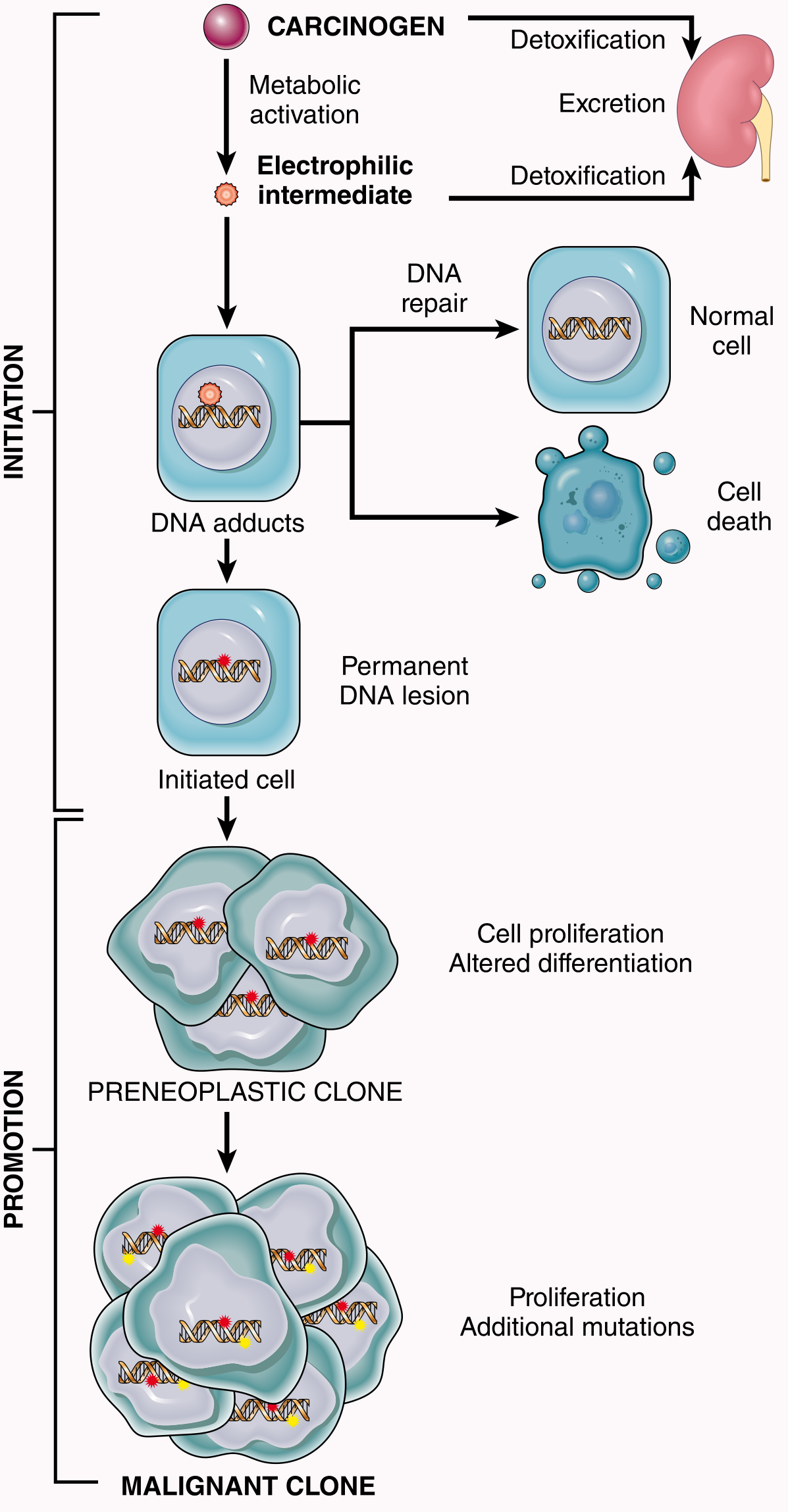
- Carcinogen (or its activated metabolite) reacts with DNA to form DNA adducts
- If DNA repair is successful → normal cell
- If DNA is not repaired before cell division → permanent heritable mutation → initiated cell
- Initiation is:
- Irreversible
- Permanent
- Requires the cell to divide (to "fix" the mutation)
- A single exposure may be sufficient
- Initiators have no threshold dose - any dose can theoretically initiate a cell
- Initiated cells appear morphologically normal but carry mutations
Step 2 - PROMOTION
- Tumor promoters stimulate proliferation of initiated (mutated) cells
- Promoters are not mutagenic by themselves - they cannot cause cancer without prior initiation
- They cause clonal expansion of the initiated cell into a preneoplastic clone
- This increased proliferation allows accumulation of additional mutations → eventual malignant transformation
- Promotion is:
- Reversible (at least in early stages)
- Dose-dependent - requires prolonged, repeated exposure
- Has a threshold dose
Examples of tumor promoters:
- Phorbol esters (TPA/12-O-tetradecanoylphorbol-13-acetate) - classic experimental promoter on mouse skin
- Unopposed estrogen - promotes endometrial and breast cancer
- Chronic inflammation with tissue repair - inflammatory bowel disease, chronic hepatitis, Barrett esophagus, chronic lung inflammation
Molecular Targets of Chemical Carcinogens
- Most initiating agents target DNA directly and are mutagenic
- Mutations occur throughout the genome; cells that sustain damage to oncogenes (RAS) or tumor suppressor genes (TP53) gain selective advantage
- Some carcinogens interact preferentially with specific DNA sequences producing hotspot mutations:
Classic example - Aflatoxin B1:
- Produced by Aspergillus flavus mold growing on improperly stored grains and nuts
- Strong correlation with hepatocellular carcinoma incidence in parts of Africa and Asia
- Aflatoxin B1-associated HCCs characteristically carry a G:C → T:A transversion at codon 249 of TP53 (Arg → Ser substitution), impairing p53 function
- This specific mutation is rare in HCCs from areas without aflatoxin contamination - a perfect "molecular fingerprint" of exposure
Cigarette smoke and lung cancer:
- Lung cancers in smokers have a 10-fold higher mutational burden than in non-smokers
- The excess mutations are strongly skewed toward base substitutions caused by carcinogens in cigarette smoke (benzo[a]pyrene metabolites) - the "smoking gun" in molecular epidemiology
- Cancer genome sequencing has revealed dozens of mutational signatures, each reflecting specific carcinogen exposures or repair defects
Important Occupational and Environmental Chemical Carcinogens
| Carcinogen | Source/Occupation | Cancer |
|---|---|---|
| Benzo[a]pyrene | Cigarette smoke, soot, chimney sweeping (Potts' original observation) | Lung, scrotal skin |
| β-Naphthylamine | Aniline dye industry, rubber industry | Bladder carcinoma |
| Benzidine | Rubber/dye industry | Bladder carcinoma |
| Aflatoxin B1 | Contaminated grain/nuts (Aspergillus) | Hepatocellular carcinoma |
| Vinyl chloride | Plastics industry | Hepatic angiosarcoma |
| Arsenic | Pesticides, smelting | Skin, lung, liver |
| Nitrosamines | Processed/cured meats, tobacco | Gastric, esophageal, nasopharyngeal |
| Alkylating agents (chemotherapy) | Cancer treatment | Secondary AML |
| Asbestos | Construction, shipbuilding | Mesothelioma, lung |
Key Concepts (Summary)
- Chemical carcinogens are electrophilic → attack nucleophilic sites in DNA → mutations
- Direct-acting agents: Active as-is (e.g., alkylating agents)
- Indirect-acting agents (procarcinogens): Require metabolic activation by CYP enzymes → ultimate carcinogen (e.g., benzo[a]pyrene, aflatoxin, aromatic amines)
- CYP polymorphisms determine individual susceptibility
- Initiation: Permanent, irreversible DNA mutation; single exposure possible
- Promotion: Clonal expansion of mutated cells; reversible, requires repeated exposure; not mutagenic alone
- Progression: Accumulation of additional mutations → full malignant phenotype
- Specific carcinogens leave mutational signatures in cancer genomes (e.g., aflatoxin → TP53 codon 249; smoking → C→A transversions in lung cancer)
(Robbins, Cotran & Kumar Pathologic Basis of Disease, 10th ed., Ch. 7, pp. 287-305)
Chemical carcinogenesis etipathogenesis based on robins testbook
The full answer is already in the previous response. Here it is reformatted cleanly for a 7-mark exam answer:
Chemical Carcinogenesis - Etiopathogenesis
(Robbins Pathologic Basis of Disease)
Definition & Introduction
Chemical carcinogenesis is a multistep process in which chemical agents cause permanent DNA mutations leading to cancer. All initiating chemical carcinogens are highly reactive electrophiles (electron-deficient) that react with nucleophilic (electron-rich) sites in DNA, producing mutations.
The concept of initiation and promotion was first established in classic mouse skin experiments.
Types of Chemical Carcinogens
1. Direct-Acting Carcinogens
- Do not require metabolic activation
- Generally weak carcinogens
- Examples: alkylating agents (cyclophosphamide, chlorambucil, nitrosoureas), β-propiolactone, dimethyl sulfate
- Important note: anticancer alkylating drugs can cure lymphoma/leukemia but may later induce secondary AML
2. Indirect-Acting Carcinogens (Procarcinogens)
- Require metabolic activation by cytochrome P-450 monooxygenases to form the ultimate carcinogen
- Since CYP genes are polymorphic, individual susceptibility to chemical carcinogenesis varies
| Class | Examples | Cancer Caused |
|---|---|---|
| Polycyclic aromatic hydrocarbons | Benzo[a]pyrene (cigarette smoke, soot, grilled meats) | Lung, skin |
| Aromatic amines & azo dyes | β-Naphthylamine, benzidine, butter yellow | Bladder, liver |
| Natural microbial products | Aflatoxin B1 (Aspergillus on grains/nuts) | Hepatocellular carcinoma |
| Nitrosamines | Nitrite food preservatives + food amines | Gastric, esophageal |
| Others | Vinyl chloride, arsenic, nickel, chromium | Angiosarcoma, lung |
Mechanism of Chemical Carcinogenesis
STAGE 1 - INITIATION
- Carcinogen (or activated metabolite) reacts with DNA → forms DNA adducts
- If not repaired before cell division → permanent heritable mutation → initiated cell
- Initiated cells appear morphologically normal but carry mutations
- Initiation is:
- Irreversible and permanent
- Requires cell division to fix the mutation
- Has no threshold dose
- A single sufficient exposure can initiate a cell
STAGE 2 - PROMOTION
- Tumor promoters stimulate proliferation of initiated cells
- Cause clonal expansion → preneoplastic clone
- Increased proliferation allows accumulation of additional mutations → malignant transformation
- Promotion is:
- Not mutagenic by itself (cannot cause cancer without prior initiation)
- Reversible (especially early)
- Dose-dependent, requires prolonged/repeated exposure
- Has a threshold dose
Examples of promoters:
- Phorbol esters (TPA) - classic experimental promoter
- Unopposed estrogen - endometrium and breast
- Chronic inflammation with tissue repair - IBD, chronic hepatitis, Barrett esophagus
STAGE 3 - PROGRESSION
- Further mutations accumulate in the proliferating clone
- Each mutation gives a selective growth advantage
- Eventually a cell with all hallmarks of malignancy emerges
Molecular Targets
- Initiators mutate oncogenes (RAS) and tumor suppressor genes (TP53) preferentially (selective advantage)
- Some carcinogens produce characteristic hotspot mutations - mutational signatures
Aflatoxin B1 (classic example):
- From Aspergillus flavus on improperly stored grains/nuts
- Strongly associated with HCC in Africa and Asia
- Produces a specific G:C → T:A transversion at codon 249 of TP53 (Arg → Ser)
- This mutation is rarely seen in HCC from aflatoxin-free areas - a molecular "fingerprint"
Cigarette smoke:
- Lung cancers in smokers have 10-fold higher mutational burden than non-smokers
- Mutations are skewed toward base substitutions caused by benzo[a]pyrene metabolites
- Cancer genome sequencing has revealed dozens of mutational signatures linked to specific carcinogens
Key Occupational Carcinogens
| Carcinogen | Occupation/Source | Cancer |
|---|---|---|
| Benzo[a]pyrene | Chimney sweeping (Potts' 1775 observation), cigarettes | Scrotal skin, lung |
| β-Naphthylamine | Aniline dye/rubber industry | Bladder |
| Vinyl chloride | Plastics industry | Hepatic angiosarcoma |
| Asbestos | Construction, shipbuilding | Mesothelioma |
| Arsenic | Pesticides, smelting | Skin, lung, liver |
Summary
Procarcinogen
     ↓  (CYP450 metabolic activation)
Ultimate Carcinogen (electrophile)
     ↓  (reacts with DNA)
DNA Adducts
     ↓  (if not repaired before cell division)
INITIATION → Permanent mutation (oncogenes/TSGs)
     ↓  (+ tumor promoters → clonal expansion)
PROMOTION → Preneoplastic clone
     ↓  (+ additional mutations)
PROGRESSION → MALIGNANT TUMOR
(Robbins, Cotran & Kumar Pathologic Basis of Disease, 10th ed., Ch. 7, pp. 287-304)
Viral carcinogenesis etipathogenesis and examples
Finding Sources
Finding Sources
Finding Sources
Finding Sources
Reading File
The microbial carcinogenesis section starts at line 67. Let me read from there:
Reading File
Reading File
The content I need (HPV, EBV, HBV, H. pylori) was captured in the first large read. Let me also check the Robbins Basic Pathology section on viral oncogenesis:
Finding Sources
I have all the content I need from the prior reads. Let me now write the complete answer:
Viral Carcinogenesis - Etiopathogenesis
(Robbins, Cotran & Kumar Pathologic Basis of Disease)
Introduction
Many RNA and DNA viruses are oncogenic in animals, but only a few viruses are firmly linked to human cancer. A common theme in viral carcinogenesis is that the infection triggers cell proliferation - initially polyclonal - but with time it becomes monoclonal due to acquisition of driver mutations in the rapidly dividing infected cells.
Viruses implicated in human cancer:
- RNA viruses: HTLV-1
- DNA viruses: HPV, EBV, HBV, Merkel cell polyomavirus, HHV-8
- Non-DNA virus: HCV (RNA virus, linked to HCC)
- Bacterium: H. pylori (discussed separately)
A. ONCOGENIC RNA VIRUSES
Human T-Cell Leukemia Virus Type 1 (HTLV-1)
- The only human retrovirus firmly implicated in human cancer
- Causes Adult T-Cell Leukemia/Lymphoma (ATLL)
- Endemic in: Japan, Caribbean basin, South America, Africa
- ~15-20 million people worldwide are infected
- Tropism for CD4+ T cells (same as HIV)
- Transmission: sexual intercourse, blood products, breastfeeding
- Only 3-5% of infected individuals develop leukemia
- Long latent period: 40-60 years after infection
Mechanism of transformation:
- HTLV-1 genome contains tax and HBZ genes (in addition to gag, pol, env)
- Tax protein: Stimulates transcription of viral RNA; alters host cell gene transcription and signaling pathways promoting growth and survival
- HBZ: A transcription factor that contributes to malignant transformation
- Tax and HBZ together: induce genomic instability, inhibit senescence, activate proliferative signaling
- HTLV-1 does not contain a recognizable oncogene and does not integrate next to a proto-oncogene
- In leukemic cells, proviral integration sites are clonal (identical in every cell within one tumor) - proving infection preceded transformation
- The mechanism is inefficient, explaining the long latency and low penetrance
B. ONCOGENIC DNA VIRUSES
1. Human Papillomavirus (HPV)
- >70 genetically distinct HPV types identified
- Low-risk types (6, 11): Cause benign squamous papillomas (warts) - no malignant potential
- High-risk types (16, 18, 31, 33): Strongly associated with malignancy
Cancers caused:
- Cervical carcinoma (most important - HPV 16 and 18 account for ~70% of cases)
- Oropharyngeal, anal, vulvar, vaginal, penile carcinomas
Mechanism of HPV-induced carcinogenesis:
HPV life cycle:
- HPV infects basal epithelial cells of the cervix (squamocolumnar junction)
- In benign lesions (condylomas): HPV exists as a circular episome (extrachromosomal)
- In malignant lesions: HPV DNA is integrated into the host genome - this is the critical step
Two key oncoproteins - E6 and E7:
| Oncoprotein | Target | Effect |
|---|---|---|
| E7 | pRB (retinoblastoma protein) | Binds and inactivates pRB → releases E2F transcription factors → uncontrolled cell cycle entry (S-phase) |
| E6 | p53 | Binds p53 and promotes its ubiquitin-mediated degradation → loss of G1 arrest and apoptosis |
| E6 | Telomerase (TERT) | Activates telomerase → immortalization |
- When HPV integrates into the host genome, the E2 gene is disrupted - E2 normally suppresses E6/E7; its disruption leads to overexpression of E6 and E7
- Result: inactivation of both RB and p53 + telomerase activation → hallmarks of cancer
Key point: HPV infection alone is not sufficient for full malignant transformation - other cofactors (smoking, immunosuppression, coinfection) and additional mutations are needed. This explains why only a minority of HPV-infected women develop cervical cancer.
Cervical cancer prevention:
- HPV vaccines (Gardasil 9 - covers HPV 6, 11, 16, 18, 31, 33, 45, 52, 58) prevent infection with high-risk types
- Pap smear screening detects premalignant cervical intraepithelial neoplasia (CIN)
2. Epstein-Barr Virus (EBV)
- A herpesvirus; infects B lymphocytes via CD21 (complement receptor)
- In immunocompetent hosts, primary infection causes infectious mononucleosis (self-limited)
- In susceptible individuals, EBV drives B-cell immortalization
Cancers associated:
| Cancer | Mechanism |
|---|---|
| Burkitt lymphoma | Endemic form in Africa; t(8;14) MYC translocation in EBV-driven B cells |
| Hodgkin lymphoma (EBV+) | EBV in Reed-Sternberg cells |
| B-cell lymphomas in immunocompromised (HIV, post-transplant PTLD) | EBV drives polyclonal B-cell proliferation → malignant transformation |
| Nasopharyngeal carcinoma | EBV in all tumor cells; endemic in southern China |
| Gastric carcinoma (~10%) | EBV in epithelial cells |
| Smooth muscle tumors (rare, in immunocompromised) |
Mechanism:
-
EBV infects B cells via gp350 binding to CD21
-
EBV genome persists as circular episome in latently infected cells
-
Key viral proteins in latency:
- LMP-1 (Latent Membrane Protein 1): Acts as a constitutively active CD40 receptor mimic → activates NF-κB and JAK/STAT pathways → B-cell survival, proliferation
- LMP-2A: Mimics BCR (B-cell receptor) signaling → survival signals
- EBNAs (EBV Nuclear Antigens): Maintain viral genome; EBNA-2 activates MYC and cyclin D
- EBER RNAs (non-coding): Inhibit interferon responses and apoptosis
-
EBV-infected B cells are normally kept in check by CD8+ cytotoxic T cells
-
In immunocompromised patients (HIV/AIDS, post-transplant), loss of T-cell surveillance allows uncontrolled EBV-driven B-cell proliferation → Post-Transplant Lymphoproliferative Disorder (PTLD) or AIDS-related lymphoma
In Burkitt lymphoma:
- EBV drives polyclonal B-cell proliferation; rapidly dividing cells acquire t(8;14) MYC translocation → MYC oncogene activation → monoclonal malignant lymphoma
3. Hepatitis B Virus (HBV) and Hepatitis C Virus (HCV)
- 70-85% of hepatocellular carcinomas (HCC) worldwide are associated with HBV or HCV infection
Mechanisms:
HBV (DNA virus):
- Causes chronic hepatitis → cirrhosis → HCC
- HBV DNA can integrate into the host genome (though integration sites are not consistent near oncogenes)
- HBV encodes HBx protein - a transcriptional activator that activates various growth-promoting pathways (Ras-MAPK, NF-κB)
- HBx also inhibits p53 function
- The major mechanism: chronic hepatocyte injury → regenerative proliferation → increased DNA replication errors → accumulation of mutations in rapidly dividing cells
HCV (RNA virus):
- HCV does not integrate into the host genome
- Causes carcinogenesis primarily through chronic hepatitis → cirrhosis → regeneration cycle (same mechanism as HBV)
- HCV core protein may also directly activate growth signaling pathways
Common theme for HBV and HCV: Chronic inflammation → repeated cycles of hepatocyte death and regeneration → accumulation of mutations → HCC (often after 20-30 years of chronic infection)
4. Human Herpesvirus 8 (HHV-8) / Kaposi Sarcoma Herpesvirus (KSHV)
- Associated with Kaposi sarcoma (vascular neoplasm) - particularly in HIV/AIDS patients and elderly men of Mediterranean/African origin
- Also associated with Primary Effusion Lymphoma and Multicentric Castleman Disease
- HHV-8 encodes viral homologs of human growth-promoting genes: v-cyclin D, v-FLIP (inhibits apoptosis), v-IL-6
5. Merkel Cell Polyomavirus (MCPyV)
- Identified in Merkel cell carcinoma (aggressive skin cancer)
- MCPyV T-antigen (large T) inactivates RB and contributes to tumor development
C. BACTERIAL CARCINOGENESIS: Helicobacter pylori
Although not a virus, H. pylori is the most important microbial carcinogen:
Associated cancers:
- Gastric adenocarcinoma (most important)
- Gastric MALT lymphoma (extranodal marginal zone B-cell lymphoma)
Mechanism for gastric adenocarcinoma:
- H. pylori colonizes gastric mucosa → chronic active gastritis
- Repeated cycles of mucosal damage and regeneration
- CagA protein (virulence factor) is injected into gastric epithelial cells via a type IV secretion system → activates RAS-MAPK, PI3K/AKT, and NF-κB pathways → proliferation
- Chronic inflammation → intestinal metaplasia → dysplasia → carcinoma
- ROS from inflammatory cells cause DNA mutations
Mechanism for MALT lymphoma:
- H. pylori infection → chronic gastric inflammation → polyclonal B-cell proliferation (reactive)
- Continued antigen stimulation → B cells acquire driver mutations → monoclonal MALT lymphoma
- Early MALT lymphomas often regress after H. pylori eradication with antibiotics (proving that continued antigen drive is needed)
Summary Table
| Virus/Agent | Cancer | Key Mechanism |
|---|---|---|
| HTLV-1 (RNA) | Adult T-cell leukemia/lymphoma | Tax/HBZ proteins → T-cell proliferation, genomic instability |
| HPV 16, 18 (DNA) | Cervical, oropharyngeal carcinoma | E6 degrades p53; E7 inactivates RB; E6 activates telomerase |
| EBV (DNA) | Burkitt lymphoma, Hodgkin lymphoma, nasopharyngeal ca, PTLD | LMP-1 mimics CD40; EBNA-2 activates MYC; immune evasion |
| HBV (DNA) | Hepatocellular carcinoma | HBx activates growth pathways; chronic inflammation → regeneration |
| HCV (RNA) | Hepatocellular carcinoma | Chronic inflammation → cirrhosis → regeneration → mutations |
| HHV-8/KSHV (DNA) | Kaposi sarcoma, PEL | Viral cyclins; v-FLIP; v-IL-6 |
| MCPyV (DNA) | Merkel cell carcinoma | Large T antigen inactivates RB |
| H. pylori | Gastric adenocarcinoma, MALT lymphoma | CagA → cell proliferation; chronic inflammation → mutations |
Key Principles of Viral Carcinogenesis
- Viral infection alone is usually not sufficient for cancer - additional mutations and cofactors are needed
- The common theme is viral-driven cell proliferation (polyclonal) followed by accumulation of somatic mutations → monoclonal malignancy
- Oncogenic DNA viruses typically encode proteins that inactivate RB and p53 (the two master tumor suppressors)
- Integration of viral DNA into the host genome is often a critical step (HPV, HBV)
- Immunosuppression (HIV, transplant) dramatically increases the risk of EBV-driven lymphomas
- Vaccines against oncogenic viruses (HPV, HBV) are the most powerful cancer prevention tools available
(Robbins, Cotran & Kumar Pathologic Basis of Disease, 10th ed., Ch. 7, pp. 305-315)
I need a 7 marks answer
All the content is already in the previous response. Here it is trimmed to a clean 7-mark answer:
Viral Carcinogenesis - Etiopathogenesis
(Robbins Pathology)
Introduction
Only a few viruses are firmly linked to human cancer. A common theme is that infection triggers cell proliferation - initially polyclonal - which over time becomes monoclonal as rapidly dividing infected cells accumulate driver mutations.
A. ONCOGENIC RNA VIRUS
HTLV-1 → Adult T-Cell Leukemia/Lymphoma (ATLL)
- The only human retrovirus causing human cancer
- Tropism for CD4+ T cells
- Endemic in Japan, Caribbean, South America
- Only 3-5% of infected individuals develop ATLL after a 40-60 year latent period
- Transmission: sexual intercourse, blood products, breastfeeding
Mechanism:
- Genome contains tax and HBZ genes
- Tax protein stimulates viral transcription and alters host cell signaling → drives T-cell proliferation, inhibits senescence, causes genomic instability
- HTLV-1 does not carry its own oncogene; proviral integration sites are clonal in tumor cells (proving infection preceded transformation)
B. ONCOGENIC DNA VIRUSES
1. HPV (Human Papillomavirus) → Cervical & Oropharyngeal Carcinoma
- >70 types identified
- Low-risk types (6, 11): Benign warts only
- High-risk types (16, 18): Account for ~70% of cervical carcinomas; also anal, vulvar, penile, oropharyngeal cancers
Mechanism:
- In benign lesions: HPV exists as episome (extrachromosomal)
- In malignancy: HPV integrates into host genome → disrupts E2 gene → E2 normally suppresses E6/E7 → loss of E2 → overexpression of E6 and E7
| Oncoprotein | Target | Effect |
|---|---|---|
| E7 | pRB | Inactivates RB → releases E2F → uncontrolled S-phase entry |
| E6 | p53 | Promotes p53 ubiquitin-mediated degradation → no G1 arrest, no apoptosis |
| E6 | Telomerase (TERT) | Activates telomerase → immortalization |
- HPV alone is insufficient - cofactors and additional mutations needed for full malignancy
- Prevention: HPV vaccine (Gardasil 9) + Pap smear screening
2. EBV (Epstein-Barr Virus) → Multiple Cancers
- Infects B lymphocytes via CD21 receptor
- Cancers caused:
- Burkitt lymphoma (endemic, Africa)
- Hodgkin lymphoma
- Nasopharyngeal carcinoma (endemic in southern China)
- B-cell lymphomas in immunocompromised (HIV/AIDS, post-transplant PTLD)
- ~10% of gastric carcinomas
Mechanism:
- EBV genome persists as circular episome in latently infected B cells
- LMP-1 (Latent Membrane Protein-1): Mimics constitutively active CD40 receptor → activates NF-κB and JAK/STAT → B-cell survival and proliferation
- LMP-2A: Mimics BCR signaling → survival signals
- EBNA-2: Activates MYC and cyclin D expression
- EBER RNAs: Non-coding; inhibit interferon responses and apoptosis
- Normally, CD8+ CTLs keep EBV-infected B cells in check
- In immunosuppression (HIV, transplant): loss of T-cell surveillance → uncontrolled EBV-driven B-cell proliferation → lymphoma (PTLD)
- In Burkitt lymphoma: EBV-driven proliferation creates conditions for t(8;14) MYC translocation → monoclonal malignancy
3. HBV and HCV → Hepatocellular Carcinoma (HCC)
- HBV + HCV together account for 70-85% of HCC worldwide
HBV mechanism:
- HBx protein: Transcriptional activator; activates RAS-MAPK, NF-κB; inhibits p53
- HBV DNA can integrate into host genome (though not consistently near oncogenes)
- Main mechanism: Chronic hepatocyte injury → repeated regeneration cycles → DNA replication errors → mutation accumulation → HCC
HCV mechanism:
- HCV does not integrate into host genome
- Causes HCC primarily via chronic inflammation → cirrhosis → regeneration cycle
- HCV core protein may directly activate growth signaling
4. HHV-8 (Kaposi Sarcoma Herpesvirus) → Kaposi Sarcoma
- Causes Kaposi sarcoma (especially in HIV/AIDS patients)
- Also: Primary Effusion Lymphoma, Multicentric Castleman Disease
- Encodes viral homologs: v-cyclin D (drives proliferation), v-FLIP (blocks apoptosis), v-IL-6
C. BACTERIAL CARCINOGENESIS: H. pylori
Cancers: Gastric adenocarcinoma + Gastric MALT lymphoma
Mechanism:
- CagA protein injected into gastric epithelial cells → activates RAS-MAPK, PI3K/AKT, NF-κB → proliferation
- Chronic gastritis → intestinal metaplasia → dysplasia → carcinoma
- Inflammatory ROS cause direct DNA mutations
MALT lymphoma: Chronic H. pylori antigen stimulation → polyclonal B-cell proliferation → mutation accumulation → monoclonal MALT lymphoma. Early MALT lymphoma regresses with H. pylori eradication.
Summary Table
| Virus | Cancer | Key Mechanism |
|---|---|---|
| HTLV-1 | ATLL | Tax/HBZ → T-cell proliferation, genomic instability |
| HPV 16/18 | Cervical, oropharyngeal ca | E6 destroys p53; E7 inactivates RB; E6 activates telomerase |
| EBV | Burkitt lymphoma, nasopharyngeal ca, PTLD | LMP-1 mimics CD40; EBNA-2 activates MYC |
| HBV | HCC | HBx protein; chronic regeneration |
| HCV | HCC | Chronic inflammation → cirrhosis → regeneration |
| HHV-8 | Kaposi sarcoma | v-cyclin D, v-FLIP |
| H. pylori | Gastric ca, MALT lymphoma | CagA; chronic inflammation |
Key Principles
- Viral infection alone is not sufficient - additional mutations and cofactors are needed
- Oncogenic DNA viruses typically encode proteins that inactivate RB and p53
- Integration of viral DNA is a critical step for HPV and HBV
- Immunosuppression dramatically increases EBV-driven lymphoma risk
- Vaccines against HPV and HBV are the most powerful cancer prevention tools available
(Robbins, Cotran & Kumar Pathologic Basis of Disease, 10th ed., Ch. 7)
This is a shared conversation. Sign in to Orris to start your own chat.