
Fetal circulation 10 marks pg obgy exam
Reading File
Fetal circulation is a common 10-mark exam topic in OB/GYN. It covers the unique pathways, shunts, and changes at birth. Let me pull the detailed content from the medical library.
Searching sources...
Finding Sources
Searching Images
fetal circulation diagram shunts ductus arteriosus foramen ovale
Reading File
Reading File
Reading File
Reading File
Reading File
Loading Image
I now have all the content needed for a detailed 10-mark answer. Let me also fetch the neonatal circulation diagram for the "changes at birth" section.
Loading Image
Here is a complete, exam-ready 10-mark answer on fetal circulation:
Fetal Circulation
Introduction
Fetal circulation is specially adapted to serve prenatal needs - the lungs do not provide gas exchange in utero, so the fetus relies entirely on the placenta for oxygenation and nutrition. Three unique shunts allow blood to bypass the liver and lungs: the ductus venosus, the foramen ovale, and the ductus arteriosus.
Diagram
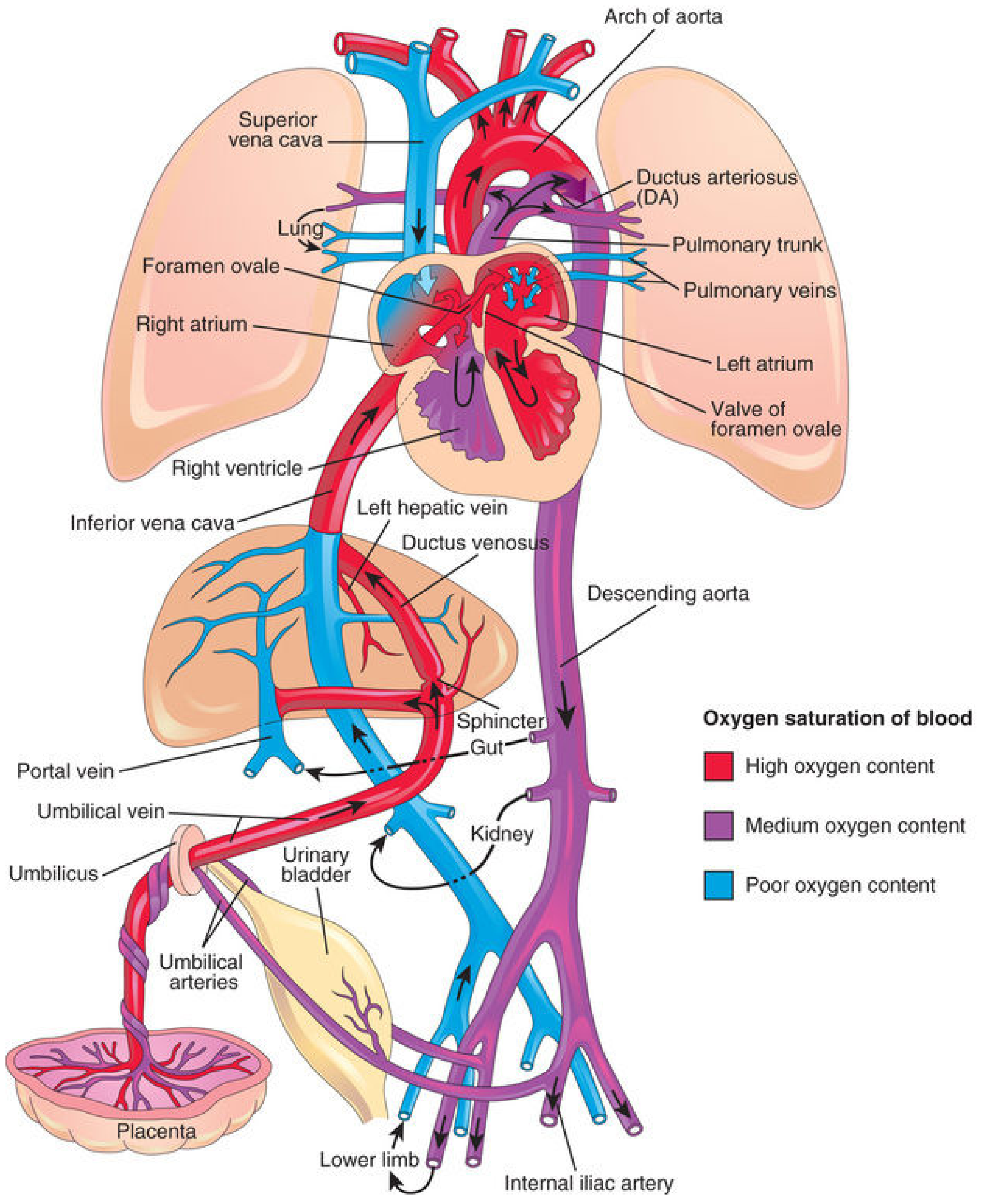
Fig. 13.46 - The Developing Human (Moore & Persaud). Red = high oxygen; purple = medium oxygen; blue = poor oxygen.
Features of Fetal Circulation
1. Gas Exchange Organ: Placenta (not Lungs)
- The placenta acts as the lung, kidney, and gut for the fetus
- Fetal blood is oxygenated at the placenta via the extensive arteriocapillary-venous network within the chorionic villi
- Maternal and fetal blood do NOT normally mix; gas exchange occurs across the placental membrane
2. Umbilical Vessels
- 1 umbilical vein: carries oxygenated blood (~80% saturated) from placenta to fetus
- 2 umbilical arteries: carry deoxygenated blood (~58% saturated) from fetus back to placenta
- These are contained in the umbilical cord
The Three Fetal Shunts
Shunt 1: Ductus Venosus (Bypasses the Liver)
- Highly oxygenated, nutrient-rich blood enters the fetus via the umbilical vein
- On approaching the liver, approximately 50% passes directly into the ductus venosus - a short vessel connecting the umbilical vein to the inferior vena cava (IVC)
- This blood bypasses the hepatic sinusoids entirely
- The remaining 50% enters the portal vein and liver sinusoids, then drains into the IVC via hepatic veins
- A physiologic sphincter near the umbilical vein regulates flow - during uterine contractions, it prevents cardiac overload
Shunt 2: Foramen Ovale (Right Atrium → Left Atrium, Bypasses Lungs)
- Blood arriving in the IVC (now mixed, but still relatively well-oxygenated) enters the right atrium
- The crista dividens (inferior border of septum secundum) deflects most of this blood through the foramen ovale directly into the left atrium
- In the left atrium, it mixes with a small amount of poorly oxygenated pulmonary venous blood
- Blood then flows: Left atrium → Left ventricle → Ascending aorta
- Since the coronary and carotid arteries are the first branches off the ascending aorta, the heart and brain receive the best-oxygenated blood in the fetus
Shunt 3: Ductus Arteriosus (Pulmonary Trunk → Aorta, Bypasses Lungs)
- Deoxygenated blood from the SVC returns to the right atrium
- It mixes with a small amount remaining from the IVC stream and passes to the right ventricle → pulmonary trunk
- Pulmonary vascular resistance is very HIGH in fetal life (lungs are fluid-filled and collapsed)
- Therefore most of this blood bypasses the lungs via the ductus arteriosus - connecting the pulmonary trunk to the descending aorta
- It mixes with blood from the proximal aorta
- Blood in the descending aorta then supplies the lower body and returns to the placenta via the two umbilical arteries
Sites of Mixing of Oxygenated and Deoxygenated Blood (Exam Favourite)
Five sites where mixing occurs:
| Site | Description |
|---|---|
| I. Liver | Umbilical venous blood mixes with portal venous blood |
| II. IVC | Mixes with deoxygenated blood from lower limbs, kidneys, pelvis |
| III. Right atrium | Mixes with deoxygenated blood from SVC (head/upper limbs) |
| IV. Left atrium | Mixes with small amount of pulmonary venous return |
| V. Descending aorta | Ductus arteriosus blood mixes with blood from proximal aorta |
Circulatory Changes at Birth
At birth, two major events trigger circulatory transition:
- Clamping of the umbilical cord - stops placental blood flow, drops right atrial pressure
- First breath - lungs expand, pulmonary vascular resistance falls dramatically, pulmonary blood flow increases, raising left atrial pressure
Closure of Shunts
| Fetal Structure | Trigger for Closure | Adult Remnant |
|---|---|---|
| Ductus venosus | Loss of umbilical blood flow | Ligamentum venosum |
| Foramen ovale | LA pressure > RA pressure; septum primum pressed against septum secundum | Fossa ovalis (functional closure at birth; anatomical by ~3 months) |
| Ductus arteriosus | Rise in PaO₂ (oxygen causes smooth muscle contraction); fall in prostaglandins | Ligamentum arteriosum (functional closure within days; anatomical by 12 weeks) |
| Umbilical vein | Cord clamping | Ligamentum teres hepatis (round ligament of liver) |
| Umbilical arteries | Cord clamping | Medial umbilical ligaments (proximal parts → superior vesical arteries) |
Diagram: Neonatal Circulation After Birth
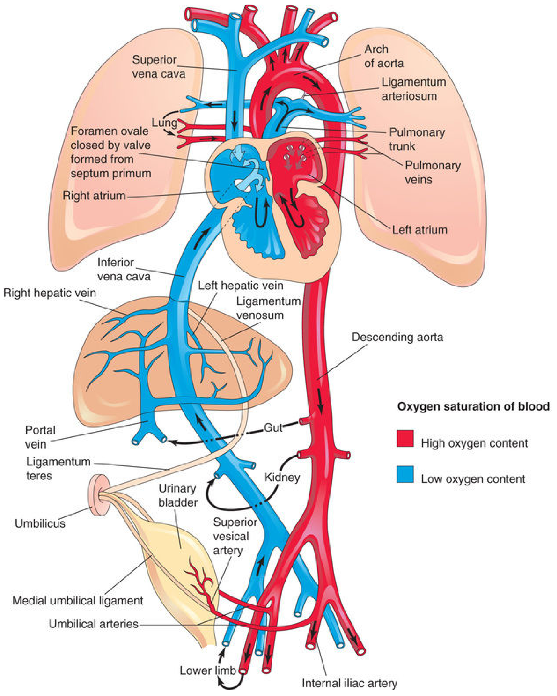
Fig. 13.47 - The Developing Human. After birth, pulmonary and systemic circulations become fully separated.
Clinical Correlates (Bonus Points)
- Patent Ductus Arteriosus (PDA): Failure of DA to close, common in preterm neonates and in congenital rubella. Treated with indomethacin (PG synthesis inhibitor) or surgical ligation
- Patent Foramen Ovale (PFO): Present in ~25% adults; usually asymptomatic but can cause paradoxical embolism
- Persistent Fetal Circulation (Persistent Pulmonary Hypertension of Newborn - PPHN): High pulmonary vascular resistance persists after birth; shunts remain open; presents with cyanosis
- Erythroblastosis fetalis: Anti-D antibodies cross the placenta (the only immunoglobulin that does so); causes hemolytic disease of the newborn
Summary Points for Exam
- Umbilical vein carries oxygenated blood; umbilical arteries carry deoxygenated blood (one vein, two arteries - rule of odd is oxygenated)
- Three shunts: ductus venosus (bypasses liver), foramen ovale (bypasses lungs - right to left), ductus arteriosus (bypasses lungs - pulmonary trunk to aorta)
- Heart and brain get the best-oxygenated blood via ascending aorta
- Liver gets intermediate oxygenation (receives both umbilical venous and portal blood)
- Lower body and placenta receive the least oxygenated blood via descending aorta
- At birth: lungs expand → PVR falls → LA pressure rises → foramen ovale closes; PaO₂ rises → ductus arteriosus constricts
Sources: The Developing Human: Clinically Oriented Embryology (Moore & Persaud); Langman's Medical Embryology
This is a shared conversation. Sign in to Orris to start your own chat.