
Anterior & Posterior uveitis & Endophthal mitis detailed
anterior uveitis hypopyon keratic precipitates slit lamp
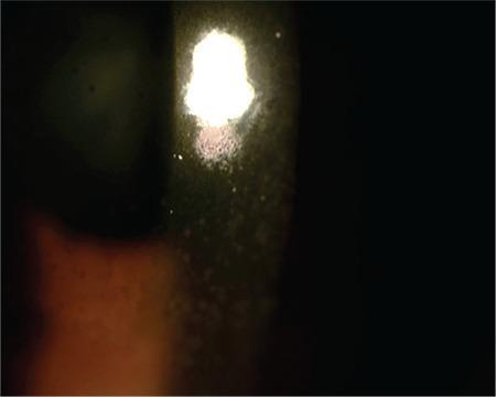
This clinical photograph shows a slit-lamp examination of the anterior segment of a human eye, focusing on the cornea. The image demonstrates a characteristic clinical finding of anterior uveitis: keratic precipitates (KP). These appear as diffuse, medium-sized, brownish-gray inflammatory cellular deposits adhered to the posterior surface of the cornea (the corneal endothelium). The precipitates are distributed across the illuminated area, contributing to visible endothelial haziness. The light reflex from the slit lamp is scattered and irregular, indicating a disruption of corneal clarity and the presence of inflammatory material. This finding is highly relevant for diagnosing intraocular inflammation and monitoring the response to therapy in conditions like viral or idiopathic uveitis.
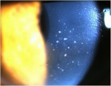
This clinical photograph, taken via slit lamp microscopy, displays the anterior segment of a human eye with a focus on corneal pathology. The image reveals diffuse keratic precipitates (KPs) distributed primarily across the central area of the corneal endothelium. These precipitates are characterized by a 'mutton-fat' appearance, manifesting as whitish, waxy, and irregularly shaped inflammatory deposits. Such findings are indicative of granulomatous uveitis, often associated with conditions like Cytomegalovirus (CMV) hypertensive anterior uveitis or sarcoidosis. The slit lamp beam highlights the depth of these deposits on the posterior corneal surface. Additionally, a mild inflammatory reaction is noted within the anterior chamber, though cell and flare are not individually resolvable at this magnification. This visual evidence serves as a critical diagnostic marker for differentiating types of intraocular inflammation and monitoring treatment response in patients with secondary glaucoma or viral uveitis.
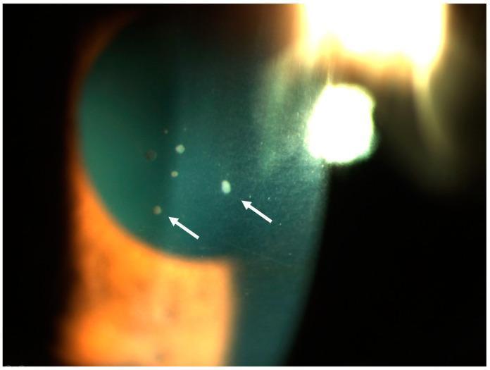
A close-up clinical photograph of the anterior segment of a human eye using slit-lamp biomicroscopy. The image demonstrates active acute granulomatous anterior uveitis. Key findings include multiple keratic precipitates (KPs) on the posterior corneal surface (retrodescemetic area), indicated by white arrows. These precipitates vary in morphology from small granular spots to larger, more opaque deposits. The distribution is localized across the lower portion of the corneal endothelium, which is characteristic of inflammatory cellular accumulation in the anterior chamber. The visual evidence of these 'mutton-fat' style precipitates serves as a hallmark for granulomatous inflammation, often associated with systemic conditions such as sarcoidosis or tuberculosis. The image also displays typical slit-lamp lighting effects, including corneal reflections and a visible beam showing the depth of the anterior chamber.
endophthalmitis fundus vitreous inflammation
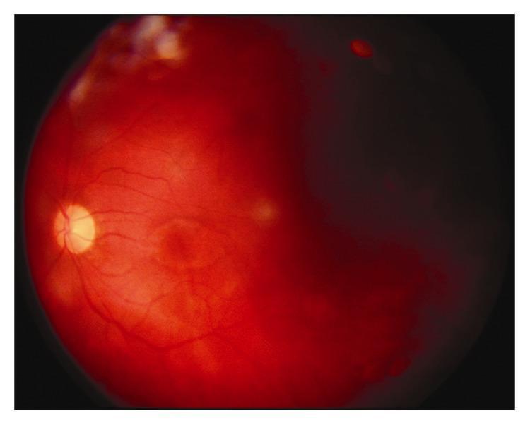
A color fundus photograph of a human eye demonstrating acute clinical features of endophthalmitis. The image shows significant vitreous haze (vitritis), which markedly reduces the clarity of the retinal details. The optic disc is visible as a yellowish-white structure, but its margins appear blurred. The retinal background exhibits a diffuse, deep reddish hue, indicative of severe inflammation and congestion. Key pathological findings include multifocal white retinal infiltrates scattered throughout the posterior pole and visible retinal hemorrhages, particularly in the superior peripheral quadrant. The retinal vasculature is partially obscured by inflammatory exudates and the hazy vitreous media. This image serves as an educational example of the funduscopic presentation of severe intraocular infection, often following penetrating trauma or surgery, where inflammatory debris prevents a sharp view of the underlying retina.
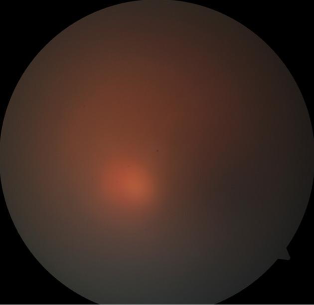
This diagnostic clinical photograph displays a fundus view of the human eye, characterized by extreme vitreous haze (Grade 4+) and reduced media clarity. The image demonstrates the classic presentation of endophthalmitis, where internal ocular inflammation severely obscures retinal landmarks. The optic disc, macula, and retinal vasculature are clinically invisible due to the dense, diffuse opacification of the vitreous humor. Scattered throughout the hazy medium are subtle, non-discrete vitreous exudates, appearing as irregular opacities that further degrade the image quality. This visual finding is a critical sign of intraocular infection or severe inflammation, often associated with a precipitous drop in visual acuity. The diagnostic image serves to illustrate the physical limitations of fundus examination in the presence of advanced vitritis, necessitating alternative diagnostic modalities like B-scan ultrasonography for assessing retinal attachment.
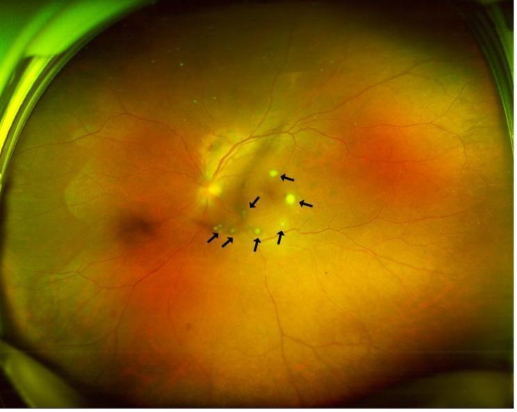
This clinical photograph is an ultra-widefield fundus image of a human left eye, illustrating features of endogenous endophthalmitis. The fundus exhibits a generalized orange-yellow hue with a hazy appearance, consistent with significant vitreous inflammation (vitritis). Multiple, small, yellowish-white, circumscribed chorioretinal lesions are scattered across the retina, primarily concentrated in the macular and peripapillary regions. Several of these lesions are specifically highlighted by black arrows, demonstrating their distinct, slightly elevated morphology. These clinical findings represent multifocal abscesses or 'puff ball' lesions, characteristic of fungal infections such as Candida albicans that have infiltrated from the choroid into the retinal layers. The image serves as an educational example for diagnosing ocular manifestations of systemic infections, particularly in patients with recent surgical or urological interventions. The visibility of the optic disc and retinal vasculature is slightly obscured by the overlying vitreous haze, emphasizing the severity of the intraocular inflammatory response.
posterior uveitis retinitis choroiditis fundus photograph
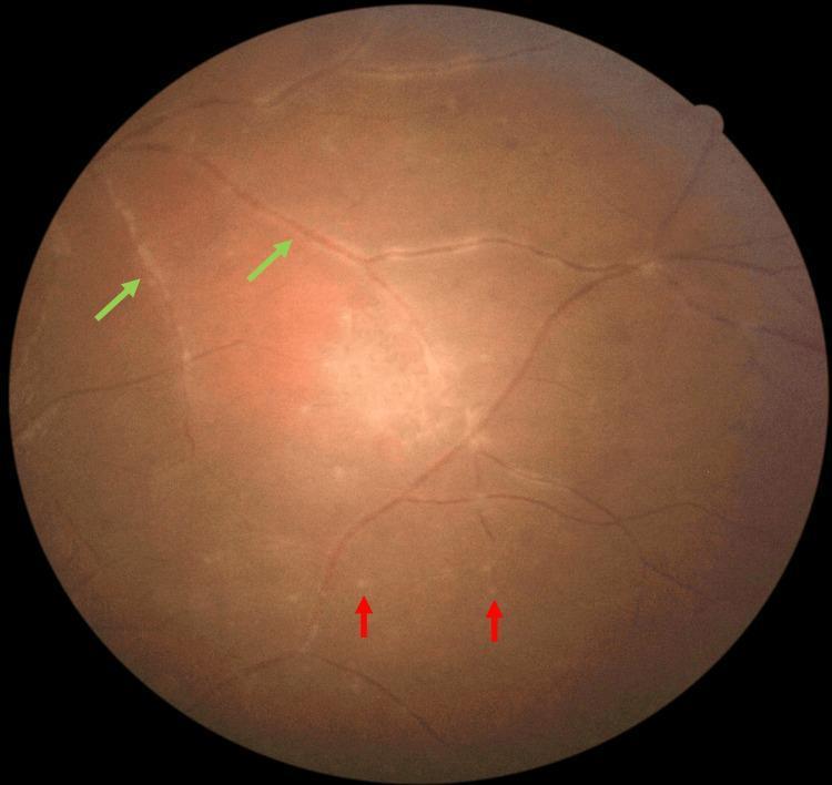
This clinical photograph displays a fundus examination of the peripheral retina, illustrating diagnostic signs of posterior uveitis, specifically associated with ocular toxoplasmosis. The image captures two primary pathological features: retinal vasculitis and punctate choroiditis. Retinal vasculitis is highlighted by green arrows, showing evidence of perivascular inflammation and vascular sheathing where the retinal vessels appear thickened and slightly obscured by inflammatory exudate. Multiple punctate choroiditis lesions are indicated by red arrows; these appear as small, discrete, yellowish-white, round-to-oval spots scattered predominantly across the inferior retinal periphery. The lesions are localized deep within the retina at the level of the choroid, representing focal inflammatory foci. This visual evidence supports the educational objective of identifying peripheral manifestations of panuveitis, including the secondary vascular and choroidal responses that occur alongside primary retinitis. These findings are critical for differentiating infectious etiologies like toxoplasmosis from systemic autoimmune conditions.
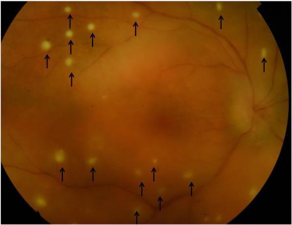
This fundus color photograph illustrates clinical manifestations of posterior syphilitic uveitis. The image shows widespread, small, circular, whitish-yellowish pre-retinal lesions (indicated by black arrows) scattered across the retina. These punctate lesions, sometimes referred to as pre-retinal precipitates, are seen overlying areas of active retinitis and often represent aggregates of inflammatory cells. A generalized haziness is visible across the fundus, characteristic of vitritis (inflammation of the vitreous humor), which obscures the clarity of the underlying retinal vessels and landmarks. The optic disc is visible in the nasal periphery with relatively preserved margins. This presentation is a classic hallmark of ocular syphilis, often termed 'the great mimicker,' where inflammatory cells migrate across the retina into the vitreous. This visual material serves as an educational resource for identifying inflammatory ocular phenotypes and distinguishing pre-retinal precipitates from intra-retinal foci in the context of infectious uveitis.
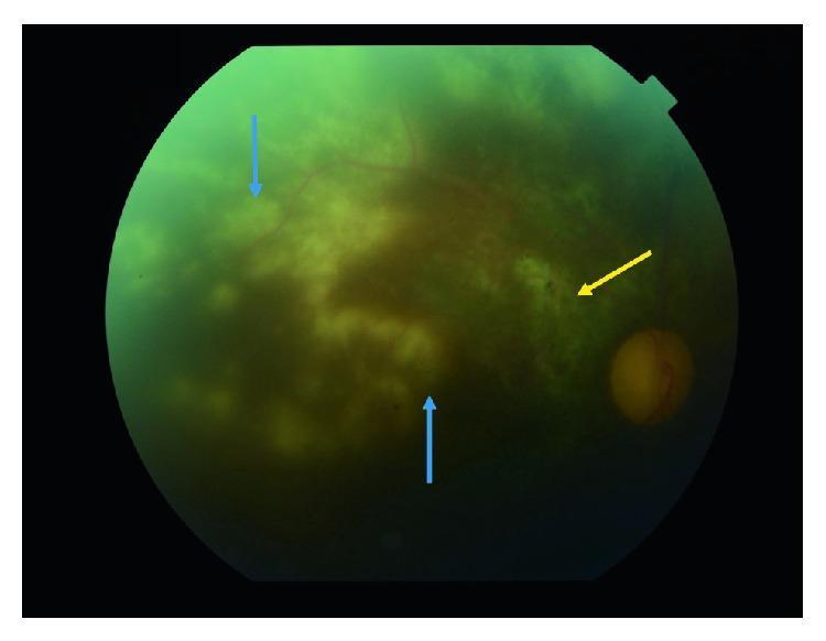
This clinical photograph is a fundus image of the right eye, demonstrating hallmark features of severe posterior uveitis, specifically cytomegalovirus (CMV) retinitis in an immunocompromised patient. The image shows significant optic disc pallor and generalized vitreous haze, which obscures the fine detail of the retinal vasculature. Multiple large, confluent, white-to-off-yellow granular exudates (noted by blue arrows) are visible, concentrated around the macular area and extending into the peripheral retina, consistent with active necrotizing retinitis. A distinct area of pigmentation and thinning, indicated by a yellow arrow, represents an old, atrophic choroidal scar. The presence of these chorioretinal lesions alongside the optic nerve pallor illustrates the extensive retinal destruction and secondary optic neuropathy often seen in progressive viral infections of the retina. The educational focus is on identifying active infectious lesions versus chronic scarring and assessing the degree of vitritis and optic nerve involvement in inflammatory eye disease.
Uveitis (Anterior & Posterior) and Endophthalmitis
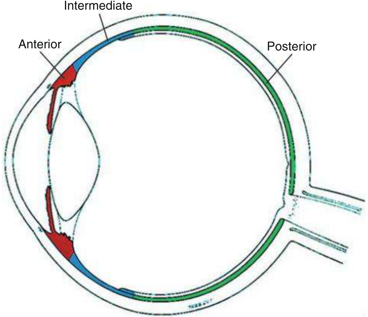
Anatomical Classification of Uveitis
| Type | Structures Involved |
|---|---|
| Anterior | Iris, anterior ciliary body (pars plicata) |
| Intermediate | Vitreous, pars plana, peripheral retina |
| Posterior | Retina, choroid (behind vitreous base) |
| Panuveitis | All segments |
PART 1: ANTERIOR UVEITIS
Definition & Epidemiology
- Iritis = iris alone
- Iridocyclitis = iris + anterior ciliary body (clinically indistinguishable from iritis)
Types
| Acute AU (AAU) | Chronic AU (CAU) | |
|---|---|---|
| Onset | Sudden | Insidious or acute |
| Laterality | Usually unilateral | More often bilateral |
| Duration | < 3 months | Persistent; relapse < 3 months after stopping treatment |
| Associated systemic disease | Less common | More common |
| Granulomatous signs | Uncommon | Often present |
| Prognosis | Generally good | More variable |
Aetiology & Systemic Associations
| Category | Conditions |
|---|---|
| Idiopathic | ~50% of all cases |
| HLA-B27 related | ~20% of AAU; ankylosing spondylitis (25% develop AU), reactive arthritis, psoriatic arthritis, IBD-related arthritis |
| Infectious | VZV (herpes zoster ophthalmicus), HSV, CMV, TB, syphilis, Lyme disease |
| Non-infectious | JIA, sarcoidosis, Behçet disease, SLE, MS, TINU syndrome, drug-induced |
| Masquerade | Lymphoma, anterior segment melanoma, juvenile xanthogranuloma |
Clinical Features
Symptoms
- Rapid-onset unilateral pain, photophobia, redness, watery discharge, visual blur
- Often preceded by mild discomfort for a few days
- CAU may be asymptomatic until complications develop (cataract)
- History of prior episodes is common (recurrent AAU)
Signs
| Grade | Cells in 1×1 mm field |
|---|---|
| 0 | < 1 |
| 0.5+ | 1-5 |
| 1+ | 6-15 |
| 2+ | 16-25 |
| 3+ | 26-50 |
| 4+ | > 50 |
- Usually in Arlt's triangle (inferior, triangular distribution, apex up)
- Non-granulomatous (AAU): small, fine KPs
- Granulomatous (CAU): medium-to-large "mutton-fat" KPs (large, greasy, composed of epithelioid and multinucleated cells)
- Fuchs uveitis syndrome: diffusely distributed, stellate/filamentous ("stellate KPs")
- Old KPs: pigmented or "ground-glass" appearance
- Busacca nodules: in iris stroma - granulomatous uveitis
- Koeppe nodules: at pupillary margin - site of PS formation
- Roseolae (syphilis), iris crystals/Russell bodies (chronic uveitis, Fuchs syndrome)
- Diffuse stromal atrophy: Fuchs uveitis syndrome
- Patchy/sectoral: herpetic uveitis (HSV and VZV)
- AAU: usually normal/low IOP (ciliary shutdown) but can rise via trabecular obstruction by inflammatory cells, trabeculitis
- CAU: trabecular scarring/sclerosis, synechial angle closure - more sustained elevation
Investigations
- Single, mild-moderate, non-granulomatous AAU with no systemic features
- Systemic diagnosis already confirmed
- Typical features of a specific entity (e.g. Fuchs syndrome)
- Recurrent, severe, or bilateral AAU
- Persistent/chronic AU resistant to treatment
- Granulomatous signs
- Associated intermediate/posterior uveitis
- Ocular/systemic features suggesting underlying disease
- HLA-B27 tissue typing (chromosome 6, MHC; present in 6-8% of White population; strongly associated with AAU, AS, reactive arthritis, psoriatic arthritis)
- ACE, chest X-ray/CT (sarcoidosis)
- VDRL/TPHA (syphilis)
- Mantoux/IGRA (TB)
- ANA, anti-dsDNA (SLE)
- RF, ANA (JIA in children)
- FBC, ESR, CRP
Treatment
1. Topical Corticosteroids (mainstay of AAU treatment)
- Hourly × 3 days → every 2 hours × 3 days → QID × 1 week → TDS × 1 week → BD × 1 week → OD × 1 week (total ~5-6 weeks)
- CAU: aim for complete suppression (zero cells AND flare); more gradual taper + maintenance
2. Cycloplegic Agents
- Cyclopentolate (12-24 h duration)
- Homatropine (3 days)
- Atropine (7-10 days)
- Phenylephrine 2.5% or 10% (supplementary, to break PS)
- Mydricaine No. 2 (subconjunctival injection of adrenaline + atropine + procaine): for fresh PS resistant to drops
3. Periocular / Intravitreal Steroids
4. Systemic Therapy
| Class | Drugs |
|---|---|
| Antimetabolites | Methotrexate, mycophenolate mofetil, azathioprine |
| Calcineurin inhibitors | Cyclosporin A, tacrolimus (T-cell critical uveitis: VKH, birdshot) |
| Alkylating agents | Cyclophosphamide, chlorambucil |
| Anti-TNF biologics | Infliximab, adalimumab, etanercept, golimumab, certolizumab (effective in neutrophil/macrophage-critical: Behçet, sarcoidosis) |
| IL-6 inhibitors | Tocilizumab |
| Anti-CD20 | Rituximab (B-cell critical, e.g. ANCA+ scleritis) |
PART 2: POSTERIOR UVEITIS
Definition
- Retinochoroiditis (originates in retina, extends to choroid)
- Chorioretinitis (originates in choroid, extends to retina)
Clinical Presentation
- Peripheral lesion → floaters
- Macular lesion → impaired central vision
- Vitritis can accompany any form and causes diffuse visual haziness
Patterns of Inflammation
Retinitis
- Focal, multifocal, geographic, or diffuse
- Active: whitish retinal opacities with indistinct borders (surrounding oedema)
- Resolving: borders become better defined, lesion scars
Choroiditis
- Focal, multifocal, geographic, or diffuse
- Does not usually cause vitritis without concurrent retinal involvement
- Active: round, yellow nodule in the choroid
Retinal Vasculitis
- Primary or secondary (adjacent to retinitis focus)
- Venous involvement (periphlebitis) more common than arterial (periarteritis)
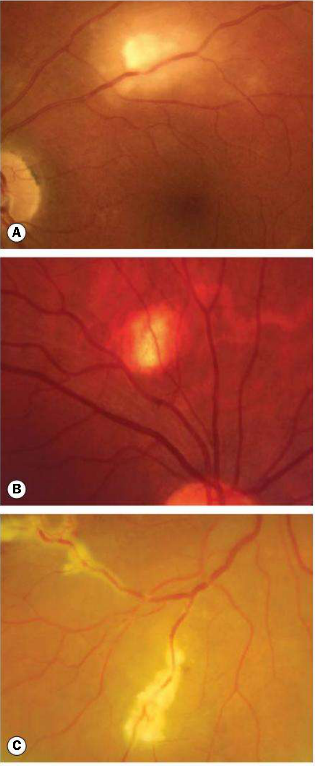
- Active: yellowish/grey-white patchy perivascular cuffing, sometimes with haemorrhage
- Quiescent: perivascular scarring (do not mistake for active disease)
Important Causes of Posterior Uveitis
| Infectious | Non-infectious |
|---|---|
| Toxoplasmosis (most common infectious cause worldwide) | Sarcoidosis |
| CMV retinitis (immunocompromised, HIV) | Birdshot retinochoroidopathy (HLA-A29) |
| HSV/VZV acute retinal necrosis (ARN) | Serpiginous choroidopathy |
| Tuberculosis | Vogt-Koyanagi-Harada (VKH) syndrome |
| Ocular histoplasmosis (OHS) | Sympathetic ophthalmia |
| Toxocariasis | Multifocal choroiditis and panuveitis (MCP) |
| Syphilis | Punctate inner choroidopathy (PIC) |
| Candida (endogenous) | MEWDS, APMPPE, AZOOR |
Investigations (Multimodal Imaging)
- Detects lipofuscin/melanin in RPE cells
- Hyperfluorescence in: MEWDS, serpiginous choroidopathy, AZOOR
- Macular oedema, vitreomacular disease, structural damage
- Inner retinal infiltrates: Behçet disease, viral infections
- Outer retinal changes: PIC, MEWDS, APMPPE, serpiginous choroiditis
- Choroidal granulomas: sarcoidosis, TB, VKH
- Active inflammatory retinal foci: early hypofluorescence → late hyperfluorescence
- Retinal vasculitis, optic disc swelling, subretinal fluid
- "Window defect" (RPE loss) vs. masking (pigment clumping)
- Investigation of choice for predominantly choroidal inflammation
- Choriocapillaris inflammation: patchy hypofluorescent areas (APMPPE, serpiginous)
- Stromal choroiditis: focal hypofluorescence → late diffuse hyperfluorescence (VKH, sympathetic ophthalmia)
- A compatible systemic diagnosis is already confirmed
- Obvious clinical pattern suggests specific entity
Treatment
- T-cell critical uveitis (VKH, birdshot retinochoroidopathy): calcineurin inhibitors (cyclosporin, tacrolimus)
- Neutrophil/macrophage critical uveitis (Behçet, sarcoidosis): anti-TNF agents (infliximab, adalimumab)
- B-cell critical (scleritis with ANCA+): rituximab
- Infectious causes require specific antimicrobial therapy first (e.g. pyrimethamine + sulfadiazine + folinic acid for toxoplasmosis; ganciclovir for CMV; aciclovir for HSV/VZV ARN)
PART 3: ENDOPHTHALMITIS
Definition
Classification by Route of Entry
| Type | Route | Common Causes |
|---|---|---|
| Exogenous (post-surgical) | Breach in eye wall post-op | Staph. epidermidis, Staph. aureus, Strep. sp., gram-negatives |
| Exogenous (post-traumatic) | Penetrating injury | Bacillus cereus, gram-negatives (often virulent, rapid) |
| Bleb-related | Infected filtering bleb | Streptococcus sp., Haemophilus influenzae, Staph. sp. |
| Endogenous (metastatic) | Haematogenous spread | Candida (IVDA, immunocompromised), Klebsiella (diabetics, East Asia), Aspergillus |
Common Organisms
| Virulence | Organisms | Course |
|---|---|---|
| High | Toxin-producing gram-positives (Strep. sp., Staph. aureus), gram-negatives (Pseudomonas, Bacillus cereus) | Rapidly destructive |
| Low (indolent) | Propionibacterium acnes, Staph. epidermidis | Insidious, chronic, less destructive |
Clinical Features
- Rapid decrease in vision (often the first symptom)
- Ocular pain (follows visual loss)
- Redness, photophobia, lid swelling
- Anterior uveitis (AC cells and flare) - often the first sign on slit lamp
- Hypopyon (whitish inferior AC level) - substantial in endophthalmitis
- Vitritis/vitreous exudate - impairment of red reflex
- White/milky vitreous opacity (vitreous abscess in severe cases)
- Chemosis and periorbital oedema
- In bleb-related: white milky bleb + severe injection
- Corneal oedema
Diagnosis
- Gram stain + culture + sensitivity
- Vitreous culture is more sensitive than aqueous alone
- Anterior chamber tap (aqueous aspirate) also performed
- Performed when media opacification prevents fundal view
- Shows vitreous echoes (inflammatory exudate) and helps assess retinal detachment
Treatment
1. Intravitreal Antibiotics (immediate, cornerstone of treatment)
- Vancomycin 1 mg/0.1 mL (gram-positive cover) +
- Ceftazidime 2-2.25 mg/0.1 mL (gram-negative cover)
- Both injected simultaneously into the vitreous cavity
2. Topical Antibiotics
3. Corticosteroids
- Topical steroids started 48 hours after intravitreal antibiotics (not immediately)
- Intravitreal dexamethasone 400 mcg may be given with antibiotics in some regimens
- Oral prednisolone as adjunct
4. Oral Fluoroquinolones
5. Pars Plana Vitrectomy (PPV)
- Immediate vitrectomy is superior to vitreous tap alone when presenting visual acuity = light perception (LP) or worse
- When vision is better than LP, vitreous tap + intravitreal antibiotics gives equivalent results
- Vision LP or worse
- No improvement after 48 hours of intravitreal antibiotics
- Bleb-related endophthalmitis (PPV considered at 48 hours)
- Fungal endophthalmitis
- Dense vitreous opacity / inability to visualize retina
6. Antifungals (for fungal endophthalmitis)
- Systemic: voriconazole or amphotericin B (systemic and intravitreal)
- Intravitreal: amphotericin B 5-10 mcg/0.1 mL
Special Case: Bleb-Related Endophthalmitis
- Vitreous + aqueous samples obtained immediately
- Intravitreal vancomycin 2 mg + ceftazidime 2 mg
- Repeat at 48 hours; consider PPV
- Oral fluoroquinolones 10-14 days
- Topical steroids after 48 hours
Prophylaxis Against Endophthalmitis
- Preoperative povidone-iodine (5%) instillation to conjunctival sac
- Perioperative topical antibiotics
- Intracameral antibiotic injection (e.g. cefuroxime) at end of cataract surgery - most effective prophylactic measure
Quick Comparison: Uveitis vs. Endophthalmitis
| Feature | Anterior Uveitis | Posterior Uveitis | Endophthalmitis |
|---|---|---|---|
| Primary location | Anterior chamber | Retina/choroid | Vitreous + AC |
| Pain | Moderate | Variable (often painless) | Severe |
| Vision loss | Mild-moderate | Central if macula involved | Rapid, severe |
| Cells | AC cells + flare | Vitreous cells, retinal lesions | AC + vitritis + hypopyon |
| Red reflex | Usually preserved | Usually preserved | Impaired/absent |
| KPs | Yes | No | Can be present |
| Hypopyon | HLA-B27, Behçet | Rare | Prominent |
| Aetiology | Immune/infectious | Immune/infectious | Almost always infectious |
| Urgency | Urgent (same day) | Urgent | Ophthalmic emergency |
| Core treatment | Topical steroids + cycloplegics | Systemic immunosuppression / antimicrobials | Intravitreal antibiotics ± PPV |
- [Kanski's Clinical Ophthalmology, 10th ed.](Kanski's Clinical Ophthalmology, ch. 12 - Uveitis) - primary reference throughout
- [Goldman-Cecil Medicine, International Ed.](Goldman-Cecil Medicine, Vol. 2, Ch. 391 & 457) - systemic associations and endophthalmitis management
- Robbins & Cotran Pathologic Basis of Disease - pathological basis
- Tintinalli's Emergency Medicine - emergency management principles