
Trigger 4: Hyperkalemia and Renal Failure Emergency * Identify the ECG changes and clinical presentation associated with severe hyperkalemia. * Formulate an acute management plan for severe hyperkalemia, including immediate cardiopulmonary stabilization
Reading File
Reading File
Finding Sources
Finding Sources
Finding Sources
Reading File
Reading File
Searching PubMed
hyperkalemia AND management
Hyperkalemia and Renal Failure Emergency
1. ECG Changes and Clinical Presentation
Hyperkalemia (K+ > 5.5 mEq/L) causes increased membrane excitability followed by progressive conduction delay as levels rise. The Miller's Anesthesia staged progression is the clearest framework:
| Serum K+ | ECG Finding |
|---|---|
| 5.5-6.5 mEq/L | Tall, peaked (tented), symmetric T-waves; shortened QT |
| 6.5-7.5 mEq/L | Prolonged PR interval, flattened/widened P-wave, ST depression |
| >7.5 mEq/L | Widened QRS complex |
| >9.0 mEq/L | Sine-wave pattern, bradycardia, ventricular tachycardia/fibrillation, asystole/cardiac arrest |
- Miller's Anesthesia, p. 3806-3816 (block17)
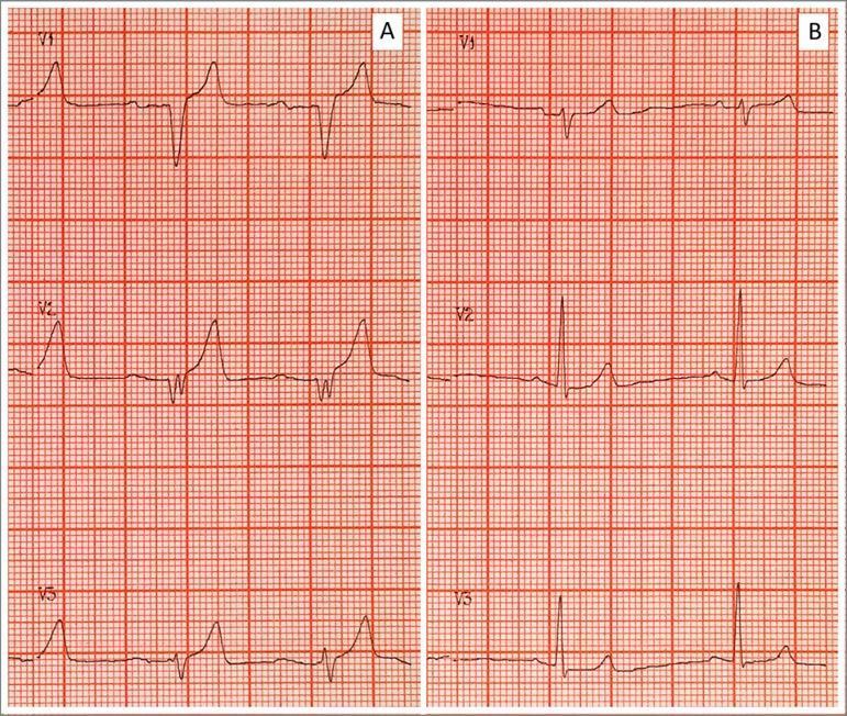
Some patients also develop a Brugada-type pattern (pseudo-right bundle branch block with ST elevation in V1-V2) with severe hyperkalemia - Harrison's Principles of Internal Medicine, 22E.
Clinical presentation beyond the ECG:
- Neuromuscular: ascending muscle weakness, paresthesias, hyporeflexia, and in severe cases flaccid paralysis (mimics Guillain-Barre)
- Cardiac: palpitations, bradyarrhythmias, and risk of sudden cardiac arrest - often the presenting event in dialysis patients who miss sessions
- Symptoms are frequently vague or absent until the cardiac toxicity is advanced, which is why any renal failure patient with unexplained weakness or bradycardia needs an immediate ECG and potassium level
- Chronic hyperkalemia (e.g., CKD) is tolerated at higher levels than an acute rise, because the intracellular/extracellular K+ gradient has re-equilibrated - Miller's Anesthesia, p. 3816
2. Acute Management Plan
The sequence follows three goals: stabilize the myocardium, shift K+ intracellularly, then remove K+ from the body - with any ECG change or K+ >6.5 mEq/L treated as an emergency.
Step 0 - Stabilization/monitoring
- Continuous cardiac monitor and 12-lead ECG immediately; IV access x2, telemetry bed/ICU-level care
- Airway/breathing support as needed (severe hyperkalemia can cause respiratory muscle weakness); have advanced airway and defibrillator/ACLS drugs at bedside given risk of VF/pulseless arrest
- Draw stat BMP, ionized calcium, magnesium, VBG, and send a repeat potassium to confirm (rule out hemolyzed sample) without delaying treatment if ECG changes are present
Step 1 - Membrane stabilization (antagonize cardiac toxicity) - give first if ECG changes present
- IV calcium gluconate 10%, 10 mL (1 g) over 2-3 minutes, or calcium chloride 10% 5-10 mL if central access available (more caustic peripherally); repeat in 5-10 minutes if ECG changes persist
- Calcium does not lower serum K+, it raises the threshold potential to directly counteract membrane excitability - Rosen's Emergency Medicine, p. 4589; Schwartz's Principles of Surgery, p. 4034
- Caution: flush the line before/after if the patient is on digoxin (calcium can precipitate digoxin toxicity)
Step 2 - Shift potassium intracellularly
- Regular insulin IV 10 units (or 0.1 units/kg) with dextrose (25-50 g D50, or D10 infusion if glucose is not already elevated) - drives K+ into cells via Na+/K+-ATPase; onset ~15-30 min, lasts 4-6 hours. Monitor glucose for delayed hypoglycemia.
- Nebulized albuterol 10-20 mg (higher than the standard bronchodilator dose) - beta-2 agonism shifts K+ intracellularly, additive to insulin
- Sodium bicarbonate (IV, ~1 mEq/kg or an ampule) - most useful when concurrent metabolic acidosis is present; less reliable as a standalone potassium-lowering agent in isolation but used adjunctively - Rosen's Emergency Medicine, p. 4589-4590; Miller's Anesthesia, p. 3135-3139
Step 3 - Remove potassium from the body
- Loop diuretics (furosemide) if the patient has residual renal function and is not anuric, to promote kaliuresis
- Potassium binders: newer agents (patiromer, sodium zirconium cyclosilicate) or older sodium polystyrene sulfonate (Kayexalate) - onset is hours, so these are adjuncts, not first-line for the acute crisis
- Hemodialysis is the definitive treatment for renal failure patients, especially with K+ >6.5 mEq/L, anuria, refractory hyperkalemia, or ongoing ECG changes despite medical therapy - Miller's Anesthesia, p. 3816; Goldman-Cecil Medicine, p. 2887-2895
Ongoing care: recheck K+ and glucose within 1-2 hours, keep continuous ECG monitoring until normalization, identify and treat the underlying cause (medication review for ACEi/ARB/MRA/NSAIDs, assess for tissue breakdown/rhabdomyolysis/tumor lysis, arrange urgent nephrology consult for dialysis access if the patient is not already established on renal replacement therapy).
A recent 2025 systematic review and meta-analysis (Jessen et al., Resuscitation, PMID 39761907) evaluated pharmacological interventions for acute hyperkalemia treatment; it is worth checking for updated comparative efficacy data on insulin/albuterol dosing regimens, as this postdates the textbook chapters cited above and could refine relative drug sequencing recommendations.
This is a shared conversation. Sign in to Orris to start your own chat.