anterior uveitis related to sarcoidosis and what should i know before my neet pg exam

Reading File
Finding Sources
Finding Sources
Searching Images

anterior uveitis sarcoidosis mutton fat keratic precipitates slit lamp

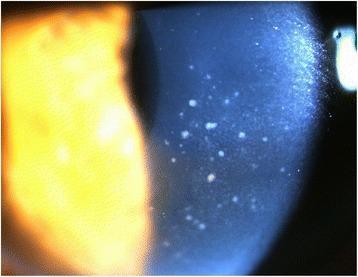
This clinical photograph, taken via slit lamp microscopy, displays the anterior segment of a human eye with a focus on corneal pathology. The image reveals diffuse keratic precipitates (KPs) distributed primarily across the central area of the corneal endothelium. These precipitates are characterized by a 'mutton-fat' appearance, manifesting as whitish, waxy, and irregularly shaped inflammatory deposits. Such findings are indicative of granulomatous uveitis, often associated with conditions like Cytomegalovirus (CMV) hypertensive anterior uveitis or sarcoidosis. The slit lamp beam highlights the depth of these deposits on the posterior corneal surface. Additionally, a mild inflammatory reaction is noted within the anterior chamber, though cell and flare are not individually resolvable at this magnification. This visual evidence serves as a critical diagnostic marker for differentiating types of intraocular inflammation and monitoring treatment response in patients with secondary glaucoma or viral uveitis.

This clinical photograph, taken via slit lamp microscopy, displays the anterior segment of a human eye with a focus on corneal pathology. The image reveals diffuse keratic precipitates (KPs) distributed primarily across the central area of the corneal endothelium. These precipitates are characterized by a 'mutton-fat' appearance, manifesting as whitish, waxy, and irregularly shaped inflammatory deposits. Such findings are indicative of granulomatous uveitis, often associated with conditions like Cytomegalovirus (CMV) hypertensive anterior uveitis or sarcoidosis. The slit lamp beam highlights the depth of these deposits on the posterior corneal surface. Additionally, a mild inflammatory reaction is noted within the anterior chamber, though cell and flare are not individually resolvable at this magnification. This visual evidence serves as a critical diagnostic marker for differentiating types of intraocular inflammation and monitoring treatment response in patients with secondary glaucoma or viral uveitis.

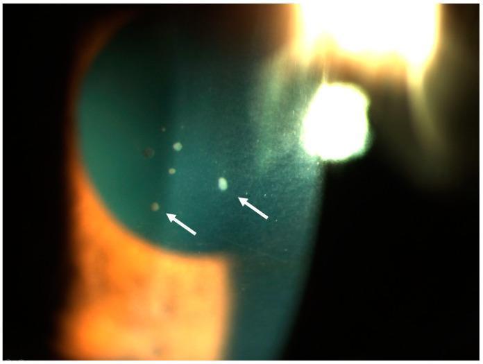
A close-up clinical photograph of the anterior segment of a human eye using slit-lamp biomicroscopy. The image demonstrates active acute granulomatous anterior uveitis. Key findings include multiple keratic precipitates (KPs) on the posterior corneal surface (retrodescemetic area), indicated by white arrows. These precipitates vary in morphology from small granular spots to larger, more opaque deposits. The distribution is localized across the lower portion of the corneal endothelium, which is characteristic of inflammatory cellular accumulation in the anterior chamber. The visual evidence of these 'mutton-fat' style precipitates serves as a hallmark for granulomatous inflammation, often associated with systemic conditions such as sarcoidosis or tuberculosis. The image also displays typical slit-lamp lighting effects, including corneal reflections and a visible beam showing the depth of the anterior chamber.

A close-up clinical photograph of the anterior segment of a human eye using slit-lamp biomicroscopy. The image demonstrates active acute granulomatous anterior uveitis. Key findings include multiple keratic precipitates (KPs) on the posterior corneal surface (retrodescemetic area), indicated by white arrows. These precipitates vary in morphology from small granular spots to larger, more opaque deposits. The distribution is localized across the lower portion of the corneal endothelium, which is characteristic of inflammatory cellular accumulation in the anterior chamber. The visual evidence of these 'mutton-fat' style precipitates serves as a hallmark for granulomatous inflammation, often associated with systemic conditions such as sarcoidosis or tuberculosis. The image also displays typical slit-lamp lighting effects, including corneal reflections and a visible beam showing the depth of the anterior chamber.

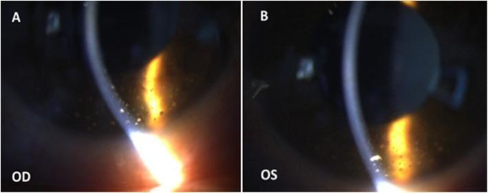
Clinical photograph utilizing slit-lamp biomicroscopy of the right (OD) and left (OS) eyes, demonstrating a granulomatous anterior uveitis. The images focus on the corneal endothelium and anterior chamber, employing a narrow slit beam to assess transparency and structural integrity. Notable pathology includes multiple large, waxy, 'mutton-fat' keratic precipitates (KPs) distributed primarily across the inferior corneal endothelium. These precipitates are variable in size and cause visible light scattering as the slit beam passes through the corneal layers. The presence of these large inflammatory cell aggregates is a hallmark clinical sign of granulomatous inflammation, often associated with systemic conditions such as sarcoidosis, tuberculosis, or specific parasitic infections. The surrounding cornea remains relatively clear, but the inferior concentration of KPs suggests a typical Arlt's triangle distribution. This visual material is intended for ophthalmology education, focusing on the diagnostic features of inflammatory eye disease and the technique of slit-lamp examination.

Clinical photograph utilizing slit-lamp biomicroscopy of the right (OD) and left (OS) eyes, demonstrating a granulomatous anterior uveitis. The images focus on the corneal endothelium and anterior chamber, employing a narrow slit beam to assess transparency and structural integrity. Notable pathology includes multiple large, waxy, 'mutton-fat' keratic precipitates (KPs) distributed primarily across the inferior corneal endothelium. These precipitates are variable in size and cause visible light scattering as the slit beam passes through the corneal layers. The presence of these large inflammatory cell aggregates is a hallmark clinical sign of granulomatous inflammation, often associated with systemic conditions such as sarcoidosis, tuberculosis, or specific parasitic infections. The surrounding cornea remains relatively clear, but the inferior concentration of KPs suggests a typical Arlt's triangle distribution. This visual material is intended for ophthalmology education, focusing on the diagnostic features of inflammatory eye disease and the technique of slit-lamp examination.

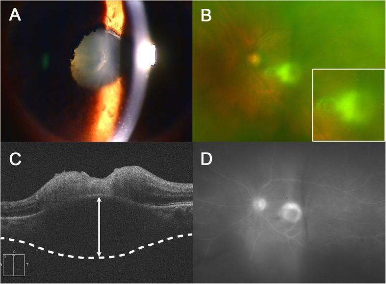
Multi-modal ophthalmic imaging of a patient with granulomatous uveitis and posterior segment involvement. (A) Slit-lamp anterior segment photograph showing numerous mutton-fat keratic precipitates on the corneal endothelium, a fibrinous pupillary membrane, and 360-degree posterior synechiae. (B) Wide-field fundus photography showing yellow-white macular infiltrates and prominent vitreous haze. (C) Spectral-domain optical coherence tomography (SD-OCT) cross-section demonstrating intraretinal hyperreflectivity, disorganization of retinal layers, and retinal pigment epithelium (RPE) elevation; a white double-headed arrow highlights significant choroidal thickening beneath the active lesion. (D) Wide-field fluorescein angiography (FA) illustrating hyperfluorescence of the active macular lesion and late-phase optic disc leakage. The images collectively demonstrate a severe inflammatory process, such as ocular toxoplasmosis or sarcoidosis, characterized by both anterior chamber reaction and active chorioretinitis.

Multi-modal ophthalmic imaging of a patient with granulomatous uveitis and posterior segment involvement. (A) Slit-lamp anterior segment photograph showing numerous mutton-fat keratic precipitates on the corneal endothelium, a fibrinous pupillary membrane, and 360-degree posterior synechiae. (B) Wide-field fundus photography showing yellow-white macular infiltrates and prominent vitreous haze. (C) Spectral-domain optical coherence tomography (SD-OCT) cross-section demonstrating intraretinal hyperreflectivity, disorganization of retinal layers, and retinal pigment epithelium (RPE) elevation; a white double-headed arrow highlights significant choroidal thickening beneath the active lesion. (D) Wide-field fluorescein angiography (FA) illustrating hyperfluorescence of the active macular lesion and late-phase optic disc leakage. The images collectively demonstrate a severe inflammatory process, such as ocular toxoplasmosis or sarcoidosis, characterized by both anterior chamber reaction and active chorioretinitis.

Reading File
Finding Sources
Reading File
Reading File
Reading File
Reading File
Reading File
I now have all the key information needed. Let me compile a comprehensive NEET PG-oriented answer.

Anterior Uveitis in Sarcoidosis - NEET PG Notes

The Big Picture

Sarcoidosis is a chronic granulomatous disorder of unknown cause characterized by non-caseating granulomas. It is one of the most common systemic associations of uveitis. Granulomatous anterior uveitis is its most common ocular manifestation. Ocular involvement occurs in 25-70% of sarcoid patients (varies by ethnicity).
  • Black people are affected 10x more than White people
  • More common in colder climates
  • Uveitis can precede the diagnosis of sarcoidosis by decades

Anterior Uveitis Pattern in Sarcoidosis

FeatureDetail
TypeGranulomatous anterior uveitis (most common)
AcuityBoth AAU (acute-onset sarcoidosis) and CAU (chronic - more common; tends to affect older patients with chronic pulmonary disease)
LateralityBilateral (usually)
KP type"Mutton-fat" KPs - large, waxy, greasy deposits on corneal endothelium
KP distributionArlt's triangle - apex near central cornea, base at inferior limbus

The 7 IWOS Signs (NEET High Yield!)

The International Workshop on Ocular Sarcoidosis (IWOS, 2009) identified 7 key signs for intraocular sarcoidosis diagnosis. Memorize these:
  1. Mutton-fat KPs and/or small granulomatous KPs and/or iris nodules (Koeppe and/or Busacca)
  2. Trabecular meshwork nodules and/or tent-shaped PAS (peripheral anterior synechiae)
  3. Vitreous opacities: snowballs and/or "strings of pearls"
  4. Multiple chorioretinal peripheral lesions (active/atrophic)
  5. Nodular/segmental periphlebitis ("candle-wax drippings" / en taches de bougie)
  6. Optic disc nodule(s)/granuloma(s) and/or solitary choroidal nodule
  7. Bilateral involvement

Iris Nodules - NEET Favorite

Three types of iris nodules in sarcoidosis (all are granulomas):
NoduleLocation
Koeppe nodulesPupillary border
Busacca (Bussaca) nodulesIris stroma
Berlin nodulesAnterior chamber angle
Note: Koeppe nodules are not specific to sarcoidosis - also seen in other granulomatous uveitis. But the triad with mutton-fat KPs is highly suggestive.

Slit-lamp Images of Granulomatous KPs

Mutton-fat KPs - granulomatous uveitis (sarcoidosis/CMV)
Mutton-fat keratic precipitates on the corneal endothelium - the hallmark of granulomatous uveitis such as sarcoidosis
Granulomatous anterior uveitis - large mutton-fat KPs in Arlt's triangle
Large, waxy mutton-fat KPs distributed in Arlt's triangle - inferior corneal endothelium

Structural Complications

  • Posterior synechiae (iris stuck to lens)
  • Cataract
  • Cystoid macular edema (CME)
  • Secondary glaucoma (trabecular meshwork involvement)
  • Peripheral retinal neovascularization (from occlusive periphlebitis)

Systemic Features (Correlate with Ocular)

SystemFeature
LungBilateral hilar lymphadenopathy (BHL), pulmonary fibrosis
SkinErythema nodosum (shins), lupus pernio (nose/cheeks/ears), granulomatous papules
JointsPolyarthralgia
NerveCranial nerve palsies (CN VII most common), meningitis
HeartArrhythmia, sudden death (5% clinically)
KidneyHypercalciuria
Lofgren syndrome (acute, good prognosis): Erythema nodosum + BHL + polyarthralgia (triad; seen in women)

Investigations - NEET High Yield

TestSignificance
Serum ACE (Angiotensin Converting Enzyme)Elevated - most important marker
Serum lysozymeElevated
Chest X-rayBHL (abnormal in 90%)
Tuberculin skin testNegative (anergy) - strongly positive makes sarcoidosis unlikely
Serum calcium / hypercalciuriaCan be elevated
Bronchoalveolar lavage (BAL)CD4/CD8 T-cell ratio elevated (>3.5 is suggestive)
Kveim testSpecific but rarely used now
BiopsyNon-caseating granulomas (gold standard for definite diagnosis)
Conjunctival biopsyUp to 75% positive if conjunctival nodules present

Diagnostic Classification (IWOS)

CategoryCriteria
Definite ocular sarcoidosisBiopsy-supported diagnosis + compatible uveitis
Presumed ocular sarcoidosisNo biopsy BUT chest X-ray shows BHL + compatible uveitis
Probable ocular sarcoidosisNo biopsy, no BHL on CXR, but other systemic/lab evidence
Possible ocular sarcoidosisNegative systemic workup, non-caseating granuloma not found

Treatment

SeverityTreatment
Mild anterior uveitisTopical corticosteroids + cycloplegics
Moderate/severeRegional (periocular) or systemic corticosteroids
Steroid-resistant / chronicImmunosuppressives (methotrexate, mycophenolate, azathioprine)
T-cell criticalCalcineurin inhibitors (tacrolimus, cyclosporine)
RefractoryAnti-TNF biologics (infliximab, adalimumab)
Treatment activity does not always parallel systemic disease activity.

Quick-Fire NEET MCQ Points

  • Most common ocular manifestation of sarcoidosis = Granulomatous anterior uveitis
  • Most common manifestation overall = Dry eyes / lacrimal gland involvement
  • KP type = Mutton-fat (granulomatous); NOT stellate
  • Distribution = Arlt's triangle
  • Iris nodule on pupillary border = Koeppe; on stroma = Busacca; in angle = Berlin
  • Fundus finding = "Candle-wax drippings" (periphlebitis)
  • Vitreous = "Strings of pearls" or snowballs
  • Serum ACE = elevated; Tuberculin test = negative
  • Chest X-ray = BHL (bilateral hilar lymphadenopathy) in 90%
  • Biopsy shows = Non-caseating granulomas (vs TB = caseating)
  • Posterior uveitis in sarcoid = frequently associated with CNS involvement
  • Sarcoidosis can precede uveitis OR uveitis can precede sarcoidosis by decades

Differentials for Granulomatous Uveitis (Eliminate in MCQs)

ConditionDifferentiating clue
TuberculosisCaseating granuloma; positive tuberculin; iris pearls; sector PAS
SyphilisVDRL/FTA-Abs positive; can mimic anything
VKHBilateral panuveitis; Dalen-Fuchs nodules; sunset glow fundus; poliosis
Sympathetic ophthalmiaHistory of penetrating trauma/surgery
Lepra (leprosy)Iris pearls; beaded nerves
CMV uveitisImmunocompromised; sectoral corneal edema

Sources:
  • Kanski's Clinical Ophthalmology: A Systematic Approach, 10th ed., pp. 456-475 (IWOS criteria, ocular features, investigations)
  • Rheumatology, 2-Volume Set (Elsevier 2022), p. 82-93 (iris nodules, treatment principles)
  • Goldman-Cecil Medicine, pp. 981-982 (epidemiology, posterior uveitis, CNS link)
This is a shared conversation. Sign in to Orris to start your own chat.