
Cirrhosis of liver and how will you evaluate a patient pf decompensated cirrhosis
cirrhosis liver pathology complications portal hypertension
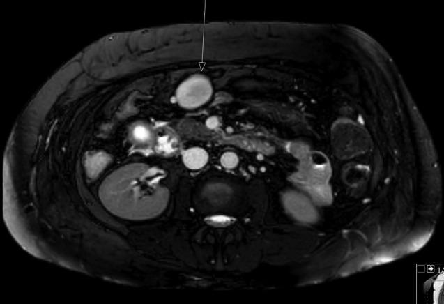
This diagnostic image is an axial T2-weighted MRI slice of the upper abdomen focusing on the hepatobiliary and pancreatic regions. The liver exhibits a nodular surface and coarse signal, characteristic of cirrhosis. Significant secondary findings of portal hypertension are visible, including multiple splenic varices and a prominent, well-defined ovoid structure located anterior to the pancreatic head. This structure, measuring approximately 40 x 28 mm, demonstrates high T2 signal intensity and is adjacent to regional vasculature. The primary educational focus of this image is the recognition of vascular complications associated with end-stage liver disease, specifically the development of varices and portal-systemic shunts. The image also captures the kidneys, aorta, and vertebral column, providing anatomical context for the pathology located in the anterior-superior abdominal quadrant. Clinical relevance includes the diagnostic workup of portal hypertension and the differentiation of vascular lesions from solid pancreatic or mesenteric masses.
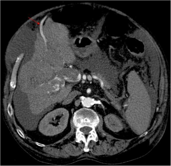
This axial multiplanar reconstruction (MPR) from an early arterial phase CT scan demonstrates advanced hepatic pathology and signs of portal hypertension. The liver parenchyma shows a nodular, heterogeneous contour consistent with cirrhosis. A primary finding is a tumoral portal vein thrombosis, appearing as a heterogeneous enhancing mass within the portal vein lumen, which suggests malignant infiltration (likely from hepatocellular carcinoma). Significant signs of portal hypertension are visible, including a prominent, repermeabilized paraumbilical vein (indicated by the red arrow) coursing anteriorly along the abdominal wall. There is also moderate ascites, visible as low-attenuation fluid surrounding the liver and filling the perihepatic space. The early enhancement of the recanalized paraumbilical vein in the arterial phase, combined with the portal thrombus, is highly suggestive of a malignant arterioportal fistula. This image serves as a clinical illustration of complications related to chronic liver disease and the secondary vascular remodeling occurring in the portal venous system.
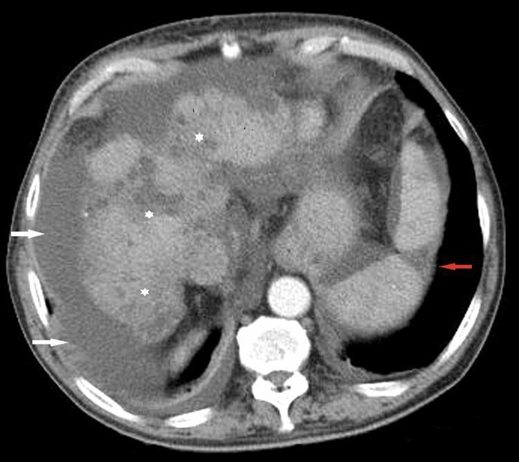
This diagnostic image is an axial contrast-enhanced CT scan of the upper abdomen, demonstrating multiple hallmarks of chronic liver disease and portal hypertension. The liver exhibits classic features of cirrhosis, including prominent surface nodularity and parenchymal heterogeneity (indicated by white asterisks), which give the organ an irregular, bumpy contour. Significant ascites is present, visualized as low-attenuation fluid collections in the perihepatic and perisplenic spaces (highlighted by white arrows). In the spleen, a well-defined, wedge-shaped hypodense area is observed (red arrow), characteristic of a splenic infarct. These findings collectively illustrate the clinical complications of advanced cirrhosis, likely associated with septic embolic events or portal venous system pathology. The image serves as an educational example of radiologic signs in gastroenterology and hepatology, specifically depicting organ morphology changes in end-stage liver disease and associated secondary vascular complications.
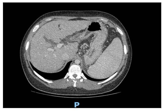
Axial non-contrast abdominal computed tomography (CT) scan illustrating advanced hepatic pathology. The liver demonstrates an inhomogeneous parenchymal appearance with multiple hyperdense regenerative cirrhotic nodules. Anatomical changes include an enlarged caudate lobe and blunt hepatic margins, characteristic of chronic liver disease. Associated secondary findings of portal hypertension are visible, specifically significant splenomegaly and an ectatic portal vein measuring approximately 17 mm. The stomach is visible with a pocket of intraluminal air (hypodense), and the posterior aspect shows the vertebral column and paraspinal musculature. This diagnostic image serves as a clinical example of cirrhosis-related morphological changes and secondary vascular complications within the hepatobiliary system.
Cirrhosis of the Liver and Evaluation of Decompensated Cirrhosis
DEFINITION
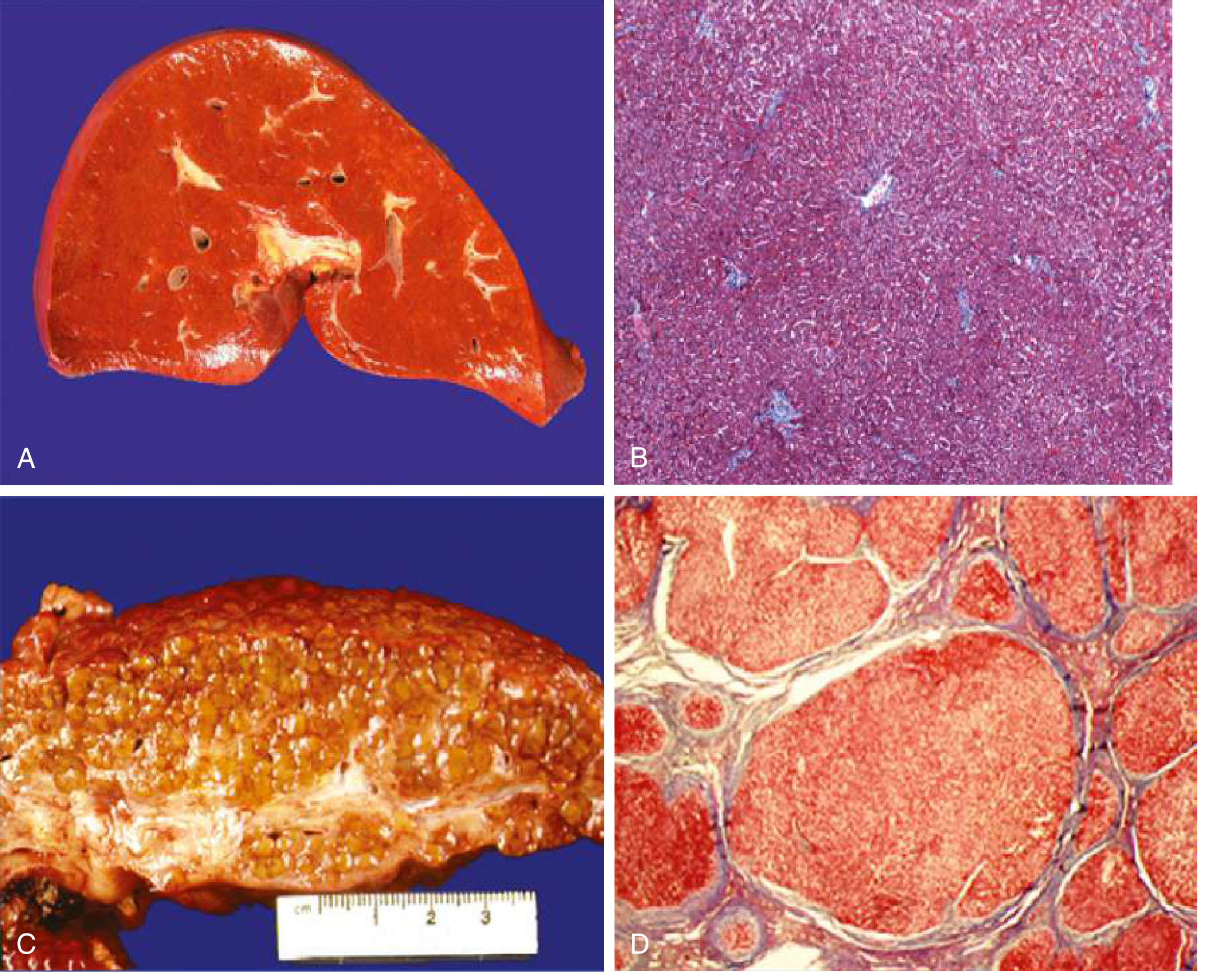
CAUSES OF CIRRHOSIS
| Category | Examples |
|---|---|
| Alcoholic liver disease | Most common in the West |
| Nonalcoholic fatty liver disease (NAFLD/NASH) | Rising globally |
| Chronic Hepatitis B | Most common worldwide |
- Chronic hepatitis C
- Cholestatic/autoimmune: Primary biliary cirrhosis, primary sclerosing cholangitis
- Metabolic: Hemochromatosis, Wilson disease, alpha-1 antitrypsin deficiency, glycogen storage diseases, porphyria
- Hepatic venous outflow obstruction: Budd-Chiari syndrome, veno-occlusive disease, right heart failure
- Biliary: Biliary atresia, cystic fibrosis
- Drugs and toxins, intestinal bypass, Indian childhood cirrhosis
PATHOGENESIS
- Located in the space of Disse between hepatocytes and sinusoidal endothelial cells
- Normally quiescent; serve as the main storage site for retinoids (vitamin A)
- In response to injury: they lose vitamin A, proliferate, develop rough endoplasmic reticulum, secrete extracellular matrix (collagen types I and III, sulfated proteoglycans, glycoproteins), and become contractile myofibroblasts
- This leads to capillarization of sinusoids - collagen deposition in the space of Disse causes defenestration of sinusoidal endothelial cells, altering hepatocyte-plasma exchange
- Fixed component: Fibrosis + compression by regenerative nodules
- Functional component: Active vasoconstriction due to intrahepatic nitric oxide (NO) deficiency + enhanced vasoconstrictors
- Paradox of NO: Intrahepatic NO deficiency causes vasoconstriction; extrahepatic NO overproduction causes splanchnic/systemic vasodilation
COMPENSATED vs DECOMPENSATED CIRRHOSIS
| Feature | Compensated | Decompensated |
|---|---|---|
| Portal pressure (HVPG) | May be <10 mmHg | >12 mmHg |
| Clinical signs | Absent or minimal | Ascites, variceal bleed, encephalopathy, jaundice |
| Median survival | >12 years | 2-5 years |
| 1-year mortality | ~1% | 20-57% depending on complication |
Ascites is the most frequent sign of decompensation, present in 80% of patients with decompensated cirrhosis. - Goldman-Cecil Medicine
EVALUATION OF DECOMPENSATED CIRRHOSIS
1. HISTORY
- Duration and etiology of liver disease
- Alcohol use history (AUDIT score)
- Prior decompensation events (ascites, GI bleeds, encephalopathy)
- Medications (NSAIDs, diuretics, nephrotoxins - avoid in cirrhosis)
- Family history (Wilson disease, hemochromatosis)
- Symptoms: abdominal distension, weight gain, hematemesis/melena, confusion, sleep inversion, jaundice, pruritus
2. PHYSICAL EXAMINATION
- Jaundice, scleral icterus
- Spider nevi (>5 is significant), palmar erythema
- Gynecomastia, loss of axillary/pubic hair, testicular atrophy
- Leukonychia, clubbing, Dupuytren's contracture
- Parotid enlargement (alcoholic)
- Caput medusae (portosystemic collaterals on abdomen)
- Ascites: Bulging flanks, shifting dullness, fluid thrill
- Splenomegaly (hypersplenism)
- Hepatomegaly (early) or small shrunken liver (late)
- Asterixis (flapping tremor) - hallmark of hepatic encephalopathy
- Fetor hepaticus
- Peripheral edema
- Signs of SBP: abdominal tenderness, rebound, guarding
3. LABORATORY INVESTIGATIONS
- AST, ALT (AST:ALT >2 suggests alcoholic; both rarely >300 in ALD)
- Bilirubin (elevated = liver insufficiency)
- Albumin (reduced = impaired synthetic function)
- Prothrombin time / INR (prolonged = impaired coagulation)
- GGT (elevated especially in ALD)
- ALP
- Thrombocytopenia (hypersplenism)
- Anemia (hemorrhage, hemolysis, nutritional)
- Leukopenia (hypersplenism)
- Serum creatinine, BUN, urine electrolytes (urine Na <10 mEq/L suggests hepatorenal syndrome)
- Serum Na (hyponatremia in ~25% of cirrhotic ascites; Na <130 mEq/L is significant)
- Urine output
- HBsAg, anti-HCV (viral hepatitis)
- Serum ferritin, transferrin saturation (hemochromatosis)
- Serum ceruloplasmin, 24-hr urine copper (Wilson disease)
- ANA, anti-smooth muscle Ab, IgG (autoimmune hepatitis)
- AMA (primary biliary cirrhosis)
- Alpha-1 antitrypsin level and phenotype
- Alpha-fetoprotein (AFP) - screen for HCC (every 6 months in cirrhosis)
- Cell count and differential: PMN >250/µL = spontaneous bacterial peritonitis (SBP)
- Culture and sensitivity (inoculate directly into blood culture bottle for best yield)
- SAAG (Serum-Ascites Albumin Gradient): >1.1 g/dL indicates portal hypertension
- Total protein, LDH, glucose (to differentiate SBP from secondary bacterial peritonitis)
- Cytology if malignancy suspected
The diagnosis of SBP is established by ascitic fluid PMN count >250/µL. Bacteria can be isolated in only 40-50% of cases even with sensitive methods. Diagnostic paracentesis should be performed when any patient with cirrhosis is admitted to hospital, regardless of reason for admission. - Goldman-Cecil Medicine
4. IMAGING
- First-line imaging
- Liver: nodularity, echogenicity, size (shrunken in late cirrhosis, enlarged caudate lobe)
- Portal vein diameter (>13 mm = portal hypertension), direction of flow
- Splenomegaly
- Ascites (as little as 100 mL can be detected)
- HCC screening (every 6 months with AFP)
- Better characterization of liver morphology, nodules
- Portal vein thrombosis, collateral vessels, varices
- HCC detection and characterization (arterial enhancement with washout)
- Splenic infarcts
- Useful for HCC characterization (LI-RADS classification)
- Hepatic fat quantification (MR-PDFF)
- Screen for gastroesophageal varices in all newly diagnosed cirrhotic patients
- Grading of varices (small vs large), red wale markings (independent predictors of bleeding)
- ~50% of newly diagnosed cirrhosis have varices; ranges from 40% (Child A) to 85% (Child C)
5. HEPATIC VENOUS PRESSURE GRADIENT (HVPG)
- Gold standard for portal pressure measurement
- Normal: <5 mmHg
- Clinically significant portal hypertension: ≥10 mmHg (varices begin to develop)
- Risk of variceal hemorrhage: ≥12 mmHg (also threshold for ascites formation)
- High-risk: ≥20 mmHg (failure to control acute variceal hemorrhage)
6. LIVER BIOPSY
- Transjugular route preferred (safer in coagulopathy/ascites)
- Confirms diagnosis when imaging is ambiguous
- Staged fibrosis (F0-F4 on METAVIR; F4 = cirrhosis)
- Mandatory in alcohol-associated hepatitis with confounding factors
- Can identify steatohepatitis, Mallory-Denk bodies, chicken-wire fibrosis (alcoholic)
SEVERITY SCORING SYSTEMS
Child-Turcotte-Pugh (CTP) Score
| Parameter | 1 point | 2 points | 3 points |
|---|---|---|---|
| Ascites | None | Grade 1-2 (easy to treat) | Grade 3-4 (refractory) |
| Hepatic encephalopathy | None | Grade 1-2 (or precipitant-induced) | Grade 3-4 (spontaneous) |
| Bilirubin (mg/dL) | <2 | 2-3 | >3 |
| Albumin (g/dL) | >3.5 | 2.8-3.5 | <2.8 |
| PT (sec above control) / INR | <4 / <1.7 | 4-6 / 1.7-2.3 | >6 / >2.3 |
- Child A: 5-6 points (1-year survival ~100%)
- Child B: 7-9 points (1-year survival ~80%)
- Child C: 10-15 points (1-year survival ~45%)
MELD Score (Model for End-Stage Liver Disease)
- Range: 6-40
- Used for prioritizing liver transplant waiting lists
- Incorporates renal function (creatinine) - reflects the hepatorenal axis
- Incorporates serum sodium to better predict mortality
- MELD >20 or Maddrey's Discriminant Function ≥32 defines severe alcohol-associated hepatitis
COMPLICATIONS EVALUATED IN DECOMPENSATED CIRRHOSIS
| Complication | Key Diagnostic Finding |
|---|---|
| Ascites | Shifting dullness; SAAG >1.1; ultrasound |
| SBP | PMN >250/µL in ascitic fluid; fever, abdominal tenderness |
| Variceal hemorrhage | Hematemesis/melena; UGI endoscopy |
| Hepatic encephalopathy | Asterixis; altered consciousness (Grade 1-4); EEG (triphasic waves) |
| Hepatorenal syndrome | Rising creatinine; urine Na <10; no improvement with albumin unless HRS-specific Rx |
| Hepatopulmonary syndrome | PaO₂ <80 mmHg; A-a gradient >15 mmHg; contrast echo showing intrapulmonary shunts |
| Portopulmonary HTN | mPAP >25 mmHg on RHC; echocardiography |
| HCC | AFP + liver ultrasound every 6 months; CT/MRI for characterization |
| Hyponatremia | Na <130 mEq/L; marker of severity and portal hypertension |
West Haven Grading of Hepatic Encephalopathy
- Grade 1: Inversion of sleep-wake pattern, forgetfulness, mild confusion
- Grade 2: Confusion, bizarre behavior, disorientation
- Grade 3: Lethargy, profound disorientation, somnolence
- Grade 4: Coma
Minimal/subclinical encephalopathy (present in up to 80% of patients with cirrhosis) is diagnosed only by psychometric tests (number connection test, digit symbol test, grooved pegboard). - Goldman-Cecil Medicine
HCC SCREENING IN CIRRHOSIS
- Ultrasound + AFP every 6 months in ALL patients with cirrhosis
- If ultrasound shows a nodule >1 cm: multiphasic CT or MRI (LI-RADS criteria)
- HCC screening is recommended regardless of etiology or Child-Pugh class