
gist
What would you like me to do?
for md pathology
Which topic in MD Pathology do you want a gist of? · What format works best for you?
everything molecular pathology diagnosis target therapy, sdh deficient gist
SDH deficient GIST succinate dehydrogenase
GIST gastrointestinal stromal tumor histology immunohistochemistry CD117 DOG1
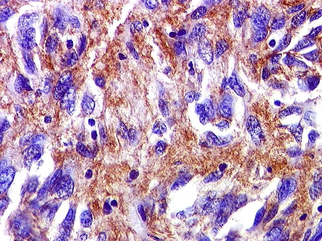
Immunohistochemical slide of a gastrointestinal stromal tumor (GIST) showing robust cytoplasmic/membranous brown staining on a hematoxylin counterstain background. The image likely represents a tissue section stained for CD117 (c-KIT) and/or DOG1, markers that confer high sensitivity and specificity for GIST. In this field, CD34 may be variably positive and SMA positivity can be focal, complicating differential diagnosis with leiomyosarcoma. The tissue is FFPE, sectioned at 4 μm, and stained with DAB chromogen to yield brown immunoreactivity. The tumor cells display spindle-cell morphology in fascicles embedded in a collagenous stroma; nuclei are ovoid to elongated with variable mitotic activity. The immunophenotype—predominant CD117 (c-KIT) and DOG1 positivity—supports the diagnosis of GIST, with DOG1 often providing superior specificity. The combined histology and IHC pattern differentiates GIST from smooth muscle and neural neoplasms. Clinically, this immunoprofile directs targeted therapy options (tyrosine kinase inhibitors such as imatinib or sunitinib) and informs mutational testing for KIT or PDGFRA to predict treatment response. This image is valuable for education in oncologic pathology, GI pathology, and diagnostic immunohistochemistry, illustrating how a mesenchymal GI tumor is identified by characteristic immunostains. It is suitable as a reference for immunophenotype, differential diagnosis, and molecular correlation in GIST research and teaching.
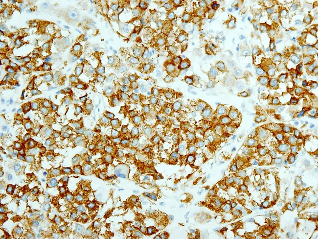
Immunohistochemistry on a formalin-fixed paraffin-embedded tumor section demonstrates strong, diffuse CD117 (c-KIT) positivity with DOG1 co-expression, consistent with a Gastrointestinal Stromal Tumor (GIST). The neoplasm shows rhabdoid features in a high-grade cellular morphology, including large polygonal tumor cells with eosinophilic cytoplasm, eccentrically placed nuclei, and conspicuous nucleoli set in a cutaneous-like solid or nested pattern and occasional spindle cells. The image, captured at high magnification with bright-field microscopy after DAB chromogen staining and hematoxylin counterstain, highlights membranous brown immunoreactivity in most tumor cells. Clinically, the background context describes a 60-year-old man with a perforated primary GIST of the cecum associated with a history of resistance to imatinib (Gleevec) and multiple metastases to liver, intercostal muscles, and diaphragm, indicating aggressive disease. The tumor’s aggressive behavior is supported by large size, necrosis, and a high mitotic index. Immunophenotype with CD117 and DOG1 positivity confirms GIST lineage and excludes many mimics (carcinoid, leiomyosarcoma, desmoid). This case underscores clonal evolution and possible secondary resistance to tyrosine kinase inhibition, with implications for therapy choices such as alternative TKIs (sunitinib, regorafenib) and consideration of surgical debulking if feasible. The finding has diagnostic and prognostic significance, guiding multidisciplinary management and genetic testing for KIT/PDGFRA mutations.
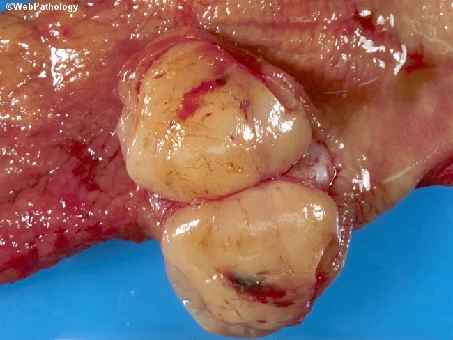
Gross pathology photograph of a resected gastric tumor illustrating the classic appearance of a gastrointestinal stromal tumor (GIST). The specimen demonstrates a solitary, well‑circumscribed, fleshy mass arising within the gastric wall, typically submucosal with a tan-yellow cut surface and occasional surface hemorrhage or focal necrosis. Gastric GISTs represent the most common mesenchymal neoplasms of the abdomen, with a predominance in the stomach (>50%), and most patients presenting in the sixth decade; pediatric cases are rare and may occur in the context of Carney triad. On gross examination, lesions are often exophytic or intraluminal and may appear as encapsulated nodules or plaque‑like thickenings. Definitive diagnosis requires microscopic evaluation and immunohistochemistry, including positivity for KIT (CD117) and DOG1, with variable spindle or epithelioid cell morphology and mitotic activity guiding risk stratification. The illustrated tumor’s solitary, well‑defined margins are consistent with a resectable, localized GIST, but differentiation from other submucosal spindle cell tumors (leiomyoma, leiomyosarcoma, schwannoma) relies on histology and immunophenotyping. Clinically, GISTs demand surgical excision and targeted therapy in select cases, and prognosis depends on size, mitotic rate, location, and rupture risk. This image is valuable for surgical pathology education, gross pathology training, and radiology–pathology correlation discussions.
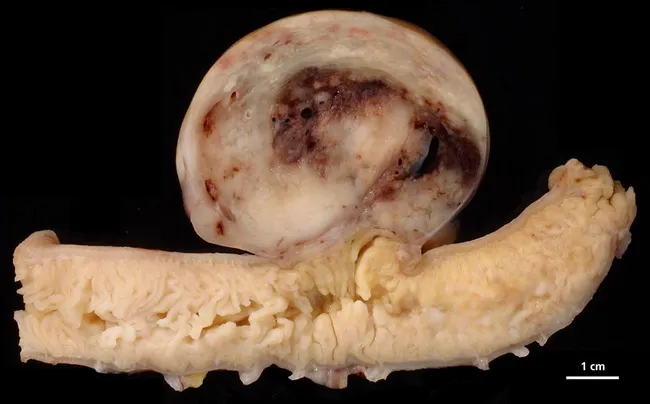
Gross pathology photograph of a resected small intestinal segment showing a subserosal/intramural, well-circumscribed, firm mass compatible with a gastrointestinal stromal tumor (GIST). The tumor appears tan-white with a nodular, rounded contour and a darker central area suggesting hemorrhagic or necrotic change; a scale bar indicates 1 cm and the lesion measures approximately 1.5–2 cm in diameter. GISTs originate from interstitial cells of Cajal within the bowel wall and often harbor activating mutations in CKIT (CD117) or PDGFRA. Clinically, GISTs present with abdominal pain, GI bleeding, or obstruction depending on size and location; prognosis correlates with tumor size, mitotic rate, and tumor location. On gross examination, these tumors are typically firm and well circumscribed within the GI tract and may show mucosal involvement or ulceration. Histology commonly reveals spindle or epithelioid cells in fascicles; immunohistochemistry shows CD117 and DOG1 positivity. Differential diagnosis includes leiomyoma, leiomyosarcoma, schwannoma, desmoid-type fibromatosis, and fibroma. Definitive diagnosis requires histopathology and mutational analysis; management centers on surgical resection and targeted therapy with tyrosine kinase inhibitors when appropriate. This image serves educational value for surgical pathology, gastrointestinal oncology, and radiology correlation, illustrating how gross features guide sampling, indicate intramural origin, and prompt targeted mutational testing for prognosis and therapy planning.
GIST - Complete Molecular Pathology, Diagnosis & Targeted Therapy Gist
1. OVERVIEW
- Every GIST has metastatic potential (no "benign" GIST)
- Behavior predicted by size + mitotic rate (except SDH-deficient type)
- Most common site: Stomach (>50%), then small intestine
2. MOLECULAR CLASSIFICATION
| Subtype | Frequency | Key Mutation | Imatinib Response |
|---|---|---|---|
| KIT-mutant | ~75-80% | KIT exons 11, 9, 13, 17 | Yes (most) |
| PDGFRA-mutant | ~5-10% | PDGFRA exon 18 (D842V most common) | Poor/Resistant |
| SDH-deficient | ~5-10% of gastric | SDHA/B/C/D loss | Resistant |
| NF1-associated | Rare | NF1 gene | Variable |
| BRAF V600E | Rare | BRAF exon 15 | Resistant |
| NTRK-fused | Very rare | ETV6-NTRK | Resistant to TKIs |
3. KIT SIGNALING PATHWAY (The Core Mechanism)
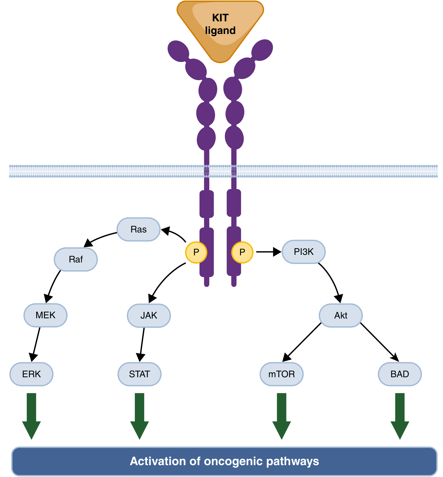
- MAPK pathway: RAS → RAF → MEK → ERK
- PI3K/AKT/mTOR pathway
- JAK/STAT pathway
KIT Exon Mutation Details
| Exon | Domain Encoded | Imatinib Response |
|---|---|---|
| Exon 11 (most common ~70%) | Intracellular juxtamembrane | Best (~85%) |
| Exon 9 | Extracellular domain | Moderate (~40-50%) |
| Exon 13 | Kinase domain | Moderate |
| Exon 17 | Kinase domain + D816V = resistance | Resistant to imatinib |
High-yield: Exon 11 deletions/stop codons = poorer prognosis. KIT and PDGFRA mutations are mutually exclusive.
4. SDH-DEFICIENT GIST - IN DEPTH
Pathogenesis
- Germline loss-of-function mutations in SDHA, B, C, or D (Carney-Stratakis syndrome)
- Epigenetic silencing - hypermethylation of SDHC promoter (Carney triad - non-hereditary)
- Somatic SDH mutations - sporadic cases
Distinguishing Features vs Conventional GIST
| Feature | Conventional GIST | SDH-Deficient GIST |
|---|---|---|
| KIT/PDGFRA mutations | Present | Absent (wild-type) |
| Patient age | Adults (6th decade) | Younger patients, pediatric |
| Sex | M = F | Female predominance |
| Location | Anywhere (stomach most common) | Stomach, almost exclusively |
| Morphology | Spindle cell (mainly) | Epithelioid, multinodular/plexiform |
| Nodal metastases | Rare | Common |
| Prognosis prediction | Size + mitotic rate | Cannot predict by size/mitotic rate |
| Clinical course | Aggressive if high risk | Indolent despite nodal mets |
| Imatinib response | Good (KIT ex11) | Resistant |
IHC Diagnosis of SDH-Deficient GIST
- Loss of SDHB staining on IHC = diagnostic hallmark
- SDHB is used as a surrogate even when SDHA, C, or D is mutated (because SDHB is destabilized in all SDH subunit defects)
- Overexpression of IGF-1R (insulin-like growth factor-1 receptor) - unique to this subtype
- Still positive for CD117 (c-KIT) and DOG1 (like all GISTs)
5. DIAGNOSIS - FULL PANEL
Histopathology Morphology
- Spindle cell type (~70%): elongated cells in fascicles
- Epithelioid type (~20%): rounded cells with clear cytoplasm
- Mixed type (~10%)
Immunohistochemistry (IHC) Markers
| Marker | Sensitivity | Notes |
|---|---|---|
| CD117 (c-KIT) | >95% | Strong diffuse cytoplasmic staining |
| DOG1 (ANO1) | ~95% | Superior specificity, especially for KIT-negative GISTs |
| CD34 | ~70% | Variable |
| SMA | ~30-40% | Focal |
| S100 | <5% | Helps exclude neural tumors |
| Desmin | Rare | Helps exclude smooth muscle tumors |
| SDHB (loss) | SDH-deficient only | Key IHC for wild-type GIST workup |
Molecular Testing (When to Do)
- KIT - exons 9, 11, 13, 17
- PDGFRA - exons 12, 18
- If KIT/PDGFRA wild-type: test for SDHx (germline + somatic), BRAF, NTRK fusions, NF1
- NGS panel now preferred over sequential testing
6. RISK STRATIFICATION
| Risk | Size | Mitotic Rate | Location |
|---|---|---|---|
| Very Low | <2 cm | <5/5mm² | Any |
| Low | 2-5 cm | <5/5mm² | Any |
| Intermediate | <5 cm or 5-10 cm | 6-10/5mm² | Gastric |
| High | >5 cm, >10 cm, or any with rupture | >5-10/5mm² | Any |
Exception: SDH-deficient GISTs - risk cannot be predicted by these criteria.
7. TARGETED THERAPY - LINE BY LINE
First-Line: Imatinib (Gleevec/Glivec)
- Mechanism: Competitive inhibitor of KIT/PDGFRA tyrosine kinase
- Best response: KIT exon 11 mutations (~85% response rate)
- Dose: 400 mg/day standard; 800 mg/day for KIT exon 9 mutations
- Used for: Metastatic/unresectable GIST, neoadjuvant, adjuvant (3 years)
- Resistance: D816V (exon 17), PDGFRA D842V, SDH-deficient
Second-Line: Sunitinib (Sutent)
- Mechanism: Multi-targeted TKI (KIT, PDGFR, VEGFR)
- Indication: Imatinib-resistant/intolerant GIST
- Some benefit in SDH-deficient and wild-type GISTs
Third-Line: Regorafenib (Stivarga)
- Mechanism: Multi-kinase inhibitor (KIT, PDGFR, VEGFR, RAF)
- Indication: After imatinib + sunitinib failure
Fourth-Line: Ripretinib (Qinlock)
- Mechanism: "Switch-control" inhibitor - stabilizes KIT/PDGFRA in inactive conformation
- Phase III: PFS 6.3 months vs 1 month placebo; OS 15 vs 6 months
- Note: Not superior to sunitinib in 2nd line; standard 4th line
Special Mutations - Precision Therapy
| Mutation | Drug | Notes |
|---|---|---|
| PDGFRA D842V (exon 18) | Avapritinib (Blueprint) | FDA-approved 1st line for D842V specifically |
| BRAF V600E | Dabrafenib + Trametinib | Combo MEK/BRAF inhibition |
| NTRK fusion | Larotrectinib / Entrectinib | TRK inhibitors |
| SDH-deficient | Sunitinib or Regorafenib (some) | No established standard; refer to specialized center; IGF-1R targeting under investigation |
8. SDH-DEFICIENT GIST - MANAGEMENT SPECIFICS
- Surgery: Selective; aggressive resection NOT recommended (indolent course)
- Manage lesions causing symptoms (bleeding, obstruction)
- Imatinib: Generally ineffective
- Sunitinib/Regorafenib: Some reported benefit for progressive disease
- Refer to specialized center - no established standard of care
- Screen for associated syndromes (see below)
9. HEREDITARY SYNDROMES WITH GIST
| Syndrome | Genetics | GIST Features | Other Features |
|---|---|---|---|
| Carney-Stratakis Dyad | Germline SDHx (A/B/C/D) | Gastric, multifocal, SDH-deficient | Paraganglioma |
| Carney Triad | Epigenetic SDHC silencing (non-germline) | Gastric, multifocal, SDH-deficient | Paraganglioma + Pulmonary chondroma |
| NF1 (Neurofibromatosis type 1) | NF1 germline | Small bowel, multifocal, KIT/PDGFRA wild-type, KIT overexpressed | Neurofibromas, cafe-au-lait spots |
| Familial GIST | Germline KIT or PDGFRA | Multifocal, any site | Skin hyperpigmentation (KIT); fibrous polyps (PDGFRA) |
Key for exam: Carney Triad = NOT hereditary (epigenetic SDHC). Carney-Stratakis = hereditary (germline SDHx). Both have SDH-deficient GISTs with lost SDHB on IHC.
10. SURVEILLANCE POST-RESECTION
- Serial imaging every 3-6 months x 3-5 years, then annual indefinitely
- Low-risk tumors: individualized follow-up
- Relapses primarily: liver and peritoneum
- SDH-deficient patients: also screen for paragangliomas (neck, chest, abdomen) and refer for genetic counseling
QUICK REVISION BOX
GIST at a glance:
- Cell of origin: ICC
- Most common location: Stomach
- KIT mutation most common: Exon 11 (best imatinib response)
- Wild-type GIST → think SDH-deficient
- SDH-deficient GIST: young females, stomach, epithelioid,
  multifocal, nodal mets, indolent, SDHB-IHC lost
- IHC: CD117 + DOG1 = GIST; SDHB loss = SDH-deficient subtype
- TKI line-up: Imatinib → Sunitinib → Regorafenib → Ripretinib
- PDGFRA D842V → Avapritinib (1st line)
- SDH-deficient → no TKI standard; sunitinib/regorafenib tried
short it
GIST - Molecular Pathology Gist (Short)
Cell of Origin
Molecular Subtypes
| Mutation | Frequency | Imatinib |
|---|---|---|
| KIT exon 11 | ~70% | Best response |
| KIT exon 9 | ~10% | Moderate (need 800mg) |
| PDGFRA D842V | ~5% | Resistant → Avapritinib |
| SDH-deficient | ~5-10% gastric | Resistant |
| BRAF/NTRK/NF1 | Rare | Resistant |
Diagnosis IHC
- CD117 (c-KIT) + DOG1 = GIST (both >95%)
- SDHB loss on IHC = SDH-deficient subtype
- Wild-type KIT/PDGFRA → do NGS (SDHx, BRAF, NTRK)
SDH-Deficient GIST - Key Points
- Who: Young females, pediatric age group
- Where: Stomach, multifocal
- Morphology: Epithelioid, multinodular/plexiform
- Unique: Nodal mets common, but indolent course
- Cannot predict behavior by size/mitotic rate
- IHC: SDHB lost; IGF-1R overexpressed
- Syndromes:
- Carney-Stratakis dyad = germline SDHx + paraganglioma (hereditary)
- Carney triad = SDHC epigenetic silencing + paraganglioma + pulmonary chondroma (NOT hereditary)
Targeted Therapy Lines
| Line | Drug | Target |
|---|---|---|
| 1st | Imatinib | KIT/PDGFRA |
| 1st (D842V) | Avapritinib | PDGFRA D842V |
| 2nd | Sunitinib | Multi-TKI |
| 3rd | Regorafenib | Multi-kinase |
| 4th | Ripretinib | Switch-control KIT/PDGFRA |
| BRAF V600E | Dabrafenib + Trametinib | BRAF/MEK |
| NTRK fusion | Larotrectinib/Entrectinib | TRK |
| SDH-deficient | No standard - sunitinib/regorafenib tried | Refer specialist |
Risk (Conventional GIST only)
SDH-deficient = excluded from this risk model
colorectal polyp and recent who classification
WHO 2019 5th edition colorectal polyp classification serrated lesions updates
colorectal polyp types adenoma tubular villous serrated histology
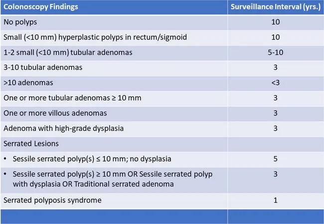
This endoscopic colonoscopy findings table summarizes colorectal polyp burden and recommended surveillance intervals based on histology and size. The patient’s mucosa is evaluated for polyps in the colon, including rectum and sigmoid segments, with findings categorized by lesion type and size. Notable entities include hyperplastic polyps (small versus diminutive), tubular adenomas (1–2 small <10 mm; 3–10 lesions; or a single large ≥10 mm lesion), and adenomas with villous features or high‑grade dysplasia. Serrated polyps are documented as sessile serrated polyps <10 mm or ≥10 mm, with or without dysplasia, and the spectrum also includes traditional serrated adenomas and the context of serrated polyposis syndrome. The surveillance interval column reflects guideline-driven risk stratification: absence of polyps generally warrants longer intervals (approximately 10 years); 1–2 small adenomas suggests 5–10 years; 3–10 adenomas or any ≥10 mm adenoma typically yields shorter intervals around 3 years; multiple lesions or villous histology often returns the most conservative intervals (<3 years). These data guide post‑colonoscopy management, need for repeat colonoscopy, and colorectal cancer risk assessment. Clinicians use it to tailor follow‑up, stratify cancer risk, and educate patients about polyp prevention strategies and lifestyle modification; it also informs research on polyp biology and serrated pathway carcinogenesis outcomes.
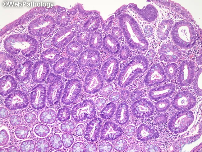
High-magnification histology image of a colonic adenomatous polyp in familial adenomatous polyposis (FAP). Modality/technique: light microscopy of a formalin-fixed, paraffin-embedded tissue section stained with Hematoxylin and Eosin (H&E). Specimen: colorectal mucosa, polypoid lesion arising from the tubular epithelium. Anatomical site: large intestine (colon), colonic mucosa with tubular adenomatous glands, showing back-to-back architecture and crowded, elongated glands with pseudostratified, hyperchromatic nuclei. Goblet cells are variably decreased; mucin depletion may be present. The epithelial cell layer exhibits dysplastic features including nuclear atypia, increased mitotic activity in the basal thirds, and loss of mucosal polarity. Overall polyp displays a tubular histology pattern with uniform, round luminal profiles and regular basement membrane. The stromal compartment is relatively dense with fibromuscular cores commonly seen in polyps; no definite invasion beyond the lamina propria is evident at this magnification. In the context of FAP, such adenomas demonstrate a high propensity for rapid development and progression toward colorectal carcinoma if not managed with surveillance and prophylactic colectomy. Clinically, this finding supports genetic testing for APC mutation and intensified colonoscopic surveillance; differential diagnoses include tubulovillous adenoma, villous adenoma, serrated lesions, or mucosal hyperplasia. Correlation with family history and prior polyp burden is critical for risk stratification and surgical planning.
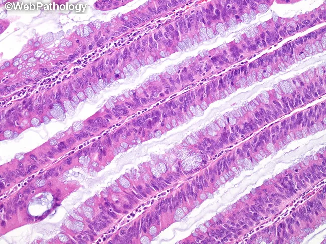
This histology image depicts colonic mucosa displaying villous adenomatous architecture with low-grade dysplasia. Prepared as a routine hematoxylin and eosin stained section, the villous processes are lined by columnar epithelium exhibiting architectural crowding, branching, and cryptal pseudostratification. There is hypercellularity with nuclear overcrowding; nuclei are moderately enlarged and elongated, hyperchromatic yet maintaining orderly polarity. Nucleoli are not prominent. Goblet cell mucin shows partial loss, contributing to a basophilic cytoplasmic appearance on H&E. Mitotic activity is not significantly increased, arguing against high-grade features. The mucosal glands demonstrate lack of maturation from crypt base to luminal surface, with vertical growth and irregular gland size; mucin depletion and basophilia accentuate the neoplastic epithelium. Overall, the lesion is an adenomatous polyp with villous architecture and low-grade dysplasia, a premalignant neoplasm in the colorectal tract. On imaging and clinical context this corresponds to a colon polyp with villous pattern, requiring complete endoscopic resection and histologic subtyping to determine malignant potential. Diagnostic significance includes risk stratification for progression to high-grade dysplasia or invasion; differential diagnoses include tubular adenoma with villous features, mixed adenoma, serrated lesions; clinical correlation: colonoscopy findings, patient age, and polyp burden inform management. Planning for surveillance is essential.
Colorectal Polyps - Complete Gist + WHO 5th Edition (2019) Classification
WHO 5th Edition (2019) - Master Classification
| Category | Types |
|---|---|
| Inflammatory | Pseudopolyps (IBD) |
| Hamartomatous | Juvenile polyp, Peutz-Jeghers polyp |
| Serrated polyps | Hyperplastic polyp (HP), Sessile serrated lesion (SSL), SSL with dysplasia, Traditional serrated adenoma (TSA), Mixed polyp |
| Adenoma | Tubular, Tubulovillous, Villous |
| Malignant polyp | Adenocarcinoma |
| Submucosal | Lipoma, GIST, Carcinoid, Lymphoid, Leiomyoma |
KEY WHO 2019 CHANGES (exam-critical)
| Old Term (WHO 4th, 2010) | New Term (WHO 5th, 2019) |
|---|---|
| Sessile serrated adenoma (SSA) | Sessile serrated lesion (SSL) |
| SSA/P (sessile serrated adenoma/polyp) | SSL - SSA and SSP terms no longer recommended |
| SSA with dysplasia (low/high grade) | SSL with dysplasia - no further LGD/HGD stratification |
| Hyperplastic polyp subtypes same | HP retained; 3 subtypes: MVHP, GCHP, Mucin-poor |
Bottom line: "Adenoma" dropped from the sessile serrated lesion name because SSLs arise via a different (non-conventional dysplasia) pathway. Clinical management guidelines for SSA/P still apply to SSL.
1. NON-NEOPLASTIC POLYPS
Hyperplastic Polyp (HP)
- Most common polyp overall (~75% of serrated polyps)
- Location: Rectosigmoid, small (<5 mm), multiple
- Morphology: Serrated/sawtooth epithelium, NO dysplasia, NO distorted crypts
- Subtypes (WHO 2019):
- MVHP (Microvesicular) - most common, precursor to SSL
- GCHP (Goblet cell) - goblet cell-rich, low malignant potential
- Mucin-poor - rare
- Molecular: BRAF mutation (MVHP) or KRAS mutation (GCHP)
- Generally benign but large (≥10 mm) proximal HPs = managed like SSL
Inflammatory / Pseudopolyps
- IBD (UC > Crohn's)
- Regenerative mucosa, NO malignant potential on their own
Hamartomatous Polyps
| Type | Syndrome | Gene | Features |
|---|---|---|---|
| Juvenile polyp | Juvenile polyposis | SMAD4/BMPR1A | Solitary = benign; syndrome = CRC risk |
| Peutz-Jeghers polyp | PJS | STK11/LKB1 | Smooth muscle core, GI + mucocutaneous pigmentation |
2. NEOPLASTIC POLYPS - ADENOMAS (Conventional Pathway)
Pathway: APC → KRAS → SMAD4/TP53 (Chromosomal Instability - CIN)
Histological Types
| Type | Frequency | Morphology | CRC Risk |
|---|---|---|---|
| Tubular | 75-85% | Branching tubular glands, usually pedunculated | Lowest (1-5%) |
| Tubulovillous | 8-16% | Mixed (25-75% villous) | Intermediate |
| Villous | 5-10% | Finger-like projections, usually sessile/broad base | Highest (40% if >2 cm) |
Rule: Villous if ≥80% villiform features. Tubular if <25% villous.
Dysplasia Grading
| Grade | Features |
|---|---|
| Low grade (LGD) | Nuclei basally oriented, mild nuclear enlargement, maintained polarity |
| High grade (HGD) | Nuclear stratification, loss of polarity, mucin depletion, gland crowding |
HGD = "carcinoma in situ" / "intraepithelial carcinoma" (old terms) - same thing
Advanced Adenoma = ANY of:
- Size >10 mm, OR
- Villous histology (≥25% villous), OR
- High-grade dysplasia
Malignant Polyp
- Invasive carcinoma into submucosa (pT1)
- Haggitt classification (pedunculated):
- Level 1: Carcinoma in head
- Level 2: Neck
- Level 3: Stalk
- Level 4: Base/submucosa (high risk)
- Level 4 or sessile with submucosal invasion = surgical resection
Size vs Malignancy Risk
- <1 cm: ~1-2% carcinoma
- 1-2 cm: ~10%
-
2 cm: ~50%
3. SERRATED PATHWAY POLYPS (WHO 2019 Focus)
Serrated Pathway: BRAF → CpG Island Methylator Phenotype (CIMP) → MLH1 silencing → MSI-H CRC
Sessile Serrated Lesion (SSL) - WHO 2019 name
| Feature | Detail |
|---|---|
| Old name | SSA, SSP, SSA/P |
| Location | Right colon (proximal), flat/sessile |
| Size | Usually >5 mm |
| Morphology | Distorted crypt architecture: L-shaped, T-shaped, horizontal crypts; crypt dilatation; goblet cells at crypt base |
| Key diagnostic criterion (WHO 2019) | Single unequivocal architecturally distorted serrated crypt is sufficient for diagnosis |
| NOT sufficient alone | Crypt dilatation, occasional branching, goblet cells at base |
| Molecular | BRAF mutation, CIMP-high, MLH1 methylation |
| Malignant potential | Yes - leads to MSI-H CRC |
SSL with Dysplasia (SSLD)
- WHO 2019: No LGD/HGD stratification - just "SSL with dysplasia"
- Conventional adenomatous, TSA-like, or serrated-type dysplasia all recognized
- ~5% of SSLs develop dysplasia
- Highest CRC risk among serrated polyps
Traditional Serrated Adenoma (TSA)
| Feature | Detail |
|---|---|
| Location | Distal colon, rectum |
| Morphology | Polypoid, often pedunculated |
| Diagnosis (WHO 2019) | Requires 2 of 3 features: (1) Slit-like serration, (2) Ectopic crypt foci, (3) Tall columnar cells with eosinophilic cytoplasm |
| Molecular | KRAS or BRAF mutation, CIMP-low |
| Frequency | Rarest serrated type (~5%) |
4. SERRATED POLYPOSIS SYNDROME (SPS) - WHO 2019 Updated Criteria
| Criterion | Threshold |
|---|---|
| Criterion 1 | ≥5 serrated lesions/polyps proximal to rectum, all ≥5 mm, with ≥2 being ≥10 mm |
| Criterion 2 | >20 serrated lesions/polyps of any size throughout the colon, with ≥5 proximal to rectum |
Key changes:
- Polyp count is cumulative over multiple colonoscopies
- Any histological subtype counts (HP, SSL, TSA all included)
- Distal/rectal polyps now included (with restrictions)
- Old criterion 2 (family history-based) removed
- CRC risk in SPS: 15-30%; first-degree relatives have 5x CRC risk
5. ADENOMA-CARCINOMA SEQUENCE vs SERRATED PATHWAY
| Feature | Conventional (CIN) | Serrated (CIMP) |
|---|---|---|
| Polyp type | Tubular/villous adenoma | SSL, TSA |
| Key mutation | APC → KRAS → TP53 | BRAF → CIMP → MLH1 loss |
| CIN/MSI | CIN (chromosomal instability) | MSI-H (microsatellite instability) |
| % of CRC | ~70-75% | ~25% |
| Location | Left colon predominant | Right colon predominant |
6. SURVEILLANCE (Post-Polypectomy)
| Finding | Repeat Colonoscopy |
|---|---|
| No polyps / HP <10mm distal | 10 years |
| 1-2 tubular adenomas <10mm, LGD | 5-10 years |
| 3-4 adenomas OR 1 adenoma ≥10mm | 3 years |
| ≥5 adenomas / ≥1 adenoma with HGD | 1-3 years |
| SSL <10mm, no dysplasia | 5 years |
| SSL ≥10mm OR SSL with dysplasia | 3 years |
| TSA | 3 years |
| Serrated polyposis syndrome | Yearly colonoscopy |
reporting protocol for colorectal carcinoma
CAP colorectal carcinoma synoptic reporting protocol 2023 2024 checklist elements
colorectal carcinoma pathology reporting protocol CAP elements pT staging CRM tumor deposits budding MMR 2024
https://documents.cap.org/documents/ColoRectal_4.3.1.0.REL.C…
Colorectal Carcinoma - Pathology Reporting Protocol
STRUCTURE OF SYNOPTIC REPORT
- Core/Required - mandatory in every report
- Conditional - required only when applicable
- Non-core/Optional (+) - recommended but not required
SECTION 1: SPECIMEN / PROCEDURE
| Element | Options |
|---|---|
| Procedure type | Right hemicolectomy, Left hemicolectomy, Sigmoidectomy, Low anterior resection (LAR), Abdominoperineal resection (APR), Total abdominal colectomy |
| Tumor site | Cecum, ascending colon, hepatic flexure, transverse colon, splenic flexure, descending colon, sigmoid, rectosigmoid, rectum |
| Macroscopic mesorectum quality | Required for rectal specimens: Complete / Nearly complete / Incomplete (Quirke grading) |
SECTION 2: GROSS FINDINGS
| Element | Detail |
|---|---|
| Tumor size | Greatest dimension in mm (3 dimensions if applicable) |
| Tumor configuration | Polypoid, ulcerating, annular/stenosing, diffusely infiltrative |
| Distance from margins | Proximal, distal, radial/CRM (in mm) |
| Perforation | Present / Not identified (pT4a + adverse prognostic indicator) |
SECTION 3: HISTOLOGICAL TYPE (WHO Classification)
| Type | Notes |
|---|---|
| Adenocarcinoma NOS | Most common (~85%) |
| Mucinous adenocarcinoma | >50% extracellular mucin; poor prognosis |
| Signet ring cell carcinoma | >50% signet ring cells; worst prognosis |
| Medullary carcinoma | Solid, abundant TILs; MSI-H association |
| Micropapillary carcinoma | Aggressive, LVI prone |
| Serrated adenocarcinoma | BRAF mutation, serrated pathway |
| Adenosquamous carcinoma | Rare |
| Undifferentiated carcinoma | No glandular differentiation |
Grade not assigned to mucinous, signet ring, medullary, or undifferentiated types - classified by definition
SECTION 4: HISTOLOGIC GRADE
| Grade | Gland Formation | Equivalent |
|---|---|---|
| G1 - Well differentiated | >95% glands | Low grade |
| G2 - Moderately differentiated | 50-95% glands | Low grade |
| G3 - Poorly differentiated | <50% glands | High grade |
| G4 - Undifferentiated | No glands | High grade |
AJCC 8th ed uses 2-tier: Low grade (G1+G2) vs High grade (G3+G4)
SECTION 5: EXTENT OF INVASION - pT STAGING (AJCC 8th Ed)
| pT | Extent |
|---|---|
| pTis | Carcinoma in situ (HGD); intramucosal carcinoma (lamina propria/muscularis mucosae) |
| pT1 | Invades submucosa |
| pT2 | Invades muscularis propria |
| pT3 | Through muscularis propria into pericolorectal tissues |
| pT3 substaging (optional) | pT3a: <1mm beyond MP; pT3b: 1-5mm; pT3c: 5-15mm; pT3d: >15mm |
| pT4a | Penetrates visceral peritoneum (serosal surface) |
| pT4b | Directly invades adjacent organs/structures |
Exam tip: pT4a = peritoneal penetration (serosa); pT4b = invasion of other organs. pTis = NO invasion through muscularis mucosae.
SECTION 6: MARGINS
| Margin | Reporting Standard |
|---|---|
| Proximal / Distal margins | Positive (tumor at ink) / Negative (distance in mm) |
| Radial / Circumferential Resection Margin (CRM) | Required for rectal cancers and retroperitoneal colon (ascending/descending); report distance in mm |
| CRM positive | Tumor ≤1 mm from CRM = positive (high local recurrence risk) |
| Serosal margin | For tumors reaching or breaching serosa (colon) |
Rectal CRM gold standard: ≤1 mm = positive → local recurrence ↑ 3-4x
SECTION 7: LYMPHOVASCULAR AND PERINEURAL INVASION
| Parameter | Notes |
|---|---|
| Lymphatic (small vessel) invasion (LVI) | D2-40 or CD31 IHC if equivocal |
| Venous invasion - intramural | Within bowel wall |
| Venous invasion - extramural (EMVI) | Elastic stain (Movat/EVG) helpful; strong independent poor prognostic marker |
| Perineural invasion (PNI) | Independent poor prognostic factor; required core element |
SECTION 8: TUMOR BUDDING (CAP 2024 - NOW REQUIRED)
| Score | Bud Count | Clinical Implication |
|---|---|---|
| Bd1 - Low | 0-4 buds | Favorable |
| Bd2 - Intermediate | 5-9 buds | Intermediate risk |
| Bd3 - High | ≥10 buds | High risk - lymph node mets ↑, worse survival |
Key rules:
- Required for conventional adenocarcinoma (well/moderately differentiated)
- NOT applicable for mucinous, signet ring, or micropapillary carcinoma
- Bd2/Bd3 in pT1 = increased risk of LN mets → consider surgery even after complete polypectomy
SECTION 9: LYMPH NODES - pN STAGING
| pN | Definition |
|---|---|
| pN0 | No regional LN metastasis (minimum 12 nodes required) |
| pN1a | 1 positive regional LN |
| pN1b | 2-3 positive regional LNs |
| pN1c | Tumor deposits without positive LNs (satellite nodules) |
| pN2a | 4-6 positive regional LNs |
| pN2b | ≥7 positive regional LNs |
Report: Total nodes examined / Total nodes positive (e.g., 3/18) Minimum harvest = 12 lymph nodes (AJCC recommendation)
SECTION 10: TUMOR DEPOSITS (Satellites)
- Discrete nodules in pericolorectal fat in the lymph drainage area
- Discontinuous from primary tumor
- No residual LN architecture, identifiable vessel, or nerve
- Do NOT add to positive LN count
- If nodes are negative + deposits present → pN1c
- If nodes positive → deposits do not change N category (but note number separately)
- Do NOT confuse with:
- Venous invasion (around a vein) → not a deposit
- Peritoneal seeding → pM1c (not a deposit)
SECTION 11: DISTANT METASTASIS - pM
| pM | Site |
|---|---|
| pM1a | 1 distant site (e.g., liver) |
| pM1b | ≥2 distant sites |
| pM1c | Peritoneal metastasis (with or without other distant sites) |
SECTION 12: TREATMENT EFFECT (Post-Neoadjuvant - Rectal Cancer)
| Grade | Description |
|---|---|
| Grade 0 - Complete response | No viable tumor cells (ypT0) |
| Grade 1 - Near complete | Single cells / small groups, no residual mass |
| Grade 2 - Partial response | Residual cancer with fibrosis |
| Grade 3 - Poor/No response | Minimal or no tumor cell kill; extensive residual |
Report: ypTNM staging (y prefix = post-treatment)
SECTION 13: ANCILLARY STUDIES (Required / Recommended)
| Test | Status | Indication |
|---|---|---|
| MMR (MLH1, MSH2, MSH6, PMS2) by IHC | Required | All CRC - Lynch syndrome screen; immunotherapy eligibility |
| MSI testing (PCR) | Conditional | If MMR IHC equivocal |
| KRAS/NRAS mutation | Required for metastatic CRC | Anti-EGFR (cetuximab/panitumumab) - only WT responds |
| BRAF V600E | Required for metastatic CRC | Prognostic; encorafenib eligibility |
| HER2 amplification | Conditional | Metastatic CRC if RAS/BRAF WT |
| NTRK fusion | Conditional | Larotrectinib/entrectinib |
SECTION 14: HIGH-RISK FEATURES - STAGE II (Drives Adjuvant Chemo Decision)
| Feature | Type |
|---|---|
| pT4a (serosal penetration) | Pathologic |
| pT4b (adjacent organ invasion) | Pathologic |
| Tumor perforation | Pathologic/Clinical |
| CRM positive or close (rectal) | Pathologic |
| LVI present | Pathologic |
| PNI present | Pathologic |
| High-grade (G3/G4) | Pathologic |
| <12 LNs examined | Pathologic |
| High tumor budding (Bd3) | Pathologic |
| EMVI present | Pathologic |
| MMR proficient (pMMR) | Molecular |
Stage II CRC + ≥1 high-risk feature → adjuvant FOLFOX typically recommended
QUICK SUMMARY CHECKLIST (Exam Format)
1. Procedure + Site
2. Tumor size + configuration
3. Histologic type
4. Grade (G1/2/3 or Low/High)
5. pT (depth of invasion)
6. Margins: proximal / distal / CRM (mm)
7. LVI (lymphatic + venous - intramural/extramural)
8. PNI
9. Tumor budding: Bd1/Bd2/Bd3
10. Lymph nodes: x/y positive; tumor deposits
11. pN stage
12. pM (if applicable)
13. pTNM overall stage
14. Treatment effect (ypTNM if post-neoadjuvant)
15. MMR IHC (MLH1/MSH2/MSH6/PMS2)
16. Molecular (KRAS/NRAS/BRAF/HER2/NTRK for metastatic)
gut microbiome and ibd
gut microbiome dysbiosis inflammatory bowel disease
PMID: 40065181
gut microbiota FMT fecal transplant IBD treatment
Gut Microbiome & IBD - Complete Gist
1. NORMAL GUT MICROBIOME - BASELINE
- ~38 trillion bacteria (≈ equal to human cells), plus fungi (mycobiome), viruses (virome), archaea
- Dominant phyla: Firmicutes (~65%), Bacteroidetes (~25%), Proteobacteria, Actinobacteria, Verrucomicrobia
- Key function: fermentation → short-chain fatty acids (SCFAs): butyrate, propionate, acetate
- Butyrate is the primary energy source for colonocytes and a key anti-inflammatory mediator
2. CORE CONCEPT: HOW MICROBIOME LINKS TO IBD
Genetic susceptibility (NOD2, ATG16L1, IL23R, FUT2...)
         +
Dysbiosis (altered microbiome composition)
         +
Mucosal barrier dysfunction (↑ intestinal permeability, "leaky gut")
         ↓
Bacterial antigen translocation
         ↓
Aberrant mucosal immune activation
         ↓
Chronic intestinal inflammation (CD / UC)
3. DYSBIOSIS IN IBD - SPECIFIC CHANGES
What is LOST (depleted) in IBD:
| Organism | Normal Function | Why Loss Matters |
|---|---|---|
| Faecalibacterium prausnitzii | Major butyrate producer; anti-inflammatory via IL-10 | Reduced in CD and UC; correlates with disease activity |
| Roseburia intestinalis | SCFA producer (butyrate) | Reduced in UC/CD |
| Akkermansia muciniphila | Mucus layer maintenance | Reduced in IBD; barrier dysfunction |
| Bifidobacterium spp. | Colonization resistance; SCFA | Depleted in active IBD |
| Lachnospiraceae family | Butyrate production, immune regulation | Consistently reduced in CD |
| Overall microbial diversity (α-diversity) | Resilience; functional redundancy | Markedly reduced in IBD |
What is GAINED (expanded) in IBD:
| Organism | Role in Disease |
|---|---|
| Enterobacteriaceae (E. coli, esp. AIEC - Adherent Invasive E. coli) | Invades ileal mucosa; key in CD; survives intracellularly in macrophages |
| Fusobacterium nucleatum | Pro-inflammatory; also linked to CRC |
| Ruminococcus gnavus | Expanded in CD; produces immunogenic polysaccharides |
| Candida albicans (fungal) | Expanded in CD; interacts with Th17 immune axis |
| Proteobacteria overall | Expand due to oxidative environment of inflamed gut |
Key exam fact: Proteobacteria thrive in inflamed (oxidative) gut - their expansion is often a consequence of inflammation, not just a cause.
4. MECHANISMS LINKING MICROBIOME TO IBD PATHOGENESIS
A. SCFA Deficiency → Loss of Colonocyte Protection
- Butyrate: inhibits NF-κB, promotes Treg differentiation, maintains tight junctions
- Depleted butyrate → ↑ mucosal inflammation + ↓ barrier integrity
B. Mucosal Barrier Breakdown
- Reduced Akkermansia + butyrate → thin mucus layer + impaired tight junctions (claudin, occludin)
- Bacterial antigens (LPS, peptidoglycans) translocate through epithelium
- Triggers innate immune response (TLR2/4, NOD2)
C. Immune Activation - CD vs UC
| Feature | Crohn's Disease | Ulcerative Colitis |
|---|---|---|
| T-cell polarization | Th1 + Th17 dominant | Th2 dominant (+ Th17) |
| Key cytokines | IL-12, IL-23, IFN-γ, TNF-α, IL-17 | IL-4, IL-13, IL-5 |
| Pattern | Transmural, granulomas | Mucosal/submucosal only |
| Microbiome driver | AIEC invasion of Peyer's patches | Dysbiosis → altered colonocyte metabolism |
D. Genetic-Microbiome Interaction (Key Genes)
| Gene | Function | Microbiome Link |
|---|---|---|
| NOD2 | Intracellular sensor for bacterial muramyl dipeptide | Loss-of-function = impaired bacterial clearance → Crohn's |
| ATG16L1 | Autophagy (Paneth cells) | Impaired autophagy → bacteria persist in macrophages |
| IL23R | IL-23/Th17 axis | Amplifies response to bacterial antigens |
| FUT2 | ABO blood group, mucus fucosylation | Affects microbial colonization; non-secretors have altered microbiome |
E. Cross-Kingdom Interactions (2025 Update)
- Mycobiome (fungi): Candida albicans + Candida tropicalis expanded in CD; interact with bacteria (e.g., F. nucleatum + C. tropicalis form biofilms in CD mouse models)
- Virome: Caudovirales phages altered in IBD; phage-bacteria dynamics influence dysbiosis
- Multi-kingdom perspective now recognized (Iliev et al., Nat Rev Microbiol 2025)
5. CD vs UC - MICROBIOME DIFFERENCES
| Feature | Crohn's Disease | Ulcerative Colitis |
|---|---|---|
| Diversity loss | Profound | Moderate |
| AIEC | Characteristic | Not typical |
| Ileal microbiome | Most disrupted | Less affected |
| Ruminococcus gnavus | ↑↑ in CD | Not specific |
| Fungal component | Candida ↑; Saccharomyces cerevisiae Ab (ASCA) positive | Less prominent |
| Antibody response | ASCA+ (Saccharomyces) | pANCA+ |
6. MICROBIOME-BASED DIAGNOSTICS
| Biomarker | Application |
|---|---|
| Fecal calprotectin | Marker of mucosal inflammation (not microbiome-specific but reflects dysbiosis effect) |
| 16S rRNA sequencing | Research: characterize dysbiosis |
| Metagenomics/Metatranscriptomics | Functional microbiome profiling |
| Faecalibacterium prausnitzii levels | Research predictor of post-surgical CD relapse |
| Mycobiome profiling | Research: Candida expansion in CD |
7. MICROBIOME-TARGETED THERAPIES
A. Fecal Microbiota Transplantation (FMT)
| Feature | Detail |
|---|---|
| UC | RCTs show remission ~24-32% vs ~5% placebo; FDA-approved FMT (Rebyota, Vowst) for recurrent C. diff, trials ongoing for UC |
| CD | Less evidence; trials underway |
| Route | Colonoscopic infusion most effective; enema, capsule also used |
| Mechanism | Restores microbial diversity; replenishes SCFA producers; suppresses pathobionts |
| Limitations | Variable donor effect; need for repeat dosing; safety concerns |
B. Probiotics
- VSL#3 / Visbiome (multi-strain): Some evidence in UC (mild-moderate); maintenance of pouchitis
- Lactobacillus/Bifidobacterium: Modest benefit in UC; little evidence in CD
- Not recommended as monotherapy for CD (Cochrane evidence insufficient)
C. Prebiotics
- Inulin, FOS: Stimulate butyrate producers; some benefit in UC
- Limited clinical trial data in IBD
D. Postbiotics
- Butyrate enemas: Topical benefit in distal UC
- SCFA supplementation: Under investigation
E. Designer/Precision Microbiome Therapy (2025 frontier)
- Rationally designed microbial consortia: e.g., VE303 (defined bacterial consortium) - Phase II trials
- Engineered bacteria: E. coli Nissle 1917 expressing IL-10 - proof of concept
- Phage therapy: Targeting specific pathobionts (AIEC phages in CD) - preclinical
- Metabolite-based: Direct butyrate pathway agonists; bile acid modulators
F. Diet as Microbiome Modifier
| Diet | Effect |
|---|---|
| Exclusive enteral nutrition (EEN) | First-line for pediatric CD induction; remodels microbiome; reduces Proteobacteria |
| Specific Carbohydrate Diet (SCD) | Reduces processed sugars → shifts microbiome; modest evidence |
| Mediterranean diet | ↑ Firmicutes diversity; anti-inflammatory |
| Emulsifiers (polysorbate-80) | Disrupt mucus layer; promote dysbiosis experimentally |
| Antibiotics | Can trigger dysbiosis; some (Metronidazole, Ciprofloxacin) used in CD perianal disease but alter microbiome |
8. QUICK REVISION TABLE: Microbiome in IBD
| Concept | Key Fact |
|---|---|
| Most studied depleted bacterium | F. prausnitzii (anti-inflammatory, butyrate) |
| Most studied pathobiont in CD | AIEC (E. coli) |
| Dominant immune axis - CD | Th1 + Th17 (IL-12, IL-23, TNF-α, IL-17) |
| Dominant immune axis - UC | Th2 (IL-4, IL-13) |
| Key gene-microbiome link | NOD2 → impaired bacterial muramyl dipeptide sensing |
| FMT evidence | Best in UC; emerging in CD |
| Multi-kingdom concept | Bacteria + fungi (mycobiome) + virome all disrupted |
| SCFAs | Butyrate most important; colonocyte fuel + anti-inflammatory |
| EEN mechanism | Microbiome remodeling (partly explains efficacy) |
robbins based short
Gut Microbiome & IBD - Robbins-Based Short Gist
CORE DEFINITION
- Two entities: Crohn disease (CD) and Ulcerative colitis (UC)
PATHOGENESIS - 4 INTERACTING FACTORS (Robbins)
1. Genetic susceptibility
2. Altered host-microbiota interactions (dysbiosis)
3. Epithelial barrier dysfunction
4. Aberrant mucosal immune response
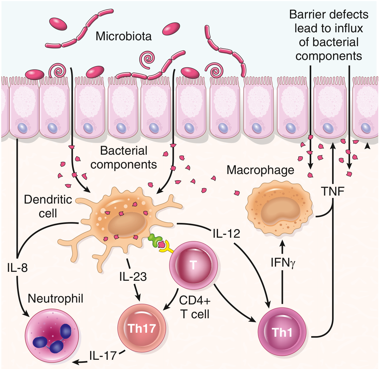
A. Microbiota Role (Dysbiosis)
- Commensal microbiota normally tolerated by the immune system
- In IBD: abnormal immune response to commensal bacteria penetrating the epithelium
- Dysbiosis of the gut microbiome supports this (evidence from lab models + clinical studies)
- Hygiene hypothesis: Reduced enteric infections early in life → inadequate regulatory immune development → susceptibility to IBD
B. Genetic Factors
| Gene | Function | Disease |
|---|---|---|
| NOD2 | Intracellular sensor for bacterial peptidoglycans → activates NF-κB | Crohn's (disease-associated form = ineffective defence → bacteria penetrate epithelium → inflammation) |
| Autophagy genes (ATG16L1) | Host response to intracellular bacteria | Crohn's only |
| IL-10 / IL-10R mutations | Regulatory T cell cytokine | Rare, severe early-onset colitis |
| >200 GWAS-identified genes | Multiple pathways | Both CD and UC |
Exam note: NOD2 polymorphisms uncommon in African/Asian descent. Autophagy gene variants not linked to UC.
C. Mucosal Immune Response
- Initial trigger: Microbial antigens presented to CD4+ T cells → differentiate into Th1 and Th17 (via IL-12 and IL-23)
- → Macrophage activation, neutrophil recruitment, TNF release
- Defective Tregs (↓ IL-10) → failure to suppress inflammation
- IL-13 (Th2): Increased in UC mucosa → mucosal damage
- Patients benefit from anti-TNF, anti-IL-12/23 therapy → confirms immune basis
D. Epithelial Barrier Dysfunction
- Defects in barrier → increased bacterial antigen translocation into lamina propria
- Exacerbated by active inflammation → vicious cycle
CROHN vs UC - ROBBINS COMPARISON TABLE
| Feature | Crohn Disease | Ulcerative Colitis |
|---|---|---|
| Location | Ileum ± colon; any GI site | Colon only |
| Rectal involvement | Sometimes | Always |
| Distribution | Skip lesions | Diffuse, continuous |
| Depth of inflammation | Transmural | Mucosa + superficial submucosa only |
| Bowel wall | Thick, fibrotic | Thin |
| Ulcers | Deep, knifelike, serpentine (cobblestone) | Superficial, broad-based |
| Granulomas | Yes (~35%) | No |
| Fistulas/sinuses | Yes | No |
| Strictures | Common | Rare |
| Creeping fat | Yes (mesenteric fat wraps serosa) | No |
| Pseudopolyps | Moderate | Marked |
| Fibrosis | Marked | Mild/none |
| Toxic megacolon | No | Yes |
| Recurrence after surgery | Common | No (colectomy is curative) |
| Vit B12 malabsorption | Yes (terminal ileum) | No |
| Perianal fistula | Yes | No |
| Malignant potential | Yes | Yes |
CROHN DISEASE - MORPHOLOGY (Robbins)
- Most common site: terminal ileum, ileocecal valve, cecum
- Skip lesions, strictures, cobblestone mucosa (edematous folds + serpentine ulcers)
- Creeping fat - mesenteric fat adherent to serosa
- Fissures → perforation / fistula tracts
- Wall thickened (edema + fibrosis + muscularis hypertrophy)
- Neutrophilic cryptitis + crypt abscesses
- Crypt distortion (branching, irregular)
- Non-caseating granulomas (~35%) - any layer, any segment, even mesenteric LN + skin
- Pseudopyloric metaplasia (gastric antral-type glands = chronic injury marker)
- Paneth cell metaplasia (left colon)
- Absence of granulomas does NOT exclude CD
ULCERATIVE COLITIS - MORPHOLOGY (Robbins)
- Rectum always involved → extends proximally, continuous
- Pancolitis = total colon involved
- Red, granular mucosa; broad-based ulcers
- Pseudopolyps (islands of regenerating mucosa projecting into lumen)
- No mural thickening, no serosal involvement
- Toxic megacolon = damaged muscularis propria → colonic dilatation → risk of perforation
- Same infiltrates as CD: neutrophils, crypt abscesses, crypt distortion
- Inflammation limited to mucosa (key difference from CD)
- No granulomas
- Mucosal atrophy after chronic disease
- May revert to near-normal histology after prolonged remission
EXTRAINTESTINAL MANIFESTATIONS (Both CD + UC)
- Migratory polyarthritis, sacroiliitis, ankylosing spondylitis
- Uveitis
- Skin: pyoderma gangrenosum, erythema nodosum
- Primary sclerosing cholangitis (PSC - more in UC)
- Pericholangitis
COLITIS-ASSOCIATED NEOPLASIA
- Both CD and UC carry increased CRC risk (colitis-associated dysplasia → carcinoma)
- Risk proportional to duration + extent of colitis
- Surveillance colonoscopy mandatory after 8-10 years of disease
QUICK EXAM BULLETS (Robbins-specific)
- "Skip lesions + transmural + granulomas + fistulas" = Crohn
- "Rectum always + continuous + no granulomas + toxic megacolon" = UC
- Monozygotic twin concordance: CD ~50% (genetic strong); UC ~20% (environmental more important)
- NOD2 = peptidoglycan sensor; mutant form = ineffective bacterial defence → CD
- Autophagy genes (ATG16L1) = intracellular bacterial handling → CD only
- IL-10/Treg defects → failure to suppress inflammation → both
- Cobblestone appearance = edematous mucosa depressed below serpentine ulcers (CD)
- Pseudopolyps = regenerating mucosal islands in UC (also seen in CD, but marked in UC)
- Creeping fat = diagnostic of CD on gross specimen