10 mark answer on le fort osteotomy of maxilla

Reading File
Finding Sources
Searching Images

Le Fort osteotomy maxilla surgical cuts
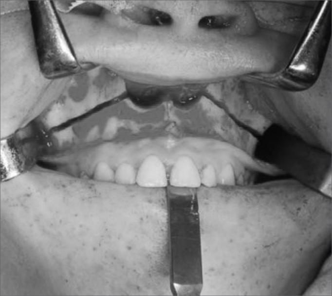
This black-and-white clinical photograph displays an intraoperative view of a maxillary Le Fort I osteotomy. The surgical site is accessed through an intraoral approach with the upper lip and labial mucosa retracted superiorly using metal surgical retractors, exposing the maxilla. A clear, horizontal osteotomy line is visible extending across the midface, superior to the apices of the maxillary teeth. The cut originates near the piriform aperture and canine pillar, traversing the lateral maxillary wall. The underlying bone is exposed, and the soft tissues are reflected to provide a clear operative field. A surgical instrument is positioned inferiorly near the midline of the maxillary incisors to stabilize or manipulate the dental arch. This procedure is a fundamental component of orthognathic surgery, often utilized here as part of a transmaxillary approach to access deeper structures such as the clivus or to correct dentofacial deformities.
This black-and-white clinical photograph displays an intraoperative view of a maxillary Le Fort I osteotomy. The surgical site is accessed through an intraoral approach with the upper lip and labial mucosa retracted superiorly using metal surgical retractors, exposing the maxilla. A clear, horizontal osteotomy line is visible extending across the midface, superior to the apices of the maxillary teeth. The cut originates near the piriform aperture and canine pillar, traversing the lateral maxillary wall. The underlying bone is exposed, and the soft tissues are reflected to provide a clear operative field. A surgical instrument is positioned inferiorly near the midline of the maxillary incisors to stabilize or manipulate the dental arch. This procedure is a fundamental component of orthognathic surgery, often utilized here as part of a transmaxillary approach to access deeper structures such as the clivus or to correct dentofacial deformities.

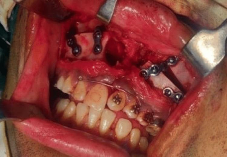
This intra-oral clinical photograph captures an active surgical field during a Le Fort I osteotomy for maxillary advancement. The image shows the maxilla with surgical retraction of the labial and buccal mucosa to expose the bone. Four internal fixation mini-plates, secured with cortical screws, are visible bridging the osteotomy sites to provide rigid stabilization of the repositioned maxillary segments. Specifically, two plates are visible on each side of the mid-face region. Orthodontic hardware, including brackets on the maxillary teeth and a stabilizing archwire, is present to facilitate dental alignment and intraoperative intermaxillary fixation if required. The surgical field demonstrates exposed alveolar bone, soft tissue hemorrhage typical of the procedure, and the use of metal surgical retractors. The image illustrates a critical phase of orthognathic surgery in a patient with a history of cleft palate repair and skeletal Class III malocclusion, focusing on the mechanical stabilization of the advanced maxilla.
This intra-oral clinical photograph captures an active surgical field during a Le Fort I osteotomy for maxillary advancement. The image shows the maxilla with surgical retraction of the labial and buccal mucosa to expose the bone. Four internal fixation mini-plates, secured with cortical screws, are visible bridging the osteotomy sites to provide rigid stabilization of the repositioned maxillary segments. Specifically, two plates are visible on each side of the mid-face region. Orthodontic hardware, including brackets on the maxillary teeth and a stabilizing archwire, is present to facilitate dental alignment and intraoperative intermaxillary fixation if required. The surgical field demonstrates exposed alveolar bone, soft tissue hemorrhage typical of the procedure, and the use of metal surgical retractors. The image illustrates a critical phase of orthognathic surgery in a patient with a history of cleft palate repair and skeletal Class III malocclusion, focusing on the mechanical stabilization of the advanced maxilla.

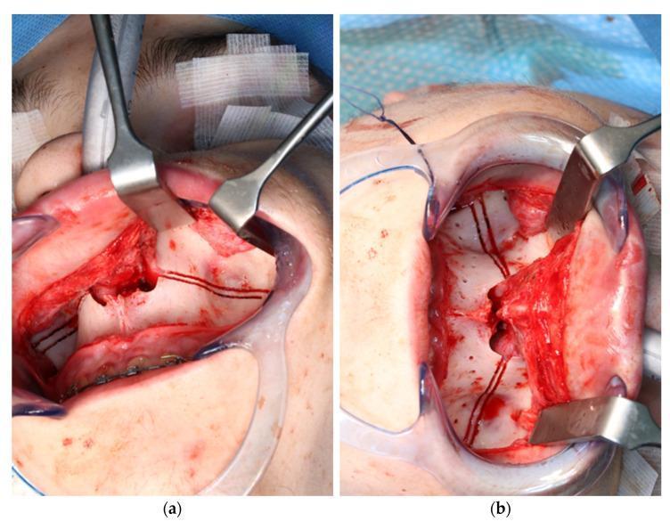
Side-by-side intraoperative clinical photographs documenting a Le Fort I osteotomy procedure using robotic laser guidance. Both images show a wide surgical exposure of the maxilla with a vestibular incision and mucoperiosteal flap elevation, maintained by surgical retractors. Image (a) demonstrates the pre-drilling phase, where the robot-guided laser (CARLO system) has accurately marked the planned osteotomy line and specific locations for patient-specific implant (PSI) drill holes, indicated by precise black thermal markings on the cortical bone. Image (b) shows the subsequent phase after a conventional rotating handpiece was used to create the physical drill holes following the laser-marked guides. The bone surface in (b) exhibits focal bleeding and structural disruption at the drill sites. This comparison illustrates the clinical application of laser-guided navigation to improve the accuracy of surgical planning and execution in orthognathic surgery for skeletal malocclusion.
Side-by-side intraoperative clinical photographs documenting a Le Fort I osteotomy procedure using robotic laser guidance. Both images show a wide surgical exposure of the maxilla with a vestibular incision and mucoperiosteal flap elevation, maintained by surgical retractors. Image (a) demonstrates the pre-drilling phase, where the robot-guided laser (CARLO system) has accurately marked the planned osteotomy line and specific locations for patient-specific implant (PSI) drill holes, indicated by precise black thermal markings on the cortical bone. Image (b) shows the subsequent phase after a conventional rotating handpiece was used to create the physical drill holes following the laser-marked guides. The bone surface in (b) exhibits focal bleeding and structural disruption at the drill sites. This comparison illustrates the clinical application of laser-guided navigation to improve the accuracy of surgical planning and execution in orthognathic surgery for skeletal malocclusion.

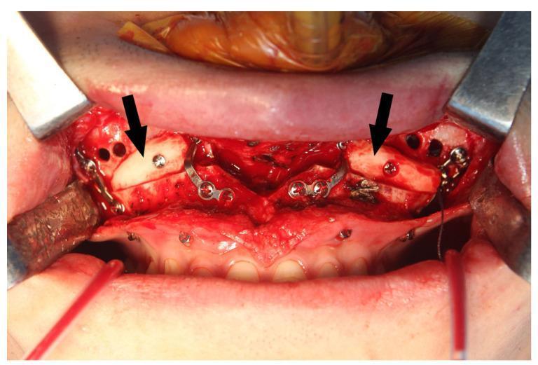
An intraoperative clinical photograph showing a Le Fort I osteotomy site in the anterior maxilla during bimaxillary orthognathic surgery. The surgical field is exposed using metal retractors, revealing the maxillary bone superior to the upper dentition. Two autogenous bone blocks (indicated by black arrows) are grafted bilaterally at the maxillary sinus wall gaps to ensure postoperative stability and facilitate sinus drainage. These pale, dense bone grafts are secured into position using titanium osteosynthesis screws. Multiple L-shaped and straight metal fixation plates are visible, bridging the osteotomy lines. Additionally, several small, circular drainage holes are drilled into the anterior maxillary wall, intended for the placement of Maxillary Sinus Vacuum Drainage (MSVD) tubes. The image demonstrates complex maxillofacial reconstruction techniques, highlighting the integration of rigid internal fixation, autogenous grafting, and sinus management protocols.

An intraoperative clinical photograph showing a Le Fort I osteotomy site in the anterior maxilla during bimaxillary orthognathic surgery. The surgical field is exposed using metal retractors, revealing the maxillary bone superior to the upper dentition. Two autogenous bone blocks (indicated by black arrows) are grafted bilaterally at the maxillary sinus wall gaps to ensure postoperative stability and facilitate sinus drainage. These pale, dense bone grafts are secured into position using titanium osteosynthesis screws. Multiple L-shaped and straight metal fixation plates are visible, bridging the osteotomy lines. Additionally, several small, circular drainage holes are drilled into the anterior maxillary wall, intended for the placement of Maxillary Sinus Vacuum Drainage (MSVD) tubes. The image demonstrates complex maxillofacial reconstruction techniques, highlighting the integration of rigid internal fixation, autogenous grafting, and sinus management protocols.

Finding Sources
Finding Sources
Reading File
Reading File
Reading File
Finding Sources
Reading File
Reading File
Finding Sources
Searching Images

Le Fort fracture classification levels I II III midface
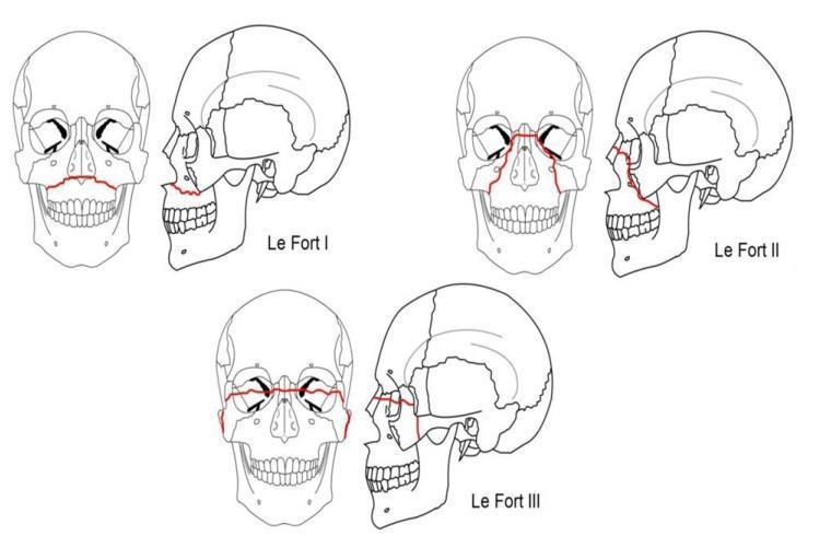
Anatomical diagram illustrating the Le Fort classification system for midface fractures, presented through frontal (anterior) and lateral (profile) line drawings of a human skull. Three distinct fracture patterns are highlighted with red lines. Le Fort I (horizontal) depicts a fracture line passing through the lower maxilla and nasal septum, effectively separating the alveolar process and palate from the rest of the facial skeleton. Le Fort II (pyramidal) shows a fracture line originating at the nasal bridge, extending through the lacrimal bones, infraorbital rim, and downward through the maxillary sinuses. Le Fort III (craniofacial dysjunction) illustrates a complete separation of the facial bones from the cranial base, with fracture lines traversing the frontonasal suture, orbits, and zygomatic arches. This diagram serves as a primary educational resource for maxillofacial surgery and emergency medicine, demonstrating the complex planes of facial trauma and the anatomical relationship between the maxilla, orbits, and zygoma.
Anatomical diagram illustrating the Le Fort classification system for midface fractures, presented through frontal (anterior) and lateral (profile) line drawings of a human skull. Three distinct fracture patterns are highlighted with red lines. Le Fort I (horizontal) depicts a fracture line passing through the lower maxilla and nasal septum, effectively separating the alveolar process and palate from the rest of the facial skeleton. Le Fort II (pyramidal) shows a fracture line originating at the nasal bridge, extending through the lacrimal bones, infraorbital rim, and downward through the maxillary sinuses. Le Fort III (craniofacial dysjunction) illustrates a complete separation of the facial bones from the cranial base, with fracture lines traversing the frontonasal suture, orbits, and zygomatic arches. This diagram serves as a primary educational resource for maxillofacial surgery and emergency medicine, demonstrating the complex planes of facial trauma and the anatomical relationship between the maxilla, orbits, and zygoma.

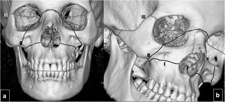
Educational 3D CT reconstruction of an adult human skull demonstrating the classic classification of midface fractures. The image consists of two panels: (a) a frontal (anterior) view and (b) an oblique view, both featuring overlaid black lines representing the three types of Le Fort fractures. 

- Le Fort I: Indicated by a horizontal fracture line passing through the maxilla, superior to the alveolar process and maxillary teeth, effectively separating the palate from the rest of the face.
- Le Fort II: Depicted as a pyramidal fracture line extending from the lateral maxilla, through the infraorbital rims and nasal bones, involving the midfacial structures.
- Le Fort III: Labeled at the superior aspect, showing complete craniofacial disjunction where the fracture line traverses the zygomatic arches, the lateral orbital walls, and the nasofrontal suture, separating the facial skeleton from the cranial base.

The visualization serves as a diagnostic algorithm for maxillofacial trauma, highlighting the anatomical involvement of the orbits, nasal cavity, and zygomaticomaxillary complex.
Educational 3D CT reconstruction of an adult human skull demonstrating the classic classification of midface fractures. The image consists of two panels: (a) a frontal (anterior) view and (b) an oblique view, both featuring overlaid black lines representing the three types of Le Fort fractures. - Le Fort I: Indicated by a horizontal fracture line passing through the maxilla, superior to the alveolar process and maxillary teeth, effectively separating the palate from the rest of the face. - Le Fort II: Depicted as a pyramidal fracture line extending from the lateral maxilla, through the infraorbital rims and nasal bones, involving the midfacial structures. - Le Fort III: Labeled at the superior aspect, showing complete craniofacial disjunction where the fracture line traverses the zygomatic arches, the lateral orbital walls, and the nasofrontal suture, separating the facial skeleton from the cranial base. The visualization serves as a diagnostic algorithm for maxillofacial trauma, highlighting the anatomical involvement of the orbits, nasal cavity, and zygomaticomaxillary complex.

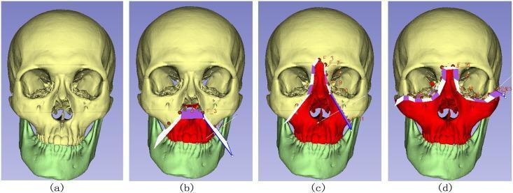
This diagnostic diagram and illustration series displays a 3D medical model of a human skull, used to demonstrate the classification of Le Fort fractures through computer-assisted clipping paths. The series includes four panels: (a) a normal skull with the cranium in pale yellow and the mandible in light green; (b) a Le Fort I fracture, indicated by a red highlight on the lower maxilla and alveolar process, separated by a horizontal plane; (c) a Le Fort II fracture (pyramidal fracture), with the red highlight extending through the nasal bridge, lacrimal bones, and infraorbital rim; and (d) a Le Fort III fracture (craniofacial dysjunction), showing the most extensive red highlight encompassing the entire midface, including the zygomatic arches and orbital floors. Overlaid white, purple, and blue geometric lines represent the implicit clipping paths used in surgical planning software to segment these complex fracture patterns. The visual serves as an educational tool for maxillofacial surgery, illustrating different levels of midfacial instability and the digital tools used for their analysis.

This diagnostic diagram and illustration series displays a 3D medical model of a human skull, used to demonstrate the classification of Le Fort fractures through computer-assisted clipping paths. The series includes four panels: (a) a normal skull with the cranium in pale yellow and the mandible in light green; (b) a Le Fort I fracture, indicated by a red highlight on the lower maxilla and alveolar process, separated by a horizontal plane; (c) a Le Fort II fracture (pyramidal fracture), with the red highlight extending through the nasal bridge, lacrimal bones, and infraorbital rim; and (d) a Le Fort III fracture (craniofacial dysjunction), showing the most extensive red highlight encompassing the entire midface, including the zygomatic arches and orbital floors. Overlaid white, purple, and blue geometric lines represent the implicit clipping paths used in surgical planning software to segment these complex fracture patterns. The visual serves as an educational tool for maxillofacial surgery, illustrating different levels of midfacial instability and the digital tools used for their analysis.

Reading File
Reading File
I now have enough comprehensive information from the textbooks and images to write a thorough 10-mark answer. Let me compile it.

Le Fort Osteotomy of the Maxilla

Introduction

Le Fort osteotomy refers to a group of surgical procedures that reproduce (or use) the classic fracture planes of the midface first described by René Le Fort in 1901. In orthognathic surgery, these osteotomies allow controlled mobilization and repositioning of the maxilla (and associated midfacial skeleton) to correct skeletal discrepancies, malocclusion, dentofacial deformities, and obstructive sleep apnea, or to provide surgical access to the skull base.

Le Fort Classification - Anatomical Basis

The three fracture levels define distinct osteotomy planes:
Le Fort fracture classification - frontal and lateral skull views showing LFI, LFII, LFIII fracture lines
LevelPlaneStructures Separated
Le Fort IHorizontal - above the apices of the maxillary teethAlveolar process + hard palate from the rest of the midface
Le Fort IIPyramidal - through nasofrontal suture, lacrimal bones, infraorbital rim, anterolateral maxillary wallsNasal-maxillary pyramid from zygomatic complex and cranium
Le Fort IIICraniofacial disjunction - through frontonasal suture, medial/lateral orbital walls, zygomatic archesEntire midface from the cranial base

Le Fort I Osteotomy (Most Common Orthognathic Procedure)

Indications

  • Skeletal Class III malocclusion (maxillary hypoplasia/retrusion) - advancement
  • Skeletal Class II - maxillary impaction or setback
  • Open bite deformity - superior repositioning (impaction)
  • Vertical maxillary excess ("gummy smile") - impaction
  • Crossbite / transverse discrepancy - with midpalatal split (segmental)
  • Obstructive sleep apnea - maxillomandibular advancement (MMA); combined Le Fort I + bilateral sagittal split osteotomy (BSSO) achieves at least 10 mm advancement, with a mean AHI reduction of ~44.8 events/hour and an 85.5% success rate (Cummings Otolaryngology)
  • Cleft palate sequelae - maxillary advancement after cleft repair
  • Skull base access - transmaxillary approach to the clivus, nasopharynx, and pterygopalatine fossa ("open-door maxillotomy")

Preoperative Planning

  • Clinical examination: occlusal analysis, facial proportions, dental midlines
  • Cephalometric and orthopantomogram radiography
  • Model surgery on dental casts with occlusal wafer fabrication
  • CT scan for surgical simulation (virtual surgical planning)
  • Orthodontic preparation (dental decompensation, usually 12-18 months)
  • Informed consent covering risks: bleeding, nerve injury, relapse, nonunion

Surgical Technique - Le Fort I

Step 1: Access Incision

A horizontal intraoral mucosal incision is made in the upper gingivobuccal sulcus, from first molar to first molar, about 5 mm above the attached mucosa. Mucoperiosteal flaps are elevated to expose the anterior and lateral maxillary walls, piriform apertures, and nasal spine.

Step 2: Osteotomy Cuts

A horizontal bone cut is made with an oscillating saw:
  • Runs from the piriform aperture, across the lateral maxillary wall (above the root apices, typically 4-5 mm above the apices of the canines), across the zygomatic buttress, and posteriorly to the pterygomaxillary junction
  • The nasal septum is separated from the maxilla using a nasal septum osteotome
  • The pterygomaxillary junction is separated bilaterally using a curved pterygoid osteotome - this is the most critical and potentially dangerous step

Step 3: Downfracture and Mobilization

The maxilla is downfractured using controlled digital pressure. Rowe disimpaction forceps are used if the maxilla is impacted. The maxilla is then mobilized to free all soft tissue and bony attachments, allowing it to be moved in three dimensions:
  • Anteroposteriorly (advancement or setback)
  • Vertically (impaction = superior movement; elongation = inferior with bone grafts)
  • Laterally (expansion or constriction)
  • Rotation (clockwise or counterclockwise)

Step 4: Fixation

The maxilla is placed in the predetermined position using the pre-fabricated occlusal wafer and intermaxillary fixation (IMF) with arch bars. Rigid fixation is achieved using:
  • L- or J-shaped titanium miniplates at the piriform rims and zygomatic buttresses bilaterally (4 plates total) using 1.5-2.0 mm screws (at least 2 screws each side of the osteotomy)
  • Alternatively, 1-mm box plates can be used at the zygomatic buttresses for additional strength
  • IMF is usually released at the end of the procedure; a palatal splint stabilizes the dentition when the palate is also split

Step 5: Bone Grafting

If the maxilla is advanced more than ~5-6 mm, autogenous bone grafts (commonly split calvarial bone) are placed at the osteotomy gaps to prevent relapse and aid osseous healing. For downward movement, interpositional bone grafts are placed to maintain vertical height.
Intraoperative Le Fort I osteotomy with four titanium miniplates and screws securing the repositioned maxilla

Le Fort II Osteotomy

Used in midface hypoplasia where the deficit involves the nasal-maxillary pyramid. The osteotomy line passes through the nasofrontal suture, across the medial orbital wall/lacrimal bone, through the infraorbital rim, down the anterior maxillary wall, and to the pterygomaxillary junction. This allows advancement of the central midface (nose + central maxilla) as a unit. Fixation is at a higher level than Le Fort I, including the infraorbital rim and sometimes the nasofrontal junction (with small microplates in this area to avoid palpability/visibility). Rotation of the fragment superiorly at the nasofrontal junction must be carefully avoided to prevent open-bite deformity (KJ Lee's Essential Otolaryngology).

Le Fort III Osteotomy (Craniofacial Dysjunction)

Indicated for syndromic craniosynostoses (Crouzon, Apert syndromes) with global midface hypoplasia. The procedure advances all midfacial structures below the frontal bone - nose, zygomas, and maxilla en bloc. It requires:
  • Bicoronal scalp flap for exposure
  • Osteotomies at: nasal root, medial orbital walls, orbital floor, lateral orbital wall (frontozygomatic suture), pterygomaxillary junction
  • Internal fixation + bone grafts, or increasingly distraction osteogenesis (rigid external distractor/RED frame) for movements >10 mm or in scarred/previously operated fields (Scott-Brown's Otorhinolaryngology, Vol 2)
A facial bipartition (Le Fort III + midline V-excision) is used when midface hypoplasia is associated with hypertelorism - the two hemifacial segments are brought medially to reduce interorbital distance.
A monobloc procedure combines Le Fort III advancement with frontal bone remodeling (FOAR) for simultaneous forehead and midface correction.

Skull Base Access Application

The Le Fort I (and modified Le Fort O) osteotomy can be used for surgical access to the skull base - clivus, nasopharynx, and cranio-cervical junction. The "open-door maxillotomy" involves:
  1. Le Fort I osteotomy
  2. Midline palatal split
  3. Lateral displacement of each maxillary half
This provides unrivalled panoramic exposure of the entire clivus, superior to the transoral transpalatine route alone (Scott-Brown's Vol 2). After tumor resection, each maxillary half is reduced and fixed with miniplates.

Complications

EarlyLate
Intraoperative hemorrhage (especially at pterygomaxillary junction - internal maxillary artery)Skeletal relapse
Devascularization of maxillary segments (necrosis if blood supply disrupted)Malocclusion
Unfavorable osteotomy (tooth root damage)Nonunion / malunion
CSF leak (rare)Plate infection/exposure requiring removal
Infraorbital nerve injury (Le Fort II)Velopharyngeal insufficiency (rare)
Nasolacrimal duct injury (Le Fort II)Temporomandibular joint problems
Blindness (Le Fort III - orbital surgery)Numbness (infraorbital nerve - V2)
Maxillomandibular advancement also carries risks of: relapse (more significant with larger advancements), nerve paresthesia, need for subsequent dental work, and TMJ dysfunction (Cummings Otolaryngology).

Summary

Le Fort osteotomies are fundamental procedures in orthognathic and craniofacial surgery. Le Fort I is the workhorse for correcting maxillary deformities and malocclusion, as well as providing skull base access. Le Fort II and III address more complex midfacial hypoplasia, increasingly performed with distraction osteogenesis for large movements. Rigid internal fixation with titanium miniplates has replaced wiring as the standard, and meticulous preoperative planning (including virtual surgical planning and orthodontic preparation) is key to achieving predictable outcomes with minimal relapse.

Sources: Cummings Otolaryngology Head and Neck Surgery (9780323612173); Scott-Brown's Otorhinolaryngology Head & Neck Surgery Vol 2 (9781138094634); KJ Lee's Essential Otolaryngology (9781260122237)
This is a shared conversation. Sign in to Orris to start your own chat.