
Supinator muscle
supinator muscle anatomy forearm
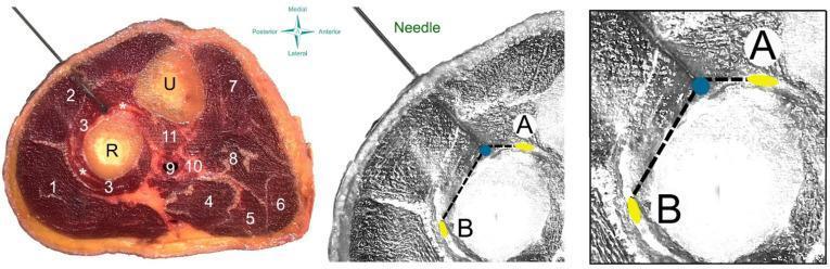
This composite educational resource illustrates a dry needling procedure targeting the supinator muscle of the forearm. The left panel shows a cadaveric cross-section of the proximal forearm with numerical labels identifying key anatomical structures: radius (R), ulna (U), brachioradialis (1), wrist extensors (2), supinator muscle (3), and various flexor compartment muscles (4–8). A needle is shown penetrating the dorsal aspect, passing through the wrist extensors into the supinator muscle belly near the radial nerve branches (*). The center and right panels provide anatomical diagrams focused on the safety profile of the procedure. They map the spatial relationship between the needle tip (blue dot) and critical neurovascular structures: the deep branch (A) and the superficial branch (B) of the radial nerve. Dashed lines represent measurement distances from the needle tip to these nerve branches, serving as a clinical guide for procedural accuracy and hazard avoidance. This material is designed for advanced musculoskeletal anatomy education and clinical training in physical therapy or sports medicine.
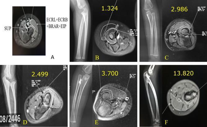
This composite educational graphic displays a series of radiographs and corresponding transverse MR images of the proximal forearm, illustrating a classification system for congenital proximal radioulnar synostosis (PRUS). Image A demonstrates the methodology for cross-sectional area measurement, targeting the supinator muscle (SUP) and a combined group of peripheral muscles (ECRL, ECRB, BRAR, and EIP). Panels B through F show a progressive spectrum of anatomical severity and their associated 'supinator ratios.' Panel B represents a normal forearm anatomy with clear separation of the radius and ulna and a ratio of 1.324. Panels C and D illustrate fibrous pseudo-synostosis (Type I), where the radial head is identifiable but articulatory dynamics are abnormal. Panels E and F demonstrate osseous synostosis (Types II and III), showing bony fusion between the radius and ulna, radial head dislocation or absence, and significantly elevated supinator ratios (up to 13.820). This visual series emphasizes the correlation between skeletal deformity and compensatory or dysplastic muscle volume changes, critical for pediatric orthopedic assessment and surgical planning.
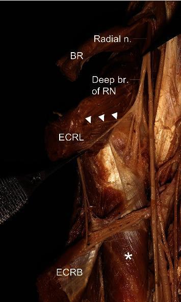
This clinical photograph displays a detailed anatomical dissection of the human forearm, focusing on the peripheral nervous system and musculature of the radial compartment. The image highlights the branching pattern of the radial nerve (Radial n.) as it descends. Key muscular structures identified include the brachioradialis (BR) positioned superiorly, the extensor carpi radialis longus (ECRL) located centrally, and the extensor carpi radialis brevis (ECRB) situated distally. The supinator muscle is visible and marked with an asterisk (*). A critical neurovascular feature demonstrated is the deep branch of the radial nerve (Deep br. of RN), with white arrowheads indicating its course along the ECRL. This anatomical specimen illustrates the relationship between the radial nerve and the muscles it innervates, which is essential for understanding radial tunnel syndrome and nerve entrapment pathophysiology. The image serves as an educational resource for medical students and surgeons studying the anatomy of the upper limb and peripheral nerve distribution.
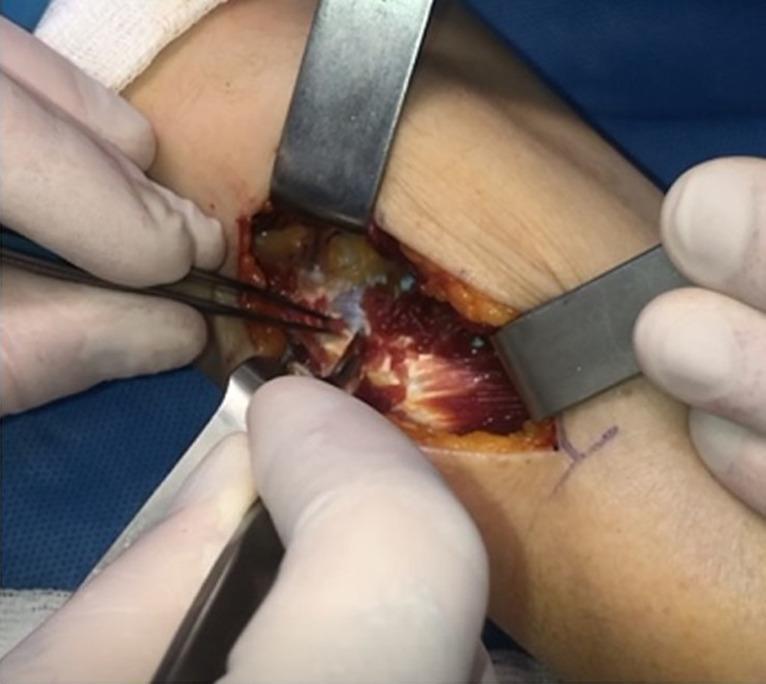
This clinical photograph captures an intraoperative view of the supinator muscle during an upper limb surgical procedure, likely a decompression of the posterior interosseous nerve (PIN). The surgical field shows a longitudinal incision distal to the lateral epicondyle of the forearm. Metallic retractors are placed at the proximal and distal margins to provide exposure, revealing the deep red, striated fibers of the supinator muscle running in an oblique orientation. The proximal portion of the muscle shows the initiation of blunt dissection, where the overlying fascia and yellowish subcutaneous adipose tissue are being separated from the muscle belly. A pair of surgical forceps is positioned to grasp and elevate the proximal tissue, while a scalpel is visible in the lower right, assisting in the dissection. The surgical field is moist and glistening, reflecting standard anatomical exposure within a controlled operative environment. This image serves as a teaching tool for identifying the supinator muscle and understanding the surgical approach for nerve decompression in the proximal forearm.
Supinator Muscle
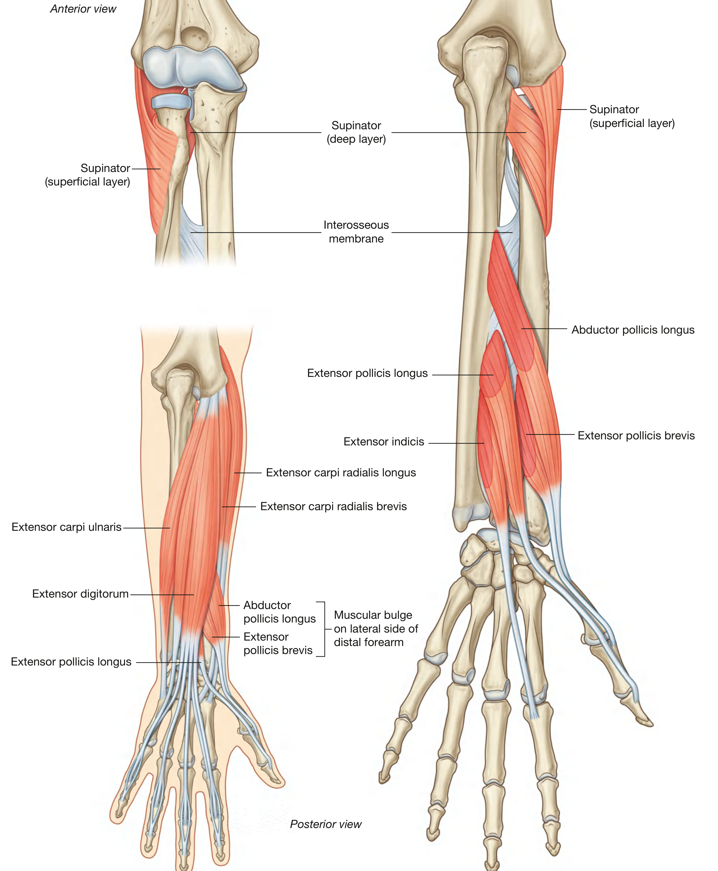
Overview
Origin
| Layer | Origin |
|---|---|
| Superficial (humeral) layer | Lateral epicondyle of the humerus, radial collateral ligament of the elbow, anular ligament of the radius |
| Deep (ulnar) layer | Supinator crest on the posterolateral surface of the ulna, olecranon |
- THIEME Atlas, p. 3290
Insertion
Action
Innervation
Blood Supply
The Arcade of Frohse (Clinical Significance)
-
No wrist drop (ECRL is innervated proximal to the supinator canal, so wrist extension is preserved - though with radial deviation because ECU is lost)
-
No sensory disturbance in the hand (the superficial sensory branch branches off before the nerve enters the canal)
-
Palsy of: extensor pollicis longus, extensor pollicis brevis, abductor pollicis longus, extensor digitorum, extensor indicis, extensor carpi ulnaris
-
THIEME Atlas, p. 4882
Relations
- The radial nerve divides just proximal to the supinator into:
- Deep (motor) branch - enters the supinator canal via the arcade of Frohse
- Superficial (sensory) branch - courses distally beneath brachioradialis
- Proximally, the supinator is covered by the superficial extensor muscles (ECRL, ECRB, brachioradialis)
Summary Table
| Feature | Detail |
|---|---|
| Location | Posterior compartment, proximal forearm |
| Layers | Superficial (humeral) + Deep (ulnar) |
| Origin | Lateral epicondyle, radial collateral lig., anular lig., supinator crest of ulna |
| Insertion | Proximal radius (radial tuberosity to pronator teres insertion) |
| Action | Supination of forearm |
| Nerve | Deep branch of radial nerve (PIN) - C6, C7 |
| Key landmark | Arcade of Frohse (PIN passes between its two heads) |
| Clinical relevance | Radial tunnel syndrome; PIN palsy |