
Nucleus pulposus and annulus fibrosis
Reading File
The nucleus pulposus is the gel-like core of an intervertebral disc, derived from the notochord, rich in proteoglycans (aggrecan) and type II collagen. The annulus fibrosus is the tough outer ring made of concentric lamellae of type I collagen fibres. Together they form the intervertebral disc.
Searching sources...
Finding Sources
Finding Sources
Good results from multiple textbooks. Let me read the most detailed sections - the anatomy atlas, Gray's, histology, and embryology sources in parallel.
Reading File
Reading File
Reading File
Reading File
Excellent content from multiple textbooks. Let me also read the fluid mechanics / biomechanics section from the anatomy atlas and the disc herniation content, and fetch key images in parallel.
Reading File
Reading File
Loading Image
Loading Image
I now have comprehensive information from multiple authoritative textbooks plus two excellent images. I have everything needed for a thorough answer.
Intervertebral Disc: Nucleus Pulposus and Annulus Fibrosus
The intervertebral disc is a fibrocartilaginous symphysis joint that lies between adjacent vertebral bodies. It has two distinct structural components - the nucleus pulposus (NP) centrally and the annulus fibrosus (AF) peripherally - plus hyaline cartilage end plates above and below.
Anatomy at a Glance
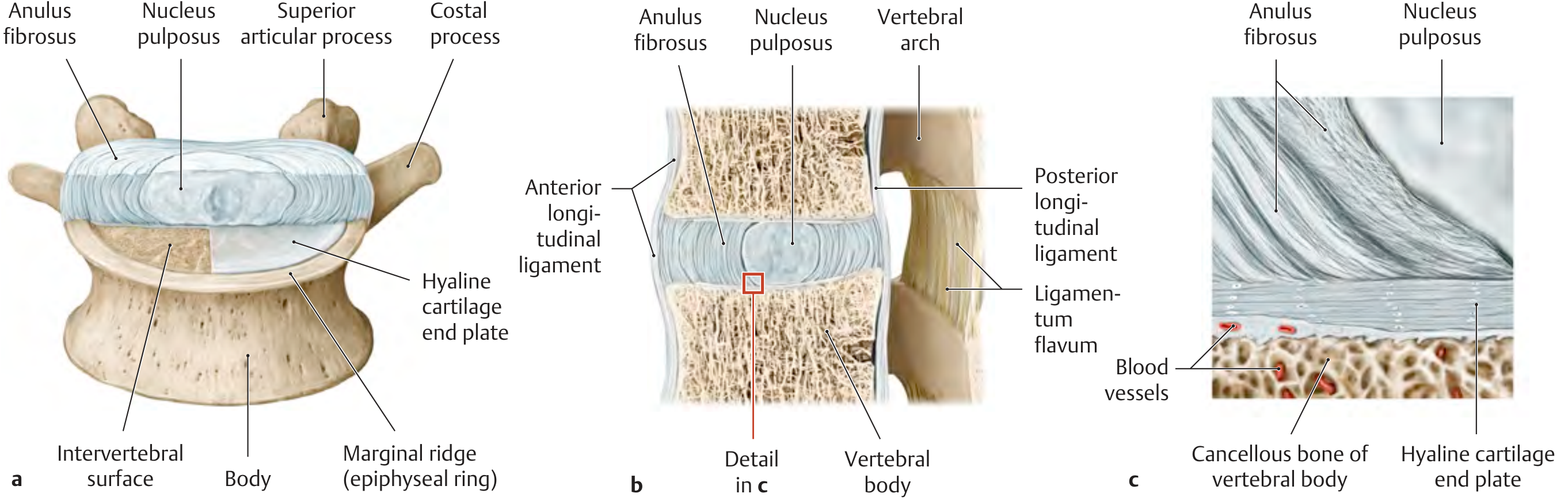
Panel (a): superior view showing AF surrounding NP with hyaline cartilage end plate. Panel (b): sagittal section showing relationships to longitudinal ligaments. Panel (c): close-up showing blood vessels supplying the disc via the end plate.
1. Embryological Origin
Both components arise from the sclerotome of somites and the notochord:
- The notochord degenerates where it is surrounded by developing vertebral bodies. Between the vertebrae, it expands to form the gelatinous core - the nucleus pulposus.
- Circularly arranged mesenchymal fibers then surround the nucleus to form the annulus fibrosus.
- Notochordal cells persist postnatally only in the NP (scattered vacuolated cells), but are progressively replaced by chondrocyte-like cells through life.
- Remnants of notochord that fail to regress can form a chordoma - a rare, slow-growing malignant tumor found at the skull base or lumbosacral region.
(The Developing Human - Clinically Oriented Embryology, p. 918)
2. Nucleus Pulposus (NP)
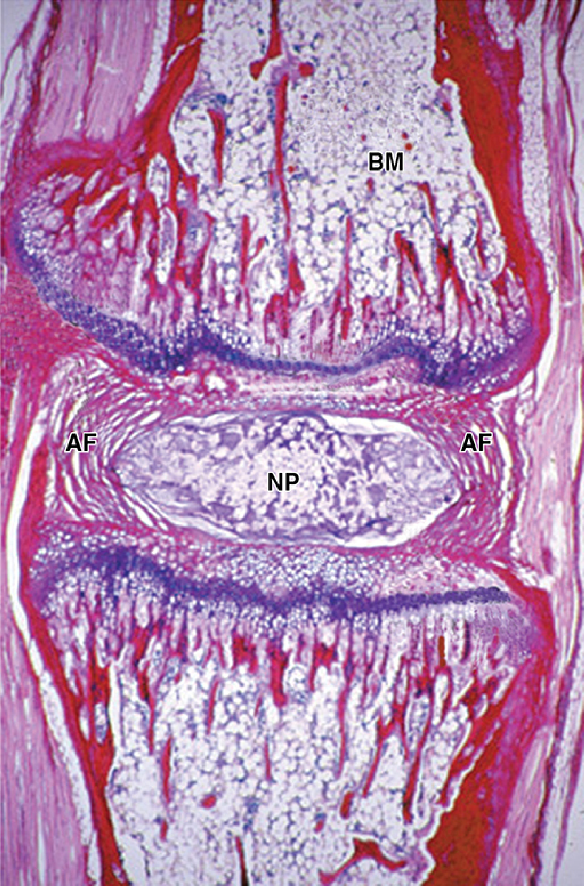
Low-power PSH section: NP (central pale zone) surrounded by concentric lamellae of AF; BM = bone marrow of adjacent vertebra.
| Feature | Detail |
|---|---|
| Location | Central core of the disc |
| Gross appearance | Soft, elastic, yellowish, gelatinous |
| Water content | 80-85% (decreases with age) |
| Matrix | Rich in hyaluronan (hyaluronic acid), aggrecan, and type II collagen fibres |
| Cells | Scattered notochordal-derived cells (vacuolated); replaced by chondrocyte-like cells with age |
| Function | Acts as a "water cushion" / hydraulic press - absorbs compressive and transient axial loads; distributes pressure uniformly over adjacent end plates |
The NP is maintained under very high hydrostatic pressure, especially under gravitational and muscular loading. It transforms compressive forces into radial tensile forces within the annulus. Because of its glycosaminoglycan-rich matrix, it reversibly binds water - under sustained load, fluid slowly seeps out through the end plates (disc height decreases ~1.5-2 cm over the course of the day). When pressure is released (e.g., during sleep), fluid is reabsorbed.
(THIEME Atlas of General Anatomy, p. 137-138)
3. Annulus Fibrosus (AF)
| Feature | Detail |
|---|---|
| Location | Outer ring surrounding the NP |
| Structure | Concentric lamellae of fibrocartilage |
| Outer zone | Dense type I collagen (Sharpey fibres anchoring into vertebral bodies and longitudinal ligaments) |
| Inner zone | Wider zone of fibrocartilage arranged in lamellar configuration |
| Fibre orientation | Oblique, at ~30° angle; adjacent lamellae run in opposite directions, forming a criss-cross meshwork |
| Function | Tension-resistant sheath; converts NP compressive forces into tensile forces; limits rotation between vertebrae |
The oblique crossed-fibre arrangement creates a very strong yet flexible structure. The outer Sharpey fibres of the AF blend with the anterior longitudinal ligament (firmly attached) and posterior longitudinal ligament (broadens at disc level to which it is firmly attached by lateral extensions).
(Grainger & Allison's Diagnostic Radiology, p. 1220; Gray's Anatomy for Students)
4. Hyaline Cartilage End Plates
The disc is sandwiched above and below by hyaline cartilage end plates attached to the vertebral bodies. The subchondral bony end plate is permeated by pores through which subchondral blood vessels supply nutrients to the avascular disc tissue (the disc has no direct blood supply in adults - nutrition is entirely by diffusion and convection through the end plates).
5. Biomechanics
The disc functions as a hydrostatic system:
- Under transient load: the NP distributes stress uniformly - acts as a shock absorber
- Under sustained load: fluid is gradually expressed through the end plates; disc height decreases
- On load release: fluid is reabsorbed; disc height recovers
- The AF converts compressive forces on the NP into circumferential tensile (hoop) stress within its lamellae
6. Clinical Relevance: Disc Herniation
Collagen loss or degenerative changes in the AF, most commonly posterolateral (where fewer collagen bundles exist and the PLL provides less support), allow the NP to herniate outward - a prolapsed / herniated disc. The posterior region is weakest because:
- Fewer AF fibre bundles posteriorly
- The PLL is narrow over the vertebral bodies and only broadens at disc levels
Herniated NP can compress spinal nerve roots in the intervertebral foramina, causing radicular pain (sciatica), sensory loss, or motor weakness in the corresponding dermatome/myotome. With ageing, the NP loses water content and becomes more fibrocartilagenous, reducing the risk of acute herniation but contributing to disc height loss and spondylosis.
(Junqueira's Basic Histology, p. 393; Gray's Anatomy for Students; Bradley and Daroff's Neurology in Clinical Practice)
Quick Comparison Table
| Nucleus Pulposus | Annulus Fibrosus | |
|---|---|---|
| Embryonic origin | Notochord | Sclerotome mesenchyme |
| Main collagen | Type II | Type I (outer), Type II (inner) |
| Main proteoglycan | Aggrecan, hyaluronan | Less proteoglycan |
| Water content | 80-85% | Lower |
| Function | Shock absorption, load distribution | Containment, limits rotation, converts compression to tension |
| Histology | Gel matrix, sparse vacuolated cells | Concentric fibrocartilage lamellae |
This is a shared conversation. Sign in to Orris to start your own chat.