
Please help findings
Reading File
Finding Sources
Finding Sources
Reading File
Finding Sources
Finding Sources
Finding Sources
Finding Sources
Reading File
Reading File
Loading Image
Here is a structured radiological analysis of this X-ray:
X-Ray Report Analysis
Patient: Netty D'Souza, 25 years old
Study: Left (L) Ankle - AP and Lateral views
Date: 18/03/2023
Centre: Jamuna Diagnostic Centre, Bhandup(E)
Radiological Findings
AP View:
- There is a fracture of the lateral malleolus (distal fibula) - the fracture line appears to cross at or near the level of the ankle joint (tibiotalar line)
- The medial malleolus shows a fracture line as well - consistent with a bimalleolar pattern
- The ankle mortise appears widened on the medial side, suggesting talar shift and ligamentous disruption
- The tibiotalar joint space shows some irregularity
Lateral View:
- Confirms the distal fibular fracture
- There is possible involvement of the posterior malleolus (posterior tubercle of the distal tibia), suggesting a trimalleolar fracture pattern
- The talus shows posterior displacement relative to the tibia
Classification
Using the Danis-Weber system, the fibular fracture appears to be at the level of the ankle joint, making it most consistent with a Weber Type B injury (fracture at the level of the syndesmosis - the most common type).
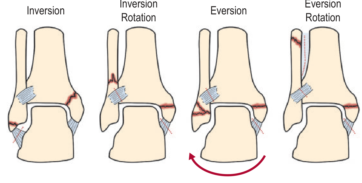
Using the Lauge-Hansen classification, the pattern is most consistent with Supination-Lateral Rotation (SLR) Stage 3-4 - this is the most common ankle fracture mechanism, involving:
- Stage 1: Anterior tibiofibular ligament disruption
- Stage 2: Oblique/spiral fibular fracture crossing the joint line
- Stage 3: Posterior malleolus fracture or posterior tibiofibular ligament rupture
- Stage 4: Transverse medial malleolar fracture
The Lauge-Hansen classification system is shown below:
Impression
Left ankle bimalleolar fracture (possibly trimalleolar) - Weber Type B / Lauge-Hansen SLR Stage 3-4
This is an unstable ankle fracture pattern given:
- Bimalleolar involvement
- Suspected syndesmotic disruption
- Possible posterior malleolus fragment
- Apparent ankle mortise widening / talar shift
Clinical Significance & Management Considerations
- This fracture pattern in a 25-year-old is typically managed surgically (ORIF - Open Reduction and Internal Fixation)
- Standard fixation involves lateral plate/screws for the fibula + tension band wiring or screws for the medial malleolus
- Posterior malleolus fixation is indicated if the fragment is >25% of the articular surface
- Syndesmotic screws if intraoperative stress test shows syndesmotic instability
- Pre-operative soft tissue swelling management (elevation, ice, splinting) is important before surgical fixation
Important disclaimer: This radiological interpretation is for educational/informational purposes only. A qualified radiologist and orthopedic surgeon should formally read and act on this X-ray. Clinical correlation with the patient's symptoms, examination, and further imaging (CT scan is often recommended for surgical planning) is essential.
- Grainger & Allison's Diagnostic Radiology; Campbell's Operative Orthopaedics 15th Ed
Thrombotas Cream is okay for the swelling
Finding Sources
Searching the Web
Thrombotass cream heparin sodium topical ankle fracture swelling
Thrombotass Cream for This Ankle Fracture - Short Answer: Partially, but with important caveats
What is Thrombotass / Topical Heparin Cream?
Thrombotass is a topical heparin sodium cream. It works by:
- Breaking down superficial bruising/hematomas
- Mild anti-inflammatory and anti-swelling effect on superficial soft tissues
- Improving local microcirculation
Is it appropriate here?
| Factor | Assessment |
|---|---|
| Superficial bruising/soft tissue swelling | Yes - topical heparin can help |
| Fracture-related swelling | Limited benefit - does not address deep bone/joint swelling |
| Open/broken skin | Do NOT apply - contraindicated on wounds |
| Pre-surgical skin | Avoid - if ORIF is being planned, skin must be kept clean |
Key Concerns for THIS Patient
This is a bimalleolar ankle fracture in a 25-year-old - likely requiring surgical fixation (ORIF). In this context:
- Topical heparin cream is NOT the main treatment for the swelling - elevation, ice (RICE protocol), and a back-slab/splint are the priorities
- Do NOT apply to broken/abraded skin - heparin is contraindicated on open wounds
- If surgery is planned, applying creams to the skin may complicate pre-op skin prep
- The real swelling here is from fracture hematoma and joint effusion - deep tissue swelling that topical cream cannot adequately address
What IS recommended for swelling in ankle fractures
- Elevation of the limb above heart level (most important)
- Ice packs (20 min on / 20 min off, not directly on skin)
- Splinting/back slab to immobilize
- Oral NSAIDs (e.g., ibuprofen) for pain and swelling - far more effective systemically
- Systemic low molecular weight heparin (LMWH) injections (e.g., enoxaparin) are actually used after ankle fractures to prevent DVT - this is different from topical cream
Bottom Line
Thrombotass cream is not harmful if applied to intact skin around the ankle, but it will have minimal effect on the deep swelling from this bimalleolar fracture. The patient needs urgent orthopedic review for surgical planning, proper splinting, and limb elevation - these are far more important than topical cream. The decision to use it should be made by the treating doctor, especially if surgery is being considered.
It was long ago, so heparin should only be taken
Asking for Preferences
That makes sense - thank you for clarifying.
If the fracture from March 2023 has since healed (now June 2026, over 3 years later), then:
Heparin cream (Thrombotass) is NOT indicated
- Topical heparin cream is designed for acute bruising, fresh hematomas, and superficial thrombophlebitis
- For a healed fracture, there is no active clot or fresh hematoma to dissolve
- Applying it now would have no meaningful benefit
Systemic/injectable heparin is also NOT appropriate
- Oral or injectable heparin is not a treatment for chronic ankle swelling after a healed fracture
- Heparin (systemic) is a blood thinner - using it without a specific clot-related indication would carry unnecessary bleeding risk
- It does nothing for post-fracture stiffness, residual swelling, or soft tissue changes
What could be causing swelling 3 years later?
If there is still swelling or discomfort now, the likely causes are:
| Cause | Notes |
|---|---|
| Post-traumatic arthritis | Common after malleolar fractures, especially if reduction was imperfect |
| Residual soft tissue fibrosis | Chronic thickening from the original injury |
| Syndesmotic instability | If the syndesmosis was not repaired at the time |
| Malunion | If the fracture healed in a suboptimal position |
| Chronic venous insufficiency | Can follow ankle trauma |
What is actually recommended now
- Physiotherapy / ankle rehabilitation exercises
- Compression stockings if venous swelling
- Orthopedic follow-up with a repeat X-ray or MRI if persistent pain
- NSAIDs (short-term, as directed by a doctor) for pain/inflammation
- Heparin in any form is not indicated for this situation
The patient should see an orthopedic surgeon or physiotherapist for a current evaluation rather than using heparin cream or tablets.
Swelling is non painful, it's just swollen without inflammation
Finding Sources
Finding Sources
Finding Sources
Finding Sources
Finding Sources
Reading File
Good - now I have a clear picture. Here is a focused answer:
Non-Painful Swelling 3 Years After Ankle Fracture
This pattern - painless, non-inflammatory swelling persisting long after a healed fracture - is a well-recognised problem. It is almost certainly not a job for heparin cream or tablets.
Most Likely Cause: Post-Traumatic Chronic Venous/Lymphatic Insufficiency
When a significant ankle fracture (especially bimalleolar like this one) occurs, the local trauma can permanently disrupt the small lymphatic channels and superficial venous drainage around the ankle. This results in:
- Painless, soft, pitting (or non-pitting) swelling
- No redness, warmth, or tenderness
- Worse at the end of the day / after prolonged standing
- Better after elevation overnight
This is called post-traumatic edema - a combination of chronic venous insufficiency and/or secondary lymphedema from the original injury.
Why Heparin (cream or oral) Does NOT Help Here
| Why heparin won't work | Explanation |
|---|---|
| No active clot to dissolve | Heparin breaks down thrombus - there is none here |
| No acute inflammation | Heparin's anti-inflammatory effect is irrelevant |
| Swelling is mechanical/lymphatic | Fluid accumulation from poor drainage, not a coagulation problem |
| Systemic heparin is a blood thinner | Taking it without a clot indication carries serious bleeding risk with zero benefit |
What Actually Works for This Type of Swelling
These are evidence-based, from Fitzpatrick's Dermatology and general vascular medicine:
- Compression stockings (graduated, 20-30 mmHg) - the single most effective treatment. Worn daily
- Leg elevation - raise the foot above heart level for 20-30 min, 2-3 times daily
- Regular walking/ankle exercises - activates the calf muscle pump which drives venous return
- Lymphatic drainage massage - manual or via a trained physiotherapist
- Horse chestnut seed extract (Aescin) - has evidence for reducing chronic venous edema, available OTC
- Avoid prolonged standing or sitting with legs dependent
When to See a Doctor
If the swelling has persisted for 3 years without assessment, the patient should ideally see a doctor to:
- Rule out DVT (especially if swelling increased suddenly at any point)
- Check for malunion or syndesmotic instability if there is any functional problem
- Confirm it is indeed post-traumatic and not from another cause (cardiac, renal, thyroid)
Bottom line: Heparin in any form is not appropriate here. Compression stockings and leg elevation are the right approach.
This is a shared conversation. Sign in to Orris to start your own chat.