Scalp
Reading File
Finding Sources
Searching Images
scalp anatomy layers
Reading File
Reading File
Reading File
Loading Image
Reading File
Reading File
Here is a comprehensive overview of the scalp — its anatomy, layers, innervation, vasculature, and lymphatic drainage.
The Scalp
The scalp extends from the superciliary arches anteriorly to the external occipital protuberance and superior nuchal lines posteriorly, and inferiorly to the zygomatic arch on each side.
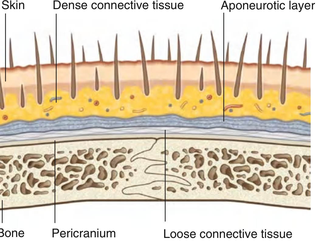
Layers — "SCALP"
The five layers are memorized by the word itself:
| Letter | Layer | Key Points |
|---|---|---|
| S | Skin | Contains hair follicles; structurally similar to skin elsewhere |
| C | Connective tissue (dense) | Anchors skin to the aponeurosis; carries arteries, veins, and nerves. Vessels do not retract when cut → profuse bleeding |
| A | Aponeurotic layer (galea aponeurotica / epicranial aponeurosis) | The tendinous sheet connecting the frontal and occipital bellies of occipitofrontalis |
| L | Loose connective tissue | A potential space; allows the scalp proper to move over the calvaria. Infections spread freely here |
| P | Pericranium | Periosteum of the outer calvaria; adherent at sutures |
The first three layers (S + C + A) are tightly bound together and move as a single unit — the "scalp proper", avulsed together in scalping injuries.
Aponeurotic Layer — Occipitofrontalis Muscle
- Frontal belly: attached to skin of eyebrows; raises eyebrows and wrinkles forehead; innervated by temporal branches of CN VII
- Occipital belly: arises from the lateral superior nuchal line and mastoid process; innervated by the posterior auricular branch of CN VII
- Epicranial aponeurosis (galea): the fibrous tendon connecting both bellies
Innervation (Sensory)
Anterior to the ears and vertex — branches of the trigeminal nerve (CN V):
| Nerve | Territory |
|---|---|
| Supratrochlear (V1) | Midline forehead |
| Supra-orbital (V1) | Forehead and vertex |
| Zygomaticotemporal (V2) | Anterior temple |
| Auriculotemporal (V3) | Temporal region up to vertex |
Posterior to the ears and vertex — cervical nerves (C2–C3):
| Nerve | Territory |
|---|---|
| Great auricular (C2–C3) | Small area posterior to ear |
| Lesser occipital (C2) | Posterior and superior to ear |
| Greater occipital (C2 posterior ramus) | Large area of posterior scalp to vertex |
| Third occipital (C3 posterior ramus) | Lower posterior scalp |
Arterial Supply
Arteries arise from the external carotid artery and ophthalmic artery (branch of internal carotid):
| Artery | Source | Territory |
|---|---|---|
| Supratrochlear | Ophthalmic a. | Anterior/superior scalp |
| Supra-orbital | Ophthalmic a. | Anterior/superior scalp to vertex |
| Superficial temporal | External carotid a. (terminal branch) | Entire lateral scalp |
| Posterior auricular | External carotid a. | Area posterior to ear |
| Occipital | External carotid a. | Large area of posterior scalp |
Clinical note: Scalp lacerations bleed profusely because (1) the dense connective tissue holds vessels open preventing retraction, and (2) in the erect position venous pressure is very low, so bleeding is predominantly arterial.
Venous Drainage
Follows arterial patterns:
- Supratrochlear + supra-orbital veins → angular vein → facial vein
- Superficial temporal vein → retromandibular vein
- Posterior auricular vein → retromandibular vein tributary
- Occipital vein → suboccipital venous plexus
Lymphatic Drainage
| Region of scalp | Primary nodes | Onward drainage |
|---|---|---|
| Occipital region | Occipital nodes (base of skull) | Upper deep cervical nodes |
| Posterior to vertex | Mastoid (retroauricular) nodes | Upper deep cervical nodes |
| Anterior to vertex | Preauricular and parotid nodes | — |
— Gray's Anatomy for Students, p. 1062–1066
This is a shared conversation. Sign in to Orris to start your own chat.