
Wht is GBS
Guillain-Barre syndrome nerve pathology ascending paralysis
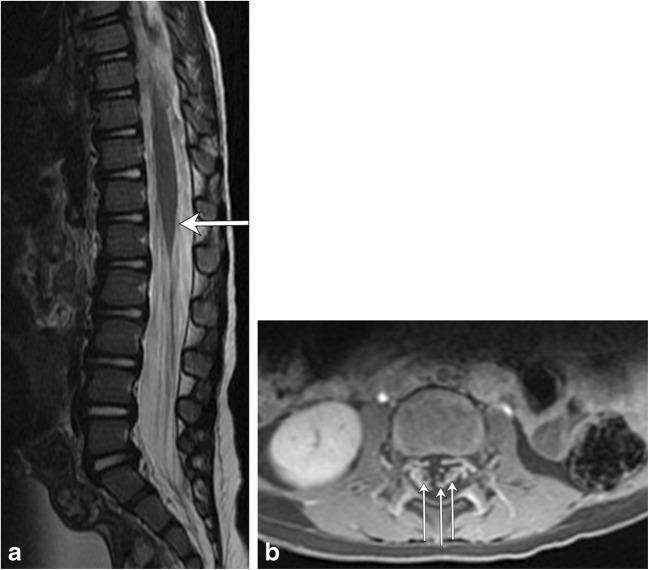
This composite diagnostic image showcases MRI findings associated with Guillain-Barré syndrome (GBS) in a pediatric patient. Figure (a) is a sagittal T2-weighted MRI of the thoracolumbar spine, demonstrating a subtle, localized area of increased signal intensity (hyperintensity) at the level of the conus medullaris (indicated by an arrow). Figure (b) provides an axial T1-weighted post-contrast MRI of the lumbar spine, revealing prominent enhancement of multiple nerve roots within the cauda equina (indicated by multiple arrows). These visual features correlate with the typical inflammatory and autoimmune pathophysiology of GBS, which affects the peripheral nervous system and nerve roots. The presence of nerve root enhancement on post-contrast imaging is a key diagnostic indicator in clinical neurology. The images serve to illustrate how MRI can support the diagnosis of GBS by identifying involvement of the lower spinal cord and emerging nerve roots in patients presenting with progressive ascending weakness or paralysis.
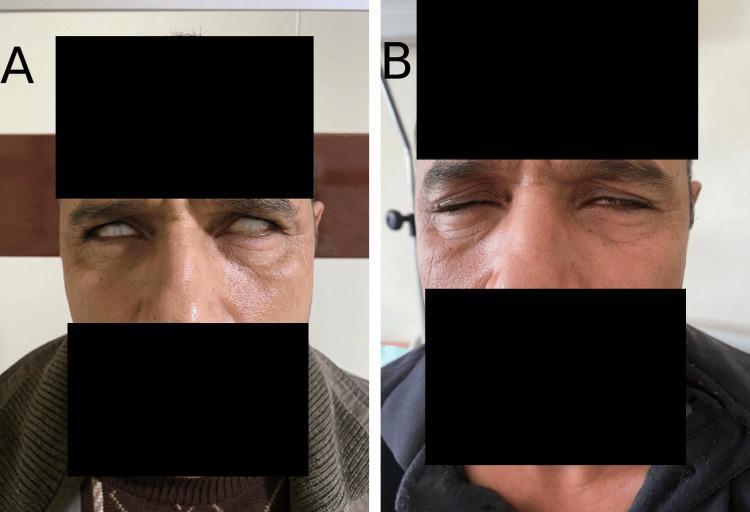
Clinical photograph presenting a side-by-side comparison (Panel A and Panel B) of a patient with facial diplegia associated with Guillain-Barré syndrome. Panel A shows the patient at admission, exhibiting bilateral peripheral facial paralysis. Key clinical signs include lagophthalmos and Bell’s phenomenon, characterized by the inability to close the eyelids and the upward rotation of the eyeballs when attempting closure. There is a lack of animation and significant periorbital strain. Panel B demonstrates clinical resolution one month after presentation and treatment. There is a marked improvement in orbicularis oculi muscle function, as evidenced by the patient's ability to partially close the eyelids and a more relaxed facial expression with reduced periorbital edema. The comparison illustrates the classic presentation and recovery phase of bilateral seventh cranial nerve (facial nerve) involvement in an inflammatory demyelinating polyneuropathy scenario.
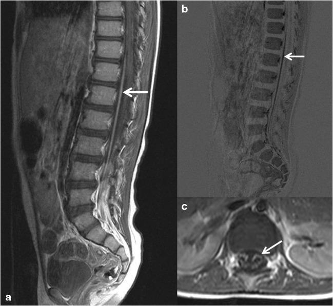
This composite diagnostic image features Magnetic Resonance Imaging (MRI) of the thoracolumbar spinal cord in a pediatric patient diagnosed with Guillain-Barré syndrome. Panel (a) is a sagittal T1-weighted contrast-enhanced MRI showing prominent enhancement of the anterior caudal nerve roots, which appear hyperintense (bright) against the relatively darker spinal cord and cerebrospinal fluid. Panel (b) presents a digital subtraction image of the sagittal T1 sequence, which minimizes background anatomical signal to highlight the contrast uptake in the ventral roots (indicated by arrows). Panel (c) is a transverse T1-weighted contrast-enhanced section demonstrating localized enhancement specifically within the anterior motor nerve roots. These radiological findings illustrate the classic breakdown of the blood-nerve barrier and inflammatory changes characteristic of acute inflammatory demyelinating polyradiculoneuropathy. Key educational features include the anatomical localization of root involvement and the utility of contrast-enhanced sequences and subtraction techniques in differentiating inflammatory neuropathies from other causes of acute flaccid paralysis.
Guillain-Barré Syndrome (GBS)
Definition
- Robbins & Kumar Basic Pathology, p. 809
- Bradley and Daroff's Neurology in Clinical Practice, p. 2663
Pathophysiology
- Approximately two-thirds of cases are triggered by a preceding infection
- The infection provokes microbe-specific T cells and antibodies that cross-react with nerve sheath antigens (molecular mimicry)
- Both T-cell-mediated and antibody-mediated responses are involved; T cells are believed to play the dominant role
- Injury is most extensive in nerve roots and proximal nerve segments, with mononuclear cell infiltrates rich in macrophages
- The classic form is multifocal inflammatory demyelination of spinal roots and peripheral nerves, producing conduction blocks and reduced velocities
Triggers / Associated Pathogens
| Pathogen | Type |
|---|---|
| Campylobacter jejuni | Bacterium (most common) |
| Epstein-Barr virus (EBV) | Virus |
| Cytomegalovirus (CMV) | Virus |
| HIV | Virus |
| Zika virus | Virus |
| SARS-CoV-2 | Virus |
Subtypes
| Subtype | Features |
|---|---|
| AIDP (Acute Inflammatory Demyelinating Polyradiculoneuropathy) | Most common in Europe & North America; demyelinating |
| AMAN (Acute Motor Axonal Neuropathy) | Pure motor axonal; common in China; summer epidemics in children |
| AMSAN (Acute Motor-Sensory Axonal Neuropathy) | Both motor + sensory axons; severe with poor recovery |
| Miller-Fisher Syndrome (MFS) | Ophthalmoplegia, ataxia, areflexia; 6% of cases in the West; up to 18% in Taiwan |
Diagnostic Criteria
- Progressive weakness of both legs and arms
- Areflexia or hyporeflexia
- Progression over days to 4 weeks
- Relative symmetry of symptoms
- Mild sensory symptoms or signs
- Bifacial palsies
- Autonomic dysfunction
- Absence of fever at onset
- Recovery beginning 2-4 weeks after progression stops
- CSF: Elevated protein with <10 cells/μL (albuminocytological dissociation)
- Electrodiagnostics: Nerve conduction slowing or block
Clinical Features (Classic Presentation)
- Ascending weakness - starts in legs, moves upward
- Areflexia - loss of deep tendon reflexes
- Sensory changes - paresthesias, mild sensory loss
- Autonomic dysfunction - BP fluctuations, arrhythmias, urinary retention
- Respiratory failure - in severe cases (~30%), requiring mechanical ventilation
- Cranial nerve involvement - facial diplegia (7th nerve most common), bulbar palsy
MRI Findings


Facial Diplegia in GBS
Treatment
| Treatment | Mechanism |
|---|---|
| IVIG (intravenous immunoglobulin) | Suppresses immune responses (exact mechanism unclear) |
| Plasmapheresis | Removes offending antibodies |
| Supportive care | Mechanical ventilation, physiotherapy, DVT prophylaxis |
Note: Steroids are NOT effective in GBS and are not recommended.
Prognosis
- Mortality dropped from ~33% (before positive-pressure ventilation) to currently 1-5%
- Most patients who survive the acute phase recover with time
- Recovery typically begins 2-4 weeks after progression stops
- AMSAN subtype has the worst prognosis
Distinguishing GBS from CIDP
| Feature | GBS | CIDP |
|---|---|---|
| Course | Acute (<4 weeks) | Chronic (>2 months) |
| Pattern | Monophasic | Relapsing-remitting or progressive |
| Triggers | Often post-infectious | Often idiopathic |
| Treatment | IVIG/plasmapheresis | Long-term steroids + IVIG |
Wht is GBS Anatomy and Physiology and signs and symptoms
peripheral nerve anatomy myelin sheath axon structure diagram
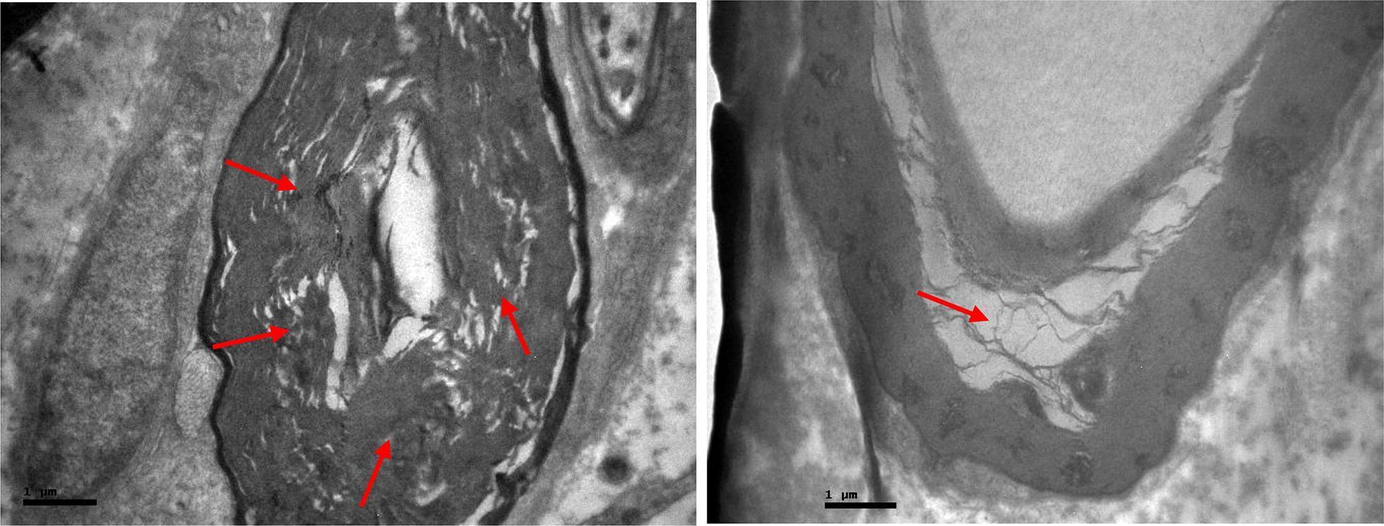
Two high-magnification transmission electron microscopy (TEM) micrographs depicting cross-sections of the femoral nerve showing advanced degenerative changes in the myelin sheath. The images demonstrate significant myelin disorganization and pathological vacuolization, highlighted by red arrows. In the left panel, the myelin sheath surrounding the axon appears thickened and fragmented, with loss of the normal compact lamellar structure. The right panel provides a closer view of focal vacuolization, where the myelin layers have separated to form clear, empty intra-myelinic spaces. These structural alterations represent demyelinating processes and nerve fiber degradation. Scale bars are provided at 1 μm. This visual content is used for neuropathology studies to illustrate pharmacological or traumatic effects on peripheral nerve integrity, specifically focusing on myelin sheath morphology and axon-myelin relationship.
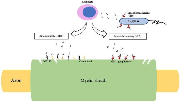
This pathophysiology diagram illustrates the cellular and molecular pathogenesis of Chronic Inflammatory Demyelinating Polyradiculoneuropathy (CIDP) and Guillain-Barr syndrome (GBS). The central focus is a peripheral nerve fiber consisting of a yellow central axon enveloped by a green myelin sheath. The diagram is divided into two mechanistic pathways originating from a central leukocyte. On the left, the 'Autoimmunity (CIDP)' pathway depicts antibodies directly targeting the paranodal and nodal proteins Neurofascin-155 (NF155) and Contactin-1 on the myelin surface. On the right, the 'Molecular mimicry (GBS)' pathway shows a C. jejuni bacterium with lipooligosaccharides (LOS) that trigger the production of cross-reactive antibodies. These antibodies are shown binding to GM-1 gangliosides located on the myelin sheath. Small 'Y'-shaped icons represent antibodies throughout the diagram, illustrating the humoral immune response against neural components. The visual highlights the distinction between direct autoimmune targeting in CIDP and the infection-triggered molecular mimicry characteristic of GBS, both resulting in demyelination and nerve damage.
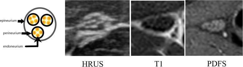
This composite educational graphic illustrates the normal internal architecture of a peripheral nerve across different imaging modalities. On the far left, an anatomical diagram depicts a transverse cross-section highlighting the nested hierarchy of nerve structures: the epineurium (outermost sheath), the perineurium (enclosing individual fascicles), and the endoneurium (surrounding internal axons/neurons, represented as orange dots). This structural organization creates a characteristic 'honeycomb pattern.' To the right, three clinical imaging panels correlate this anatomy: 1) High-Resolution Ultrasound (HRUS) shows the nerve as a speckled, hyperechoic structure relative to surrounding muscle. 2) T1-weighted MRI demonstrates the nerve with moderate signal intensity, where fascicular boundaries begin to emerge. 3) Proton Density Fat-Suppressed (PDFS) MRI provides the clearest visualization of the honeycomb architecture, showing hypointense 'dots' representing nerve fascicles surrounded by hyperintense signal from the interfascicular epineural fat. This comparison is essential for diagnosing peripheral neuropathies and identifying loss of normal fascicular patterns.
Guillain-Barre syndrome ascending paralysis clinical signs autonomic dysfunction
Clinical photograph presenting a side-by-side comparison (Panel A and Panel B) of a patient with facial diplegia associated with Guillain-Barré syndrome. Panel A shows the patient at admission, exhibiting bilateral peripheral facial paralysis. Key clinical signs include lagophthalmos and Bell’s phenomenon, characterized by the inability to close the eyelids and the upward rotation of the eyeballs when attempting closure. There is a lack of animation and significant periorbital strain. Panel B demonstrates clinical resolution one month after presentation and treatment. There is a marked improvement in orbicularis oculi muscle function, as evidenced by the patient's ability to partially close the eyelids and a more relaxed facial expression with reduced periorbital edema. The comparison illustrates the classic presentation and recovery phase of bilateral seventh cranial nerve (facial nerve) involvement in an inflammatory demyelinating polyneuropathy scenario.
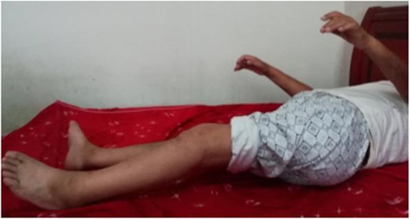
Clinical photograph of a patient in a supine position on a bed, illustrating long-term motor deficits following a case of Guillain-Barre syndrome (GBS). The patient exhibits significant limb weakness and impaired motor control, documented as Medical Research Council (MRC) grade 3 power in the upper extremities and grade 2 power in the lower extremities five years post-onset. The upper limbs are partially elevated with visible wrist drop and lack of coordinated hand extension, while the lower limbs are extended and flaccid. The image demonstrates the chronic sequelae of Landry's ascending paralysis and the limitations in functional recovery despite long-term rehabilitation. This visual serves as an educational reference for assessing chronic neuromuscular motor weakness, deep tendon reflex impairment, and the potential for persistent disability in severe GBS cases involving albuminocytological dissociation and axonal damage.
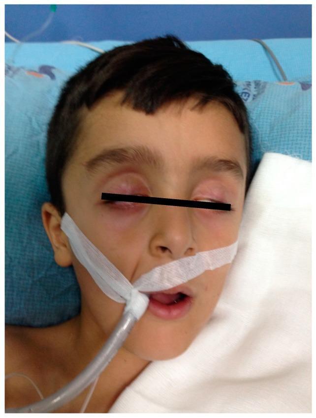
This clinical photograph shows a pediatric patient in a critical care setting demonstrating signs of bilateral peripheral facial paralysis. The patient is supine on a hospital bed, exhibiting lack of facial muscle tone and an inability to achieve full eyelid closure (lagophthalmos), which is managed by taping the eyelids shut to prevent corneal desiccation and abrasion. An orotracheal tube is placed and secured with white adhesive tape at the right corner of the mouth, indicating the need for mechanical ventilation or airway protection due to respiratory muscle weakness or neurological compromise. Monitoring cables are visible in the background, further characterizing the patient's intensive care requirements. The image serves as a clinical illustration of severe cranial nerve involvement in neuromuscular disorders such as Guillain-Barré syndrome (GBS), where bilateral facial nerve palsy can occur alongside ascending weakness or respiratory failure.
GBS: Anatomy, Physiology, and Signs & Symptoms
Part 1: Anatomy - Structures Affected in GBS
Normal Peripheral Nerve Anatomy
| Layer | What it is |
|---|---|
| Epineurium | Outermost connective tissue sheath surrounding the whole nerve |
| Perineurium | Surrounds individual fascicles (bundles of nerve fibers) |
| Endoneurium | Surrounds individual axons within a fascicle |
| Myelin sheath | Lipid-protein insulation wrapped around the axon by Schwann cells |
| Axon | The actual conducting fiber carrying electrical signals |
| Node of Ranvier | Gaps in the myelin sheath where saltatory conduction occurs |
What GBS Attacks
- Spinal nerve roots (proximal segments)
- Mixed peripheral nerves (motor + sensory)
- Cranial nerves (in ~45-75% of cases)
- Autonomic ganglia and nerve fibers (in ~65%)
Part 2: Physiology - How GBS Damages Nerves
Normal Nerve Conduction
What Goes Wrong in GBS: Molecular Mimicry
- Preceding infection (e.g., Campylobacter jejuni) stimulates the immune system
- C. jejuni has lipooligosaccharides (LOS) on its surface that mimic gangliosides (GM1, GD1b, GQ1b) found on nerve myelin - this is molecular mimicry
- The body produces cross-reactive antibodies that attack myelin sheaths and/or axons
- T cells and macrophages infiltrate nerve roots and peripheral nerves, stripping myelin
- Loss of myelin causes conduction block and slowing - signals cannot travel efficiently
- In severe cases, the axons themselves are damaged (axonal subtypes: AMAN, AMSAN)
Myelin Damage Under Electron Microscopy
- The myelin layers fragment and separate, forming vacuoles
- Normal compact lamellar structure is lost
- Macrophages intrude between the axon and myelin sheath, especially at Nodes of Ranvier
Part 3: Signs and Symptoms
- Progressive phase - worsening over days to 4 weeks
- Plateau phase - stable for days to weeks
- Recovery phase - begins 2-4 weeks after progression stops
A. Motor Symptoms (MOST PROMINENT)
| Symptom | Details |
|---|---|
| Ascending weakness | Starts in both legs, spreads upward to arms, trunk, face |
| Flaccid paralysis | Lower motor neuron pattern (floppy, not spastic) |
| Proximal > distal | Often affects thigh/hip muscles early |
| Severity range | From mild difficulty walking to total quadriplegia |
| Respiratory failure | 9-30% of patients need mechanical ventilation |
| Bilateral facial weakness | In 50% of patients - cannot close eyes, drooping face |
| Bulbar palsy | Difficulty swallowing and speaking (lower cranial nerves) |
| Ophthalmoplegia | Extraocular muscle weakness (especially in Miller-Fisher variant) |
B. Sensory Symptoms
| Symptom | Details |
|---|---|
| Paresthesias | Tingling, "pins and needles" - often first symptom, worse in hands/fingers |
| Sensory loss | Mild; mainly distal vibration sense impaired |
| Pain | Present in ~70% during acute phase - back pain, limb pain, interscapular pain |
| Dysesthetic pain | Burning/tingling of limbs |
Note: Sensory symptoms are present but motor weakness dominates - pure sensory loss is not a major feature.
C. Areflexia (Reflex Loss)
- Loss of deep tendon reflexes (DTRs) is an invariable feature - present in virtually all patients
- Knee jerks, ankle jerks, biceps/triceps reflexes all absent
- May be absent early or initially only in the legs
D. Autonomic Dysfunction (65% of patients)
| Autonomic Symptom | Mechanism |
|---|---|
| Sinus tachycardia | Sympathetic overactivity |
| Bradycardia / Heart block / Asystole | Excessive vagal (parasympathetic) activity |
| Labile hypertension / Orthostatic hypotension | Loss of vascular tone regulation |
| Tachyarrhythmias | Sympathetic-parasympathetic imbalance |
| Urinary retention | Autonomic bladder dysfunction |
| Constipation / Ileus | Gastrointestinal atony |
| Anhidrosis or excessive sweating | Sudomotor dysfunction |
| ECG changes | T-wave abnormalities, ST depression, QT prolongation |
"Vagal spells" - sudden bradycardia or asystole triggered by tracheal suctioning - are a serious ICU hazard.
E. Cranial Nerve Signs (45-75% of cases)
| Nerve | Sign |
|---|---|
| CN VII (Facial) | Bilateral facial paresis - most common; lagophthalmos, inability to close eyes |
| CN III, IV, VI | Ophthalmoplegia (especially Miller-Fisher variant) |
| CN IX, X | Bulbar palsy - dysphagia, dysphonia |
| CN XI, XII | Less common - neck/tongue weakness |
F. In Severe/Prolonged Cases
Summary Table of All Signs and Symptoms
| Category | Feature | Frequency |
|---|---|---|
| Motor | Ascending flaccid weakness | ~100% |
| Motor | Respiratory failure | 9-30% |
| Motor | Bilateral facial palsy | ~50% |
| Reflexes | Areflexia | ~100% |
| Sensory | Paresthesias (tingling) | Common |
| Sensory | Pain (back/limb) | ~70% |
| Sensory | Sensory loss | Mild, ~majority |
| Autonomic | Tachycardia/bradycardia | ~65% |
| Autonomic | BP instability | ~65% |
| Autonomic | Urinary retention | Common |
| Cranial | Facial nerve palsy | 45-75% |
| Cranial | Ophthalmoplegia (MFS) | 6% overall |
Key Lab/Diagnostic Findings That Support the Anatomy
| Test | Finding | Anatomical basis |
|---|---|---|
| CSF | High protein, <10 WBC/mL ("albuminocytological dissociation") | Protein leaks from inflamed nerve roots into CSF |
| Nerve conduction studies | Slowed velocity, conduction blocks, absent F-waves | Demyelination of peripheral nerves |
| MRI spine (contrast) | Enhancement of cauda equina nerve roots | Breakdown of blood-nerve barrier at root level |
| EMG | Denervation patterns in axonal subtypes | Motor axon damage |