
Hyperandronism in female
"hyperandrogenism" AND "female" AND "diagnosis"
hyperandrogenism female hirsutism PCOS clinical features
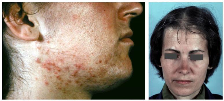
A composite clinical photograph illustrating dermatological manifestations of hyperandrogenism, specifically in the context of Polycystic Ovary Syndrome (PCOS). The left image provides a lateral view of the lower face and neck of a female patient, showing inflammatory acne vulgaris characterized by a dense distribution of erythematous papules and pustules along the jawline, mandibular area, and upper neck. Significant hirsutism is also visible, with terminal hair growth in androgen-dependent areas such as the preauricular region and chin. The right image is a frontal view of a female patient exhibiting androgenetic alopecia. Notable features include a receding frontal hairline with temporal sparing, resulting in an 'M' shaped pattern, along with diffuse thinning of the hair on the vertex and mid-scalp. Together, these images demonstrate the cutaneous 'SAHA' syndrome (seborrhea, acne, hirsutism, and alopecia) associated with systemic androgen excess, providing a key educational example for endocrine-related skin disorders.
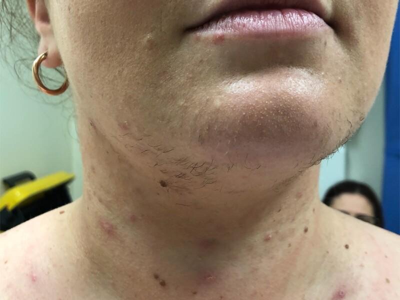
Clinical photograph of the lower face, neck, and upper chest of a female patient exhibiting signs of hyperandrogenism. The primary clinical finding is hirsutism, characterized by coarse, dark, androgenic terminal hair growth in a male-pattern distribution, specifically along the chin, jawline, and submandibular region of the neck. Accompanying dermatological features include inflammatory acne vulgaris, presenting as erythematous papules and pustules scattered across the neck and upper chest. Additionally, there are multiple pigmented macules and small nevi visible on the neck. The skin on the chin demonstrates a slightly thickened and irregular texture, consistent with follicular stimulation. This visual presentation is a key clinical indicator of endocrine dysfunction, such as that seen in polycystic ovary syndrome (PCOS), Cushing syndrome, or androgen-secreting tumors (e.g., adrenocortical carcinoma), where excess androgens stimulate the conversion of vellus hair to terminal hair in androgen-sensitive areas.
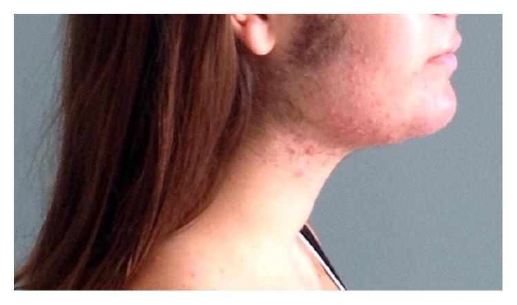
A clinical photograph of a female patient's face and neck in a lateral profile view, demonstrating clinical signs of hyperandrogenism and virilization. The image reveals prominent hirsutism, characterized by coarse, dark hair growth in an androgen-dependent distribution, specifically involving the preauricular (sideburn) area, cheeks, and along the mandibular jawline. Accompanying these findings is an inflammatory acneiform eruption, presenting as erythematous papules and pustules clustered around the jaw and upper neck. This combination of acne and hirsutism is highly suggestive of an underlying hyperandrogenic state. The educational focus of this image is the recognition of physical manifestations of excessive androgen levels in female patients, which may be associated with conditions such as polycystic ovary syndrome (PCOS) or, in this clinical context, pregnancy-related luteoma.
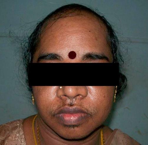
This clinical photograph displays significant dermatological and endocrine-related signs on a female patient, illustrating virilization. The primary finding is androgenic alopecia, characterized by a receding hairline and marked thinning of the hair at the frontal and temporal scalp, following a male-pattern distribution. Additionally, the patient exhibits severe hirsutism, with coarse terminal hair growth visible on the upper lip, chin, and mandibular regions. Notable stubble in these areas suggests recent hair removal efforts. The facial skin appears slightly oily with scattered blemishes on the cheeks, possibly related to hyperandrogenism. These visual features—male-pattern baldness combined with excess facial hair in a female—are classic indicators of hyperandrogenic states, such as those caused by androgen-secreting tumors or severe polycystic ovary syndrome (PCOS).
Hyperandrogenism in Females
Definition and Overview
Clinical Manifestations
| Feature | Details |
|---|---|
| Hirsutism | Excess coarse terminal hair - chin, lip, chest, inner thigh, lower back, abdomen (midline) |
| Acne | Particularly severe, jawline-distributed, sudden-onset acne |
| Androgenic alopecia | Male-pattern scalp thinning/recession |
| Menstrual irregularity | Oligomenorrhea or amenorrhea from anovulation |
| Virilization | Voice deepening, clitoromegaly, breast atrophy, increased muscle bulk - suggests tumor |
| Acanthosis nigricans | Indicates associated insulin resistance |
Causes
Ovarian (most common)
- Polycystic Ovary Syndrome (PCOS) - the single most common cause, accounting for ~70-80% of cases
- Functional ovarian hyperandrogenism / hyperthecosis
- Ovarian neoplasms (Sertoli-Leydig cell tumor, thecoma, granulosa cell tumor)
- Syndromes of extreme insulin resistance
Adrenal
- Non-classic congenital adrenal hyperplasia (CAH) - 21-hydroxylase deficiency accounts for >90% of CAH cases
- Functional adrenal hyperandrogenism
- Cushing syndrome (adrenal adenoma, carcinoma)
- Adrenal neoplasms
Other Endocrine
- Hyperprolactinemia
- Acromegaly
- Hypothyroidism
Peripheral / Idiopathic
- Obesity (increased peripheral androgen conversion)
- Idiopathic hirsutism (increased 5α-reductase activity at the pilosebaceous unit)
Drug-induced (hypertrichosis - not true hyperandrogenism)
- Phenytoin, minoxidil, cyclosporine, valproic acid, diazoxide
Pregnancy-related
- Luteoma of pregnancy, hyperreactio luteinalis (gestational theca-lutein cysts)
Polycystic Ovary Syndrome (PCOS) - The Commonest Cause
- Hyperandrogenism - clinical (hirsutism, acne, alopecia) or biochemical (elevated androgens)
- Ovarian dysfunction - oligo/anovulation (oligomenorrhea <8 cycles/year, amenorrhea)
- Polycystic ovarian morphology (PCOM) on ultrasound - ≥20 follicles (2-9 mm) in either ovary and/or ovarian volume >10 mL
- Excess LH stimulation → increased ovarian theca cell androgen production (testosterone, androstenedione)
- Insulin resistance and hyperinsulinemia → further stimulation of ovarian androgen production and reduced SHBG (raising free testosterone)
- Elevated androgens impair folliculogenesis → anovulation → further LH excess (a vicious cycle)
-
50% of PCOS patients have obesity (central/android distribution); metabolic risk is significant
- Type 2 diabetes mellitus (IR → hyperglycemia)
- Metabolic syndrome, dyslipidemia
- Cardiovascular disease risk
- Endometrial hyperplasia/carcinoma (from unopposed estrogen in anovulatory cycles)
- Infertility
Clinical Assessment and Ferriman-Gallwey Score
- Age of onset and speed of progression (rapid onset → rule out tumor)
- Menstrual pattern (oligomenorrhea → ovarian source more likely)
- Medications (exogenous androgens, progestins)
- Family history (CAH, PCOS)
- Signs of Cushing syndrome: hypertension, striae, easy bruising, central obesity
Hormonal Evaluation
| Hormone | Interpretation |
|---|---|
| Total / free testosterone | First-line; >200 ng/dL strongly suggests androgen-secreting tumor |
| DHEA-S | Elevated in adrenal source (>700 μg/dL → adrenal tumor) |
| 17-OHP (fasting, follicular phase) | Elevated in non-classic CAH (21-hydroxylase deficiency); confirm with ACTH stimulation if 17-OHP >2 ng/mL |
| LH:FSH ratio | Often >2:1 in PCOS |
| Fasting insulin / glucose | Assess insulin resistance |
| Prolactin | Exclude hyperprolactinemia |
| TSH | Exclude hypothyroidism |
| SHBG | Low in PCOS/hyperinsulinemia → high free testosterone |
- Testosterone >200 ng/dL or DHEA-S >700 μg/dL → pelvic/adrenal imaging (CT/MRI) to rule out tumor
- Rapid-onset virilization at any testosterone level → urgent imaging
- Assess ovarian morphology for PCOS
- Identifies ovarian masses
Treatment
1. Lifestyle Modification (First-line in overweight PCOS)
2. Oral Contraceptive Pills (OCP) - First-line pharmacotherapy
- Progestin suppresses LH → reduced ovarian androgen production
- Estrogen increases hepatic SHBG → lowers free testosterone
- Reduces DHEAS (partly LH/SHBG-independent)
- Estrogen inhibits 5α-reductase → less conversion of testosterone to DHT in skin
- Reduces hair growth in ~two-thirds of hirsute patients
- Improves acne by ~50%
- Hair growth improvement takes 6-12 months (full hair cycle needed)
- Preferred progestins: norgestimate, desogestrel, drospirenone (low androgenicity; avoid norgestrel/levonorgestrel)
- Drospirenone has both anti-mineralocorticoid and anti-androgenic activity
3. Anti-androgens
| Drug | Mechanism | Notes |
|---|---|---|
| Spironolactone | Androgen receptor blocker + reduces androgen synthesis | 50-200 mg/day; first choice anti-androgen; use with OCP (teratogenic in male fetus) |
| Cyproterone acetate | Potent androgen receptor blocker | Combined with ethinyl estradiol (Diane-35); widely used outside USA |
| Flutamide | Androgen receptor blocker | Risk of hepatotoxicity; less preferred |
| Finasteride | 5α-reductase inhibitor (type II) | 5 mg may benefit post-menopausal; teratogenic |
4. Insulin Sensitizers
- Metformin - reduces hyperinsulinemia → lowers ovarian androgen production; may restore ovulation; especially useful when fertility is desired or OCP is contraindicated
- Weight loss of 5-7% achieves similar or better effect
5. GnRH Analogues
- Suppress both LH and FSH → profound suppression of ovarian androgen synthesis
- Reserved for severe cases or when other treatments fail
- Require add-back estrogen therapy if used long-term
6. Glucocorticoids
- For adrenal-source hyperandrogenism (non-classic CAH)
- Low-dose dexamethasone or hydrocortisone suppresses ACTH → reduces adrenal androgen output
7. Mechanical Hair Removal
- Temporary: shaving (does NOT increase growth rate), waxing, chemical depilatories
- Permanent: electrolysis, laser, intense pulsed light (IPL)
- Laser/IPL best for large areas of dark terminal hair - works by targeting melanin (photothermolysis)
Specific Causes and Their Management
Non-classic CAH
- Autosomal recessive; 21-hydroxylase deficiency in >90%
- Basal follicular-phase 17-OHP should be measured in ALL women with hirsutism to exclude this
- Treatment: low-dose glucocorticoid (dexamethasone or hydrocortisone) + OCP for hirsutism
- Berek & Novak's Gynecology, p. 1920
Androgen-Secreting Tumors
- Ovarian tumors more common than adrenal
- Suspect if: testosterone >200 ng/dL, rapid onset, frank virilization
- Treatment: surgical resection
- Berek & Novak's Gynecology, p. 1952
Cushing Syndrome
- Suspect if: hypertension, central obesity, striae, easy bruising, proximal myopathy, moon face, buffalo hump
- ACTH-independent forms are adrenal in origin; adrenal cancers are often large at presentation
- Treatment: surgical (adrenalectomy) or medical depending on etiology
Idiopathic Hirsutism
- Normal androgen levels + regular menses + normal ovaries
- Increased end-organ sensitivity (5α-reductase activity) at the pilosebaceous unit
- Treatment: OCP + anti-androgens (spironolactone most useful) + mechanical removal
Key Points Summary
- Most common cause: PCOS (~70-80% of cases)
- Red flags for tumor: testosterone >200 ng/dL, rapid onset, virilization
- Always measure 17-OHP (follicular phase) to exclude non-classic CAH in all hirsute women
- OCP is first-line pharmacologic treatment; hair improvement takes 6-12 months
- Spironolactone is the most commonly used anti-androgen (combine with OCP due to teratogenicity)
- Weight loss of 5-7% restores ovulation in >75% of obese PCOS patients
- Metformin is indicated when fertility is desired or insulin resistance is prominent
- Hirsutism and virilization are distinct - virilization always requires urgent investigation
- A systematic review in Human Reproduction Update (2025) evaluated the diagnostic accuracy of androgen measurement in PCOS and found that free testosterone (calculated or measured by equilibrium dialysis) remains the most accurate biochemical marker of hyperandrogenism - PMID 39305127.
- A 2024 meta-analysis in European Journal of Endocrinology reported on PCOS prevalence in adolescents and highlighted that ovulatory dysfunction and hyperandrogenism must both be clearly established before diagnosing PCOS in this age group - PMID 39353075.
Evaluate
Ferriman Gallwey score hirsutism grading scale diagram
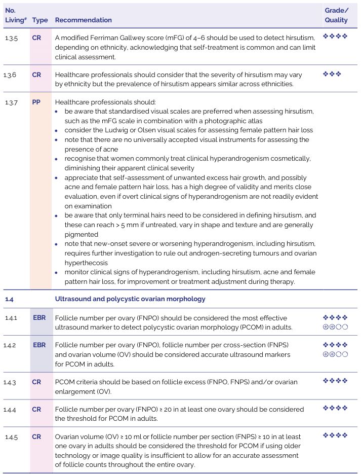
<table><thead><tr><th>No. Living*</th><th>Type</th><th>Recommendation</th><th>Grade/ Quality</th></tr></thead><tbody><tr><td>1.3.5</td><td>CR</td><td>A modified Ferriman Gallwey score (mFG) of 4–6 should be used to detect hirsutism, depending on ethnicity, acknowledging that self-treatment is common and can limit clinical assessment.</td><td>◊◊◊◊</td></tr><tr><td>1.3.6</td><td>CR</td><td>Healthcare professionals should consider that the severity of hirsutism may vary by ethnicity but the prevalence of hirsutism appears similar across ethnicities.</td><td>◊◊◊</td></tr><tr><td>1.3.7</td><td>PP</td><td>Healthcare professionals should: • be aware that standardised visual scales are preferred when assessing hirsutism, such as the mFG scale in combination with a photographic atlas • consider the Ludwig or Olsen visual scales for assessing female pattern hair loss • note that there are no universally accepted visual instruments for assessing the presence of acne • recognise that women commonly treat clinical hyperandrogenism cosmetically, diminishing their apparent clinical severity • appreciate that self-assessment of unwanted excess hair growth, and possibly acne and female pattern hair loss, has a high degree of validity and merits close evaluation, even if overt clinical signs of hyperandrogenism are not readily evident on examination • be aware that only terminal hairs need to be considered in defining hirsutism, and these can reach > 5 mm if untreated, vary in shape and texture and are generally pigmented • note that new-onset severe or worsening hyperandrogenism, including hirsutism, requires further investigation to rule out androgen-secreting tumours and ovarian hyperthecosis • monitor clinical signs of hyperandrogenism, including hirsutism, acne and female pattern hair loss, for improvement or treatment adjustment during therapy.</td><td></td></tr><tr><td>1.4</td><td colspan="3">Ultrasound and polycystic ovarian morphology</td></tr><tr><td>1.4.1</td><td>EBR</td><td>Follicle number per ovary (FNPO) should be considered the most effective ultrasound marker to detect polycystic ovarian morphology (PCOM) in adults.</td><td>◊◊◊◊ ⊕⊕⊙⊙</td></tr><tr><td>1.4.2</td><td>EBR</td><td>Follicle number per ovary (FNPO), follicle number per cross-section (FNPS) and ovarian volume (OV) should be considered accurate ultrasound markers for PCOM in adults.</td><td>◊◊◊◊ ⊕⊕⊙⊙</td></tr><tr><td>1.4.3</td><td>CR</td><td>PCOM criteria should be based on follicle excess (FNPO, FNPS) and/or ovarian enlargement (OV).</td><td>◊◊◊◊</td></tr><tr><td>1.4.4</td><td>CR</td><td>Follicle number per ovary (FNPO) ≥ 20 in at least one ovary should be considered the threshold for PCOM in adults.</td><td>◊◊◊◊</td></tr><tr><td>1.4.5</td><td>CR</td><td>Ovarian volume (OV) ≥ 10 ml or follicle number per section (FNPS) ≥ 10 in at least one ovary in adults should be considered the threshold for PCOM if using older technology or image quality is insufficient to allow for an accurate assessment of follicle counts throughout the entire ovary.</td><td>◊◊◊◊</td></tr></tbody></table>
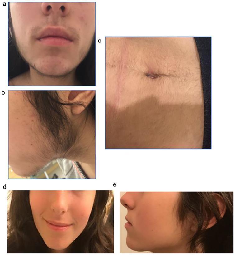
A series of clinical photographs documenting a case of severe hirsutism and its response to treatment. Panels (a) and (b) show the patient at presentation, demonstrating dense, dark terminal hair growth on the upper lip, chin, and submandibular region (jawline) extending toward the preauricular area, consistent with a high Ferriman-Gallwey score. Panel (a) also shows evidence of mild inflammatory acne on the cheeks. Panel (c) displays significant terminal hair growth on the abdomen, both supraumbilical and infraumbilical. Panels (d) and (e) provide a follow-up comparison at 6 months after pharmacological intervention, showing a dramatic clinical reduction in facial terminal hair density and improvement in skin texture. This comparison illustrates the visual manifestation of hyperandrogenism and the efficacy of hormonal management in reversing androgen-dependent hair patterns in female patients.
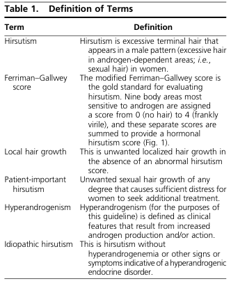
<table> <tr> <th>Table 1. Definition of Terms</th> <th></th> </tr> <tr> <th>Term</th> <th>Definition</th> </tr> <tr> <td>Hirsutism</td> <td>Hirsutism is excessive terminal hair that appears in a male pattern (excessive hair in androgen-dependent areas; <i>i.e.</i>, sexual hair) in women.</td> </tr> <tr> <td>Ferriman–Gallwey score</td> <td>The modified Ferriman–Gallwey score is the gold standard for evaluating hirsutism. Nine body areas most sensitive to androgen are assigned a score from 0 (no hair) to 4 (frankly virile), and these separate scores are summed to provide a hormonal hirsutism score (Fig. 1).</td> </tr> <tr> <td>Local hair growth</td> <td>This is unwanted localized hair growth in the absence of an abnormal hirsutism score.</td> </tr> <tr> <td>Patient-important hirsutism</td> <td>Unwanted sexual hair growth of any degree that causes sufficient distress for women to seek additional treatment.</td> </tr> <tr> <td>Hyperandrogenism</td> <td>Hyperandrogenism (for the purposes of this guideline) is defined as clinical features that result from increased androgen production and/or action.</td> </tr> <tr> <td>Idiopathic hirsutism</td> <td>This is hirsutism without hyperandrogenemia or other signs or symptoms indicative of a hyperandrogenic endocrine disorder.</td> </tr> </table>
Evaluation of Hyperandrogenism in Females
Step 1: History
Key Questions to Ask
| Domain | Points |
|---|---|
| Onset & progression | Sudden, rapid onset → suspect androgen-secreting tumor; gradual since puberty → PCOS or idiopathic |
| Menstrual history | Oligomenorrhea (<8 cycles/year) or amenorrhea → ovarian source more likely; regular cycles → idiopathic or adrenal |
| Age of onset | Teens/early 20s with onset around menarche → PCOS; adult onset with weight gain → insulin-driven PCOS |
| Cosmetic self-treatment | Waxing, shaving, threading underestimate clinical severity on examination - always ask |
| Drug history | Phenytoin, minoxidil, cyclosporine, valproic acid, diazoxide → hypertrichosis (not true HA); androgenic progestins in older OCP formulations |
| Family history | Hirsutism + infertility → non-classic CAH; PCOS in mother/sisters |
| Galactorrhea | → Hyperprolactinemia |
| Cushing features | Weight gain (central), easy bruising, striae, hypertension, proximal muscle weakness → Cushing syndrome |
| Acromegaly features | Headache, visual field defects, jaw changes, hand/foot enlargement (rare cause) |
| Fertility goals | Determines which treatment arm to pursue |
Red flag history: Sudden onset + rapid progression + virilization = androgen-secreting tumor until proven otherwise. - Harrison's 22E, p. 3183
Step 2: Physical Examination
General
- Height, weight, BMI - BMI >30 commonly associated with hirsutism (increased androgen conversion peripherally)
- Blood pressure - hypertension suggests adrenal etiology (Cushing's, adrenal tumor)
- Waist circumference - central obesity → insulin resistance
Skin Signs
| Sign | Significance |
|---|---|
| Hirsutism | Androgen-sensitive distribution (see mFG score below) |
| Acne | Comedonal/inflammatory, especially jawline; suggests androgen excess |
| Androgenic alopecia | Diffuse crown thinning (Ludwig pattern) in women; frontoparietal recession in severe cases |
| Acanthosis nigricans | Hyperpigmented, velvety skin at nape/axillae/groin → insulin resistance |
| Skin tags | Also suggest insulin resistance |
| Striae, easy bruising | → Cushing syndrome |
| Seborrhea | Oily skin → androgen-stimulated sebaceous glands |
Gynaecological / Endocrine
- Clitoromegaly, voice deepening, breast atrophy, increased muscle bulk → virilization; demands urgent tumor work-up
- Thyroid enlargement → hypothyroidism/hyperthyroidism
- Galactorrhea → hyperprolactinemia
- Abdominal/pelvic mass → ovarian tumor
The Modified Ferriman-Gallwey (mFG) Score
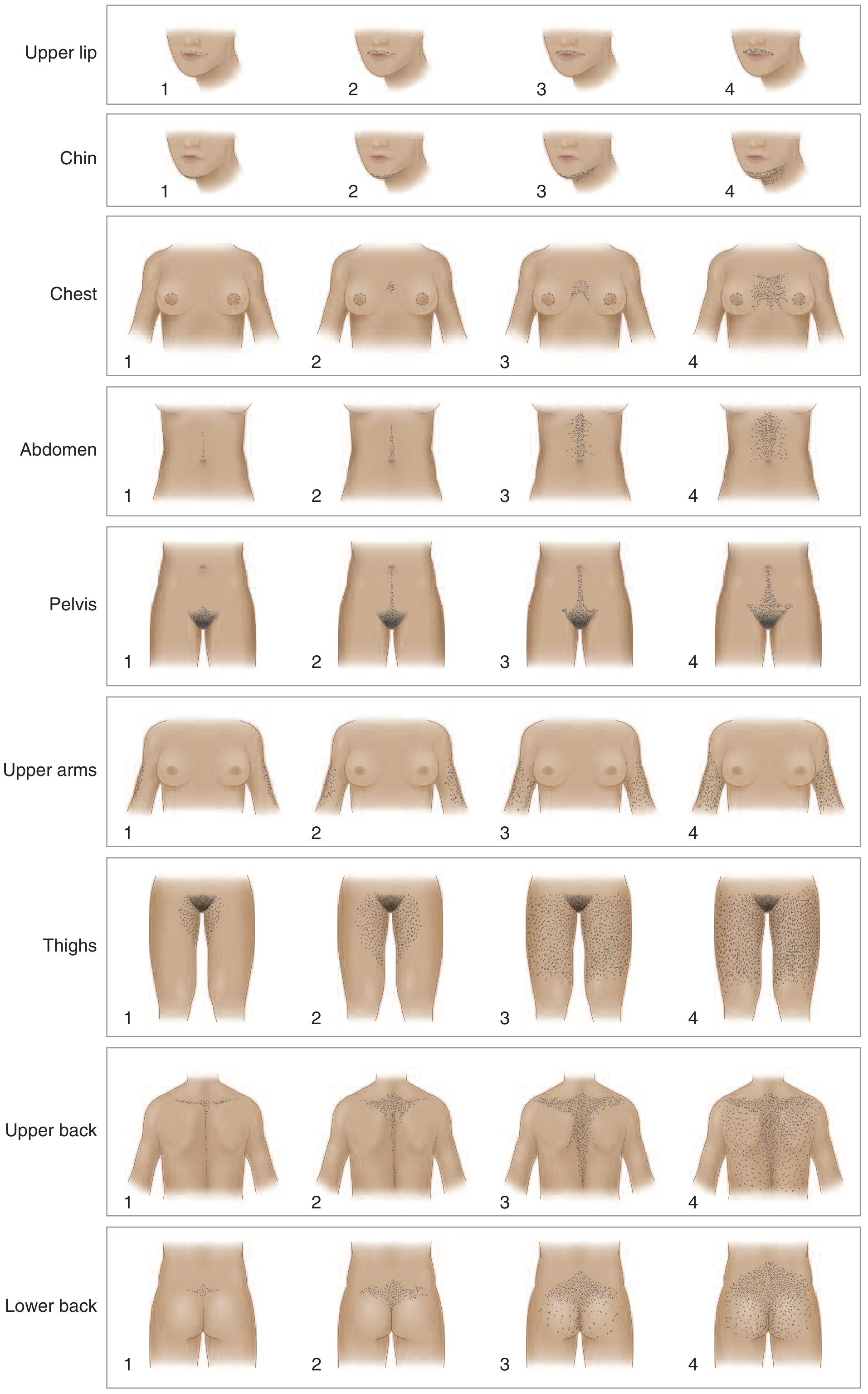
| Score | Interpretation |
|---|---|
| < 8 | Normal (non-Hispanic White, African-American) |
| ≥ 8 | Hirsutism - warrants hormonal evaluation |
| ≥ 4-6 | Threshold for some ethnicities (Endocrine Society 2018) |
Note: Asian and Native American women may score <8 but still have significant biochemical HA manifesting only as acne or scalp hair thinning. - Harrison's 22E, p. 3184
Step 3: Investigations
First-Line Blood Tests
| Test | What it Detects | Key Thresholds |
|---|---|---|
| Total testosterone (by LC-MS/MS specialty assay) | Ovarian source; PCOS; tumor | >200 ng/dL (>7 nmol/L) → strongly suggests tumor; >100 ng/dL → investigate further |
| Free / calculated free testosterone (from total T + SHBG) | Most accurate biochemical marker; elevated even when total T is normal due to low SHBG | Elevated in PCOS with hyperinsulinemia |
| SHBG | Low in insulin resistance/PCOS → increases free testosterone fraction | |
| DHEA-S | Adrenal androgen source | >7000 μg/L (>18.5 μmol/L) → adrenal tumor; modestly elevated in PCOS too |
| 17-Hydroxyprogesterone (17-OHP) - fasting, follicular phase | Screens for non-classic CAH (21-hydroxylase deficiency) | Basal >200 ng/dL → proceed to ACTH stimulation test |
| LH : FSH ratio | PCOS | Ratio >2-3 supportive of PCOS (but not required for diagnosis) |
| Fasting glucose + fasting insulin / HOMA-IR | Insulin resistance in PCOS | |
| Prolactin | Hyperprolactinemia | |
| TSH | Hypothyroidism |
Labs are most accurate when the patient is off oral contraceptives for 4-6 weeks and drawn before menses (follicular phase). - Fitzpatrick's Dermatology, Table 137-18
Additional Tests When Indicated
| Indication | Test |
|---|---|
| Testosterone >200 ng/dL or DHEA-S >7000 μg/L | CT abdomen/pelvis (adrenal) + transvaginal ultrasound (ovary) |
| Rapid-onset virilization at any testosterone level | Urgent pelvic ultrasound + CT adrenals |
| Cushing syndrome suspected | 24-hour urinary free cortisol; overnight 1 mg dexamethasone suppression test; late-night salivary cortisol |
| Non-classic CAH (basal 17-OHP 200-1000 ng/dL) | ACTH stimulation test (250 μg IV ACTH; 17-OHP >10,000 ng/dL at 60 min → confirms CAH) |
| Acromegaly suspected | IGF-1, oral glucose tolerance test with GH |
| Adolescent PCOS | Caution - diagnosis requires both persistent features of HA and ovulatory dysfunction; do not over-label |
Pelvic Ultrasound (Transvaginal preferred)
- Polycystic ovarian morphology (PCOM): ≥20 follicles (2-9 mm) in either ovary AND/OR ovarian volume >10 mL
- Identifies ovarian tumors (Sertoli-Leydig, thecoma, granulosa cell)
- Only 1 ovary needs to meet criteria for PCOM diagnosis
- PCOM alone (without HA + ovulatory dysfunction) does NOT diagnose PCOS - found in ~23% of reproductive-age women
Diagnostic Algorithm
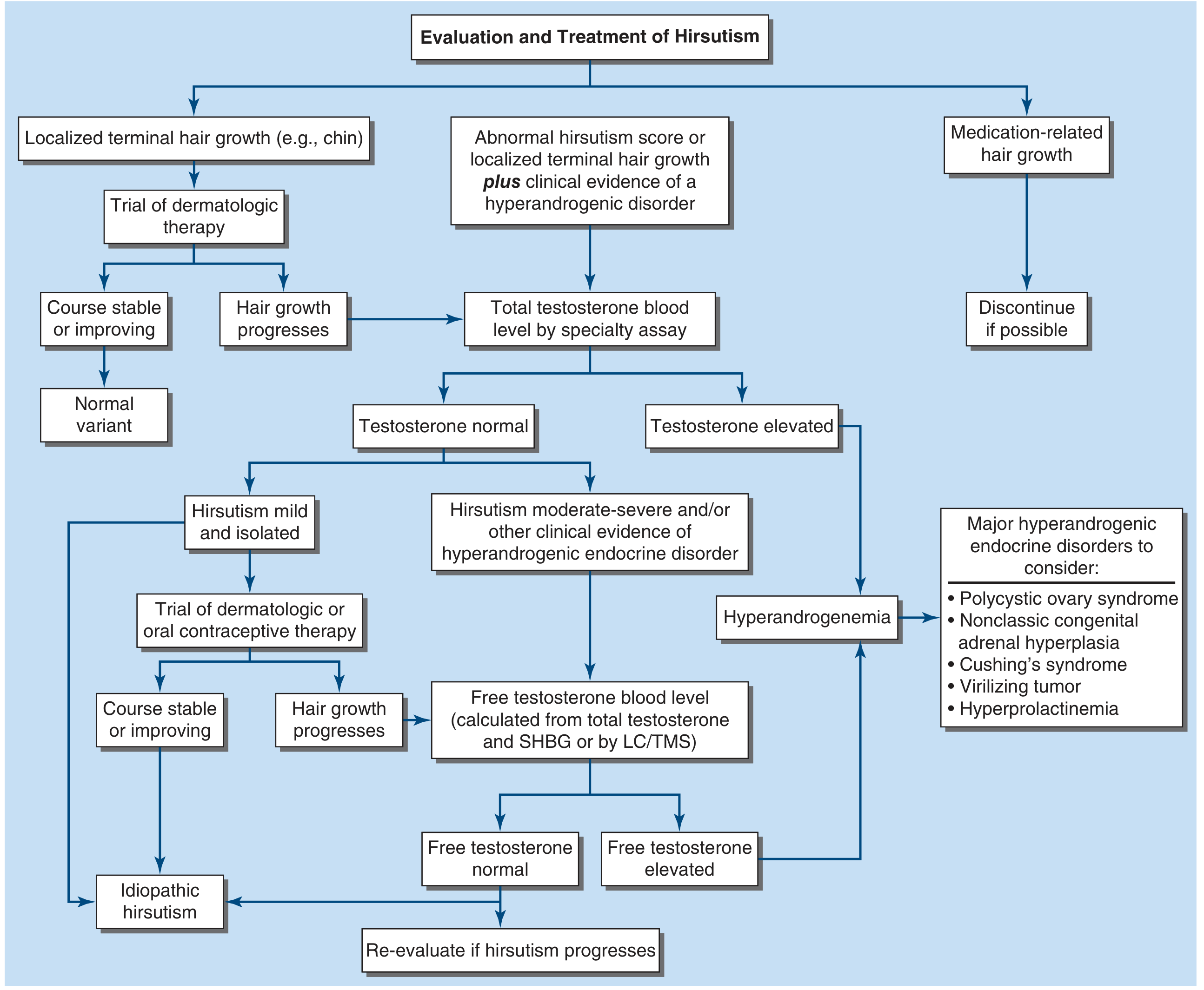
- Localized hair (e.g. chin only) → trial of dermatologic therapy; if stable, normal variant
- Abnormal mFG score or clinical evidence of HA disorder → total testosterone by specialty assay
- Testosterone normal + mild isolated hirsutism → trial of OCP or dermatologic therapy; if unchanged, idiopathic hirsutism
- Testosterone normal + moderate-severe hirsutism or other HA features → free testosterone (calculated from total T + SHBG, or by LC/MS)
- Free testosterone elevated or total testosterone elevated → Hyperandrogenemia → work up for:
- PCOS
- Non-classic CAH (17-OHP)
- Cushing syndrome
- Virilizing tumor (imaging)
- Hyperprolactinemia
- Medication-related → discontinue if possible
Differential Diagnosis Summary
| Condition | Key Clinical Clues | Key Lab Finding | Confirmatory Test |
|---|---|---|---|
| PCOS | Irregular periods + hirsutism since puberty, obesity, acanthosis nigricans | Elevated free T, low SHBG, elevated LH:FSH | Pelvic USS (PCOM) + Rotterdam criteria |
| Non-classic CAH | Hirsutism ± irregular cycles; family history; Ashkenazi/Hispanic/Slavic ethnicity | Elevated basal 17-OHP (>200 ng/dL) | ACTH stimulation test (17-OHP >10,000 ng/dL) |
| Androgen-secreting ovarian tumor | Rapid onset, virilization, severe hirsutism | Testosterone >200 ng/dL (>7 nmol/L) | Transvaginal ultrasound → MRI if needed |
| Adrenal tumor | Rapid onset, virilization | DHEA-S >7000 μg/L | CT adrenals |
| Cushing syndrome | Centripetal obesity, striae, easy bruising, hypertension, moon face | Elevated urinary free cortisol | 1 mg DST, late-night salivary cortisol, CRH test |
| Hyperthecosis | Marked virilization, postmenopausal, testosterone >200 ng/dL, normal imaging | Testosterone >200 ng/dL | Bilateral oophorectomy confirms |
| Hyperprolactinemia | Galactorrhea, amenorrhea | Elevated prolactin | MRI pituitary |
| Idiopathic hirsutism | Normal menses, normal androgens, positive mFG | All labs normal | Diagnosis of exclusion |
Investigations Warrant Urgent Attention
- Testosterone >200 ng/dL (>7 nmol/L)
- DHEA-S >7000 μg/L (>18.5 μmol/L)
- Rapid onset (<1-2 years) with progressive virilization at any testosterone level
- Virilization: clitoromegaly, voice change, breast atrophy, male-pattern hair loss with full virilization syndrome
Osteogenesis imperfecta
"osteogenesis imperfecta"[MeSH Terms] AND treatment
osteogenesis imperfecta radiograph fractures blue sclera

A multi-panel clinical and diagnostic figure illustrating manifestations of Osteogenesis Imperfecta (OI) in a pediatric patient. Panels A and B are clinical photographs showing a broad forehead and a distinctive triangular facial shape at age 1 and 2 years. Panel C provides a close-up of the eyes, highlighting blue sclera. Panels D, E, and F are serial radiographs of the lower extremities. Panel D (11 months) shows a transverse fracture of the proximal right femur, a curved left femur, slender long bones, and generalized osteopenia. Panel E (13 months) demonstrates the healing progress of the right femur fracture. Panel F (24 months) shows surgical management of recurrent fractures in the right femur, featuring an intramedullary rod used for stabilization and alignment. The combination of blue sclera, recurrent fractures, and radiographic osteopenia is characteristic of a Type I or Type IV OI phenotype, often associated with COL1A1 or COL1A2 mutations.

Two-panel clinical photograph of a pediatric patient with osteogenesis imperfecta (OI) demonstrating conservative management of neonatal fractures through immobilization. Panel (a) shows the patient as a neonate in a supine position with a white, multi-layered cotton wrap applied to the left lower extremity, extending from the mid-thigh to the ankle to provide stability and comfort for a femoral or tibial fracture. A blue pacifier is seen nearby. Panel (b) shows the same child at an older age, seated and holding a toy, with a similar soft cotton immobilization wrap on the right upper extremity, secured by a white mesh swathe/garment over the torso. The visual illustrates the educational concept of using lightweight, non-synthetic padding for neonatal fractures in OI, which avoids the risks associated with heavy plaster casts while providing sufficient stabilization and a visual reminder to caregivers to handle the infant with caution. Key clinical features of OI, such as potential blue sclera and macrocephaly, are subtly visible in the patient's facies.

This comparison clinical photograph features two frontal views focusing on the ocular region of two related individuals to demonstrate phenotypic variations in the sclera. The left panel shows a patient with distinct blue sclera, a classic clinical sign characterized by a bluish-gray discoloration of the white part of the eye, which occurs due to thinning of the collagen fibers allowing the underlying uveal pigment to show through. This finding is highly associated with Osteogenesis Imperfecta (OI) Type I. The patient also exhibits a triangular facial shape and minor skin lesions. The right panel displays the patient's father for comparison, showing a normal, white opaque sclera and typical age-related skin changes, including solar lentigines and fine rhytids. The contrast between the two images highlights the pathognomonic physical examination finding of blue sclerae in the context of hereditary connective tissue disorders. This visual is used in medical education to assist in the differential diagnosis of skeletal dysplasia and collagen synthesis defects.

A multi-panel clinical photograph illustrating the phenotypic and physical manifestations of Osteogenesis Imperfecta (OI) Type I following a traumatic hip injury. Panel (a) provides a close-up of the eyes, demonstrating the characteristic blue sclera indicative of connective tissue abnormality. Panels (b), (c), and (d) depict the patient's lower extremities in various views. The right lower limb exhibits classic clinical signs of a central hip dislocation and acetabular fracture, including visible limb shortening, external rotation, and abduction. The skin over the right hip (panel c) appears intact without evidence of significant ecchymosis or edema. These findings collectively illustrate the multisystemic nature of OI, where bone fragility and ligamentous laxity predispose patients to complex orthopedic injuries, such as the comminuted acetabular fracture and central hip dislocation noted in this case.
Osteogenesis Imperfecta (OI)
Definition
Epidemiology
- Prevalence: approximately 1 in 20,000 individuals
- Affects all ethnic groups equally
-
80% of cases result from autosomal dominant mutations in COL1A1 or COL1A2 (encoding the α1(I) and α2(I) chains of type I procollagen)
- Remaining ~20% involve at least 22 other genes affecting collagen biosynthesis, post-translational modification, trafficking, cross-linking, osteoblast differentiation, and bone mineralization - Firestein & Kelley's Rheumatology, p. 2337
Pathogenesis
| Mechanism | Effect | Typical Type |
|---|---|---|
| Quantitative defect - Nonsense/frameshift mutations → premature stop codon → haploinsufficiency → ~50% normal collagen synthesized | Too little bone matrix | OI Type I (mild) |
| Qualitative defect - Glycine substitution mutations → misfolded procollagen → dominant negative effect on wild-type chains → abnormal collagen assembly | Structurally defective bone matrix | OI Types II, III, IV (moderate-lethal) |
- The dominant negative effect means even one mutant chain disrupts assembly of all trimers containing it - markedly amplifying the pathological effect
- In recessive forms: mutations affect the CRTAP-P3H1-CyPB prolyl 3-hydroxylase complex, which hydroxylates Pro986 - a critical post-translational modification of type I collagen
- The fundamental abnormality in all forms: synthesis of too little bone → extreme skeletal fragility - Robbins Pathology; Goldman-Cecil Medicine, p. 2751
Classification (Sillence Classification - Extended)
| Type | Severity | Key Features | Inheritance | Gene/Defect |
|---|---|---|---|---|
| I | Mild | Blue sclerae; 5-15 major fractures before puberty; fractures heal without deformity; near-normal stature; hearing loss in adults; DI uncommon | AD | COL1A1 nonsense/frameshift → haploinsufficiency (50% normal collagen) |
| II | Lethal | Stillborn or neonatal death; innumerable fractures at birth; beaded ribs; crumpled long bones; small thorax → pulmonary failure | AD (de novo) / AR | COL1A1 or COL1A2 glycine substitutions |
| III | Severe, progressively deforming | Fractures from birth; progressive limb deformity; very short stature (<90-100 cm); triangular facies with frontal bossing; sclerae blue at birth → lighten to white; DI in 75%; wheelchair-dependent; restrictive lung disease; basilar invagination | AD (de novo) / AR | Single amino acid substitution in COL1A1 or COL1A2 |
| IV | Moderate | Fractures common; variable short stature; mild-moderate deformity; normal scleral hue; DI common; hearing loss variable | AD | Point mutations or exon-skipping in COL1A1/COL1A2 |
| V | Moderate (similar to IV) | No DI, no blue sclerae; distinctive hyperplastic callus formation; calcification of interosseous membrane (radius-ulna) | AD | Unknown (IFITM5 mutations identified later) |
| VI | Moderate-severe | Excess osteoid on bone biopsy; no DI, no blue sclerae, no wormian bones | AR | SERPINF1 (pigment epithelium-derived factor) |
| VII | Severe (like II/III) | Fractures at birth; rhizomelic limb shortening; coxa vara; blue sclerae; no DI | AR | CRTAP mutations |
| VIII | Severe (like II/III) | Similar to VII | AR | LEPRE1 (P3H1) mutations |
| IX | Severe (like II/III) | Similar to VII | AR | PPIB (Cyclophilin B) mutations |
Clinical Features
Skeletal
- Recurrent fractures - often from minimal or no trauma; key feature is fractures at all ages
- Bone deformity - progressive bowing of long bones from recurrent fractures; most severe in types II and III
- Short stature - mild in type I, extreme in type III (<3 feet in adults)
- Scoliosis / kyphoscoliosis - may progress to restrictive lung disease (scoliosis >60° → respiratory compromise)
- Basilar invagination - softening at skull base → brain stem compression → headache, diplopia, nystagmus, upper motor neuron signs, sleep apnea, hydrocephalus (especially type III)
- Wormian bones - microfractures/supernumerary ossification centers along cranial sutures (pathognomonic on skull X-ray)
Extra-skeletal
| Feature | Mechanism | Notes |
|---|---|---|
| Blue sclerae | Reduced collagen → thin sclerae → choroid visible through | Hallmark of type I; sclerae normalize in type III with age |
| Dentinogenesis imperfecta (DI) | Abnormal dentin → opalescent, translucent, blue-yellow teeth that chip and wear rapidly | Prevalence increases with OI severity; affects deciduous > permanent teeth |
| Hearing loss | Sensorineural + conductive (ossicular chain affected) | Usually appears in 2nd-3rd decade; mixed or predominantly conductive |
| Joint laxity / hypermobility | Defective collagen in ligaments | Contributes to deformity |
| Easy bruising | Defective collagen in skin and blood vessel walls | Common complaint |
| Aortic root dilatation | Collagen in aortic wall affected | Rare; occasional kindreds with aortic regurgitation |
Radiological Features

- Osteopenia (generalized) - most prominent in spine
- Fractures at various stages of healing; may show florid callus formation
- Gracile bones (thin, over-tubulated long bones) or deformed/shortened bones in severe forms
- Wormian bones on skull X-ray
- Vertebral fractures - biconcave/"cod-fish" vertebrae; platybasia
- Beaded ribs in type II (due to multiple healing calluses)
- Interosseous membrane calcification between radius and ulna (type V)
- Coxa vara in recessive types (VII, VIII)
- Prenatal diagnosis on 2nd trimester ultrasound: deformed/shortened limbs, relative macrocephaly, frontal bossing, flat midface - Grainger & Allison's Radiology, p. 1092
Diagnosis
Clinical Diagnosis
- Recurrent fractures (especially with minimal trauma)
- Blue sclerae
- Dentinogenesis imperfecta
- Wormian bones on skull X-ray
- Family history (often absent in de novo types)
Important Differential Diagnoses
| Condition | Distinguishing Features |
|---|---|
| Non-accidental injury (child abuse) | Normal bone mineralization; pattern of fractures usually distinct; no blue sclerae, no DI, no wormian bones |
| Hypophosphatasia | Low alkaline phosphatase; premature loss of deciduous teeth |
| Osteoporosis (juvenile/idiopathic) | May resemble type I OI; genetic overlap exists with COL1A1 mutations |
| Achondrogenesis / skeletal dysplasias | Specific radiologic patterns differ |
| Menkes syndrome | Low serum copper; kinky hair; X-linked recessive |
| Osteopetrosis | Dense sclerotic bones (not osteopenic) |
Laboratory and Molecular Testing
- Bone densitometry (DXA scan): BMD significantly reduced; T-scores frequently -2.5 to -4.0 (osteoporotic range); Z-scores used in children
- Serum alkaline phosphatase: Low → hypophosphatasia (excludes that DDx)
- Collagen synthesis study: Cultured skin fibroblasts → reduced or structurally abnormal type I collagen
- Molecular genetic analysis (COL1A1/COL1A2 sequencing + deletion/duplication analysis): definitive diagnosis; enables reproductive counseling
- Gene panel testing: For recessive forms when dominant gene testing is negative
- Prenatal: Chorionic villus sampling (if family mutation known); detailed fetal USS/MRI from 2nd trimester
In child abuse cases, OI can be excluded/confirmed by collagen production from cultured skin fibroblasts or DNA mutation analysis. - Goldman-Cecil, p. 2752
Complications
- Pulmonary insufficiency (leading cause of death in type III) - from restrictive lung disease due to kyphoscoliosis + chest wall deformity
- Basilar invagination → brainstem compression, central sleep apnea, hydrocephalus
- Fracture non-union (rare in mild forms; more common in severe)
- Hearing loss - progressive; may require hearing aids or cochlear implants
- Dental complications - from DI; need early dental referral
- Cardiovascular - aortic root dilatation; monitor with echo
- Psychological - chronic pain, impaired mobility, body image issues
Management
Pharmacological
- Pamidronate (IV cyclical) - most studied; increases BMD, reduces fracture frequency, decreases bone pain, improves mobility
- Zoledronic acid (IV annual) - increasingly preferred for convenience
- Risedronate (oral) - 2.5 or 5 mg daily; shown to increase BMD and reduce first and recurrent fractures in children
- Mechanism: inhibit osteoclast-mediated bone resorption → increased bone mass
- Long-term use: concern about oversuppression of bone turnover and atypical femoral fractures (same as in osteoporosis)
- Teriparatide (PTH 1-34, anabolic): used in adult OI; increases BMD; contraindicated in children (open growth plates)
- Denosumab (RANK-L inhibitor): used in some severe pediatric cases; risk of rebound hypercalcemia on discontinuation
- Calcitonin: limited evidence; used in some centers for acute pain management
- Calcium and Vitamin D supplementation: adjunctive
Surgical
- Intramedullary rodding (IM rods) - placed in long bones (particularly femur, tibia) to prevent deformity and allow ambulation
- Bailey-Dubow rods (telescoping/elongating) preferred in children - grow with the bone
- Non-elongating rods used in adults
- Goal: maintain ambulation → prevent disuse demineralization
- Spinal fusion - for progressive scoliosis (>40-50°)
- Basilar decompression - for symptomatic basilar invagination
- Osteotomies - corrective procedures for severely bowed limbs
Rehabilitative / Supportive
- Physiotherapy - water therapy (hydrotherapy) well tolerated; strengthens muscles without fracture risk; weight-bearing exercise when safe
- Occupational therapy - adaptive equipment, mobility aids, home modification
- Orthotics - bracing to prevent deformity
- Hearing aids / cochlear implants - for hearing loss
- Dental care - early referral for DI management; may need early deciduous tooth extraction
Genetic Counseling
- Essential for all families; type II carries risk of germinal mosaicism (documented), meaning recurrence risk is not negligible even in apparently de novo cases
- Prenatal diagnosis available via CVS or amniocentesis when mutation is known
- Molecular testing of sperm can quantify recurrence risk when de novo paternal mutation is suspected
- Goldman-Cecil, p. 2751; Firestein & Kelley's Rheumatology
Prognosis
| Type | Prognosis |
|---|---|
| Type I | Normal or near-normal lifespan; fracture frequency falls after puberty, increases again post-menopausally in women |
| Type II | Lethal - stillborn or neonatal death from pulmonary failure |
| Type III | Significantly shortened lifespan; most wheelchair-dependent; death from pulmonary insufficiency |
| Type IV | Reduced but functional lifespan; fracture frequency diminishes with adolescence |
| Types V-IX | Variable; recessive types II/III-like carry poor prognosis |
Key Memory Points
- Most common inherited connective tissue disorder; due to defective type I collagen (COL1A1/COL1A2)
- Classic triad: Fragile bones + Blue sclerae + Dentinogenesis imperfecta (not all types have all three)
- Type I = Mildest; blue sclerae; normal or near-normal stature; AD
- Type II = Lethal; beaded ribs; crumpled long bones; pulmonary failure
- Type III = Most severe non-lethal; progressive deformity; triangular facies; sclerae white in adults
- Type IV = Normal scleral hue is the key distinguishing feature from type I
- Wormian bones on skull X-ray - pathognomonic
- Bisphosphonates are the pharmacological cornerstone
- IM rods (Bailey-Dubow telescoping) prevent deformity and maintain ambulation in children
- Pulmonary insufficiency is the leading cause of death in severe forms