
Kidneys: developmental anatomy, topography, structure of nephron, developmental anamolies of kidney, their blood supply and innervation
Reading File
This covers a broad renal anatomy unit. I can prepare detailed notes on one section at a time:
- Developmental anatomy - embryologic origin, ascent, rotation, and fetal lobulation
- Topography and gross anatomy - position, relations, coverings, hilum, and internal structure
- Nephron structure - renal corpuscle, tubules, collecting system, and juxtaglomerular apparatus
- Developmental anomalies - ectopic kidney, horseshoe kidney, fusion anomalies, malrotation, agenesis, and duplex systems
- Blood supply, lymphatics, and innervation - renal arterial branches, venous drainage, segmental anatomy, and autonomic nerves
At a high level, permanent kidneys arise from reciprocal interaction between the ureteric bud and metanephric mesenchyme. They initially form in the pelvis, ascend to the upper posterior abdominal wall, and acquire sequential arterial supplies. Each kidney contains about a million nephrons that filter blood at the glomerulus and modify filtrate through specialized tubular segments. Many congenital anomalies result from abnormal renal formation, ascent, rotation, or fusion.
Which section would you like in detailed exam-note format?
Asking for Preferences
Choose the section to study first.
This covers a broad renal anatomy unit. I can prepare detailed notes on one section at a time:
- Developmental anatomy - embryologic origin, ascent, rotation, and fetal lobulation
- Topography and gross anatomy - position, relations, coverings, hilum, and internal structure
- Nephron structure - renal corpuscle, tubules, collecting system, and juxtaglomerular apparatus
- Developmental anomalies - ectopic kidney, horseshoe kidney, fusion anomalies, malrotation, agenesis, and duplex systems
- Blood supply, lymphatics, and innervation - renal arterial branches, venous drainage, segmental anatomy, and autonomic nerves
At a high level, permanent kidneys arise from reciprocal interaction between the ureteric bud and metanephric mesenchyme. They initially form in the pelvis, ascend to the upper posterior abdominal wall, and acquire sequential arterial supplies. Each kidney contains about a million nephrons that filter blood at the glomerulus and modify filtrate through specialized tubular segments. Many congenital anomalies result from abnormal renal formation, ascent, rotation, or fusion.
Which section would you like in detailed exam-note format?Please select one topic to begin: development, topography, nephron structure, congenital anomalies, or blood supply and innervation.
Explain kidney development with a labeled embryology sequence
Reading File
Finding Sources
Finding Sources
Searching Images
embryology diagram development of kidney pronephros mesonephros metanephros ureteric bud nephron
Searching PubMed
kidney development embryology
Searching the Web
kidney embryology development ureteric bud metanephric mesenchyme ascent rotation authoritative
Reading File
Reading File
Reading File
Reading Web Page
https://embryology.med.unsw.edu.au/embryology/index.php?titl…
Reading File
Reading File
Finding Sources
Kidney development: labeled embryology sequence
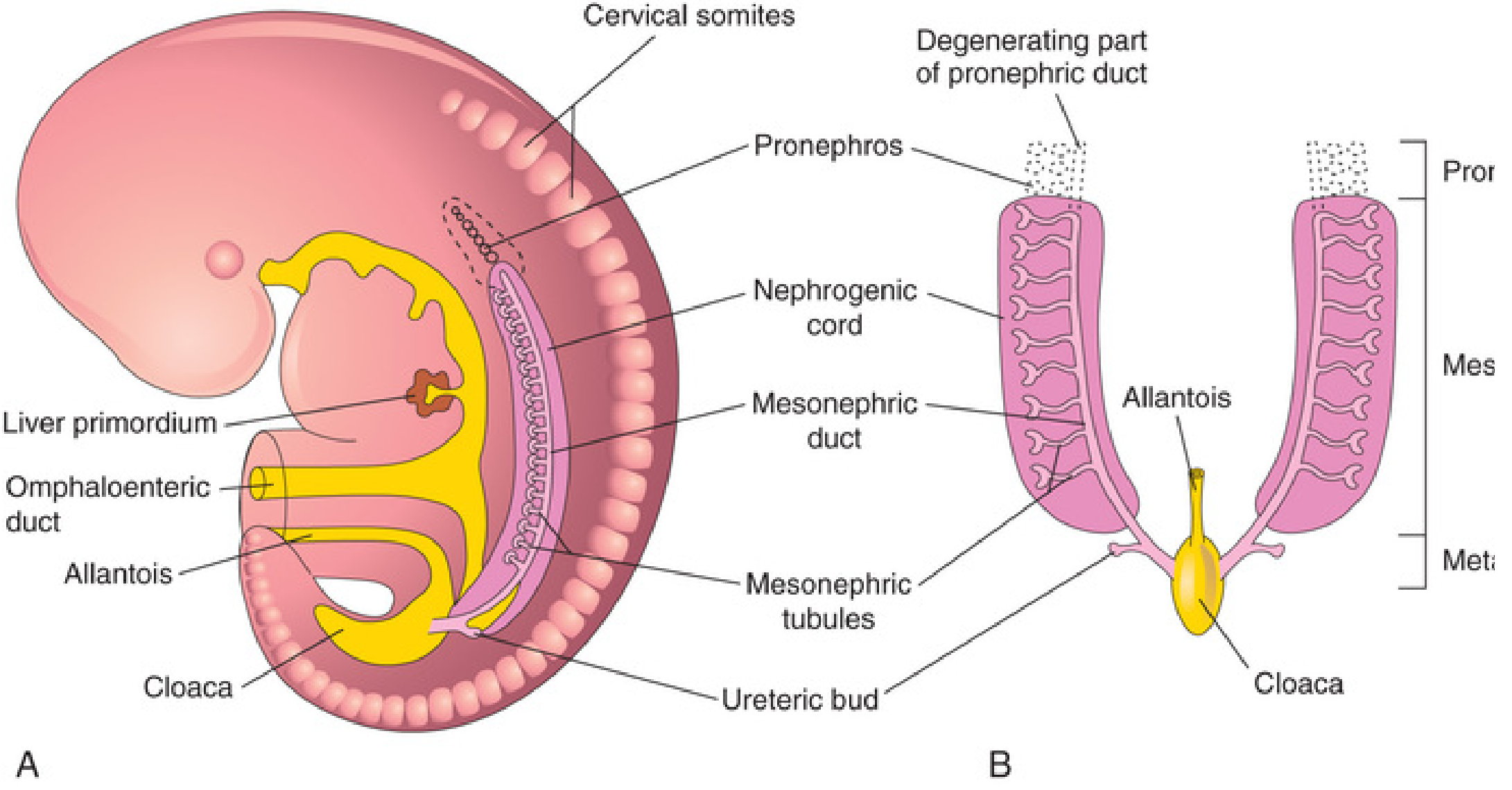
1. Origin
The urinary system develops from intermediate mesoderm, which forms a longitudinal urogenital ridge on either side of the dorsal aorta. Its urinary component is the nephrogenic cord.
Three renal systems appear in a cranial-to-caudal, partly overlapping sequence:
Intermediate mesoderm
        ↓
Nephrogenic cord / urogenital ridge
        ↓
1. Pronephros        2. Mesonephros        3. Metanephros
   transient            temporary              definitive kidney
The Developing Human: Clinically Oriented Embryology, p. 679-680
2. Sequential development
| Stage | Approximate time | Position | Fate / significance |
|---|---|---|---|
| Pronephros | Early 4th week | Cervical region | Rudimentary and nonfunctional in humans. Degenerates rapidly. Its duct persists as the mesonephric duct. |
| Mesonephros | Late 4th week to end of 2nd month | Thoracic and upper lumbar region | Functions briefly as a temporary excretory organ. Most regresses; mesonephric duct and some tubules contribute to male genital ducts. |
| Metanephros | Begins in 5th week | Initially sacral/pelvic region | Forms the permanent kidney. Begins producing urine at approximately weeks 9-10. |
Memory aid: Pro - Meso - Meta = First - Middle - Mature kidney.
The Developing Human: Clinically Oriented Embryology, p. 680-682; Langman’s Medical Embryology, p. 275-276
3. Formation of the permanent kidney: metanephros
The metanephros requires interaction between two embryological primordia:
Mesonephric duct
       │
       └── Ureteric bud  ───────────────→ Collecting system

Intermediate mesoderm
       │
       └── Metanephric mesenchyme
           (metanephric blastema) ──────→ Nephrons
A. Ureteric bud
The ureteric bud is an epithelial diverticulum from the caudal mesonephric duct, near its opening into the cloaca.
It repeatedly branches to form the collecting part of the kidney:
Ureteric bud
   ↓
Ureter
   ↓
Renal pelvis
   ↓
Major calyces
   ↓
Minor calyces
   ↓
Collecting ducts and collecting tubules
B. Metanephric mesenchyme
The metanephric mesenchyme, also called the metanephric blastema, forms the excretory part of the nephron:
Metanephric mesenchyme
   ↓
Renal vesicle
   ↓
Comma-shaped body
   ↓
S-shaped body
   ↓
Nephron
Its adult derivatives are:
Bowman's capsule
Glomerular podocytes
Proximal convoluted tubule
Loop of Henle
Distal convoluted tubule
Important boundary: The distal tubule joins a collecting tubule, creating a continuous pathway for urine flow.
4. Labeled nephron-development sequence
Terminal tip of branching ureteric bud
                 │
                 │ induces
                 ▼
Condensed metanephric mesenchyme
                 │
      mesenchymal-to-epithelial transition
                 ▼
Renal vesicle
                 ▼
Comma-shaped body
                 ▼
S-shaped body
      ┌──────────┴───────────┐
      │                      │
      ▼                      ▼
Proximal end             Distal end
invaginated by           joins collecting
capillary tuft           duct
      │
      ▼
Glomerulus + Bowman's capsule
      │
      ▼
Renal corpuscle
Formation of the glomerulus
At the proximal end of the S-shaped body, capillaries grow in and invaginate the epithelium. This produces:
- Glomerular capillary tuft
- Bowman's capsule
- Visceral layer of Bowman's capsule, formed by podocytes
- Renal corpuscle
The remainder of the S-shaped body differentiates into the proximal tubule, loop of Henle, and distal tubule.
5. Reciprocal induction
Development depends on bidirectional induction:
Metanephric mesenchyme
       └── induces growth and repeated branching of ureteric bud

Ureteric bud
       └── induces metanephric mesenchyme to form nephrons
Important signaling examples:
- GDNF from metanephric mesenchyme acts through RET receptors to promote ureteric-bud outgrowth and branching.
- WT1 helps metanephric mesenchyme respond to induction.
- PAX2, WNT4, and related pathways participate in nephron differentiation.
Failure of this interaction may cause renal agenesis, hypoplasia, or dysplasia.
6. Ascent, rotation, and changing blood supply
Initially, the metanephric kidneys lie close together in the pelvis, anterior to the sacrum, with their hila facing anteriorly.
Weeks 6-9 approximately

Pelvic kidneys
     ↓ ascent due mainly to differential body growth
Lumbar region
     ↓ medial rotation about 90°
Adult retroperitoneal position
Changes during ascent
| Feature | Early pelvic kidney | Adult kidney |
|---|---|---|
| Position | Sacral/pelvic | Upper posterior abdominal wall |
| Hilum direction | Anterior | Medial |
| Blood supply | Branches from common iliac / lower aorta | Renal arteries from abdominal aorta |
| Relationship | Close together | Separate and lateral to vertebral column |
As the kidney ascends, it receives successive temporary arterial branches from progressively higher levels of the aorta. Lower vessels normally regress. Persistence produces accessory renal arteries.
The kidneys reach their adult position at the beginning of the fetal period. Apparent ascent is mainly caused by growth of the body caudal to them. The Developing Human: Clinically Oriented Embryology, p. 685.
7. One-page examination flowchart
INTERMEDIATE MESODERM
        ↓
UROGENITAL RIDGE
        ↓
NEPHROGENIC CORD
        ↓
PRONEPHROS (early week 4)
- cervical
- rudimentary
- regresses
        ↓
MESONEPHROS (late week 4 to week 8)
- temporary kidney
- mesonephric duct persists
        ↓
METANEPHROS (week 5 onward)
        ↓
      Two sources
        ├──────────────────────────────────────────────┐
        ↓                                              ↓
URETERIC BUD                                  METANEPHRIC MESENCHYME
from mesonephric duct                         from intermediate mesoderm
        ↓                                              ↓
ureter, pelvis, calyces,                     renal vesicle → comma body
collecting ducts                              → S-shaped body → nephron
        └───────────────── join ───────────────────────┘
                           ↓
                    FUNCTIONAL PERMANENT KIDNEY
                           ↓
                ASCENT + MEDIAL ROTATION
                           ↓
              ADULT LUMBAR RETROPERITONEAL KIDNEY
Key exam line: The ureteric bud forms the collecting system, whereas the metanephric mesenchyme forms the nephron from Bowman's capsule through the distal convoluted tubule.
This is a shared conversation. Sign in to Orris to start your own chat.