
Make the notes as mind maps with different colors, arrows, icons, and boxes Salivary Glands 11 Convert this in one image Structure of Terminal Secretory Units 241 Serous cells 242 Mucous cells 244 Myoepithelial cells 244 Ducts 247 Intercalated ducts 247 Striated ducts 248 Excretory ducts 249 Connective tissue elements 250 Blood supply 250 Nerve supply and pattern of innervation 251 Classification and Structure of Human Salivary Glands 252 Major salivary glands 252 Parotid gland 252 Submandibular gland 252 Sublingual gland 252 Minor salivary glands 254 Labial and buccal glands 254 CHAPTER CONTENTS Glossopalatine glands 255 Palatine glands 255 Lingual glands 255 Von Ebner’s glands 255 Development and Growth 255 Control of Secretion 257 Composition of Saliva 258 Functions of Saliva 258 Protection of the oral cavity and oral environment 259 Digestion 259 Mastication and deglutition 259 Taste perception 259 Speech 260 Tissue repair 260 Excretion 260 Clinical Considerations 260 Summary 261 Review Questions 262 The salivary glands are a group of compound exocrine glands secreting saliva. Saliva is a complex fluid produced by the salivary glands. The saliva forms a film of fluid coating the teeth and mucosa thereby creating and regulating a healthy environment in the oral cavity. The parenchymal elements are derived from the oral epithelium and consist of terminal secretory units lead- ing into ducts that eventually open into the oral cavity. The connective tissue forms a capsule around the gland and extends into it, dividing groups of secretory units and ducts into lobes and lobules. The blood and lymph vessels and nerves that supply the gland are contained within the connective tissue. The salivary glands are compound glands as they have more than one tubule entering the main duct. A duct is a passage that allows the glandular secretion emptied directly into an anatomic location where the secretion is to be used. The salivary glands have numerous ducts associated with them hence they are exocrine glands. The architectural arrangement of the salivary glands is tubuloacinar, where acini are secretory units. These tubuloacinar units are merocrine as they release only the secretion of the cell from the secreting units. STRUCTURE OF TERMINAL SECRETORY UNITS The basic functional unit of a salivary gland is the terminal secretory unit called acini. The terminal secretory unit irrespective of size and location is made up of epithelial secretory cells, namely serous and mucous cells. The serous, mucous along with myoepithelial cells are ar- ranged in an acinus or acini (multiple) with a roughly spherical or tubular shape and a central lumen (Fig. 11.1). The cells in the acini rest on a basement membrane. They are arranged in a single layer. The intercellular spaces of the apical ends of the cells are separated from the lumen by junctional complexes which are tight (zonula occludens), intermediate junction (zonula adhe- rens), and one or more desmosomes (maculae adher- ens). The junctional complexes hold the cells together in an acinus and regulate the permeability. Tight junctions seal the adjacent secretory cells, controlling paracellular ion influx. This helps in maintaining cell polarity and tissue homeostasis. The main tight junctional proteins are claudin, occludin and junctional adhesion molecules. The myoepithelial cells are located on the surfa 242 Orban’s Oral Histology and Embryology The central lumen of each acini may have a star- shaped morphology because of extension of lumen in between the cells called intercellular canaliculi. The cen- tral lumen of the acini continue via a fine series of ducts which constantly merge with each other and grow larger eventually to merge into the main excretory duct. These ducts comprise the ductal system. The mucous acini have a larger lumen than serous acini (end piece). The secretory terminal unit in serous acini is generally made of 8–12 serous acini surrounding a central lumen (Fig. 11.1). Secretory end piece of mucous cells have a tubular con- figuration. The mucous cells are joined to each other by a variety of intracellular junctions but unlike the serous acini, they lack the presence of intercellular canaliculi. The inter- cellular canaliculi are said to be present only in acini with demilunes. Sometimes mucous acini have bonnet or cres- cent shaped covering which is made of serous cells. These are called demilunes (Fig. 11.1). The presence of demilunes is questioned. It has been shown that demilunes are as a re- sult of artifact during tissue preparation. Recent methods like rapid freezing, freeze substitution and three-dimensional reconstruction techniques have shown that serous cells align with mucous cells to surround a common lumen. Serous Cells Serous secretory cells are pyramidal with a broad base on the basement membrane, the apex faces the lumen. The serous cells have a spherical nucleus placed at the basal region. The apical cytoplasm of these cells shows accu- mulation of secretory granules. The secretory granules are 1 mm in diameter with a distinct limiting membrane. In human beings the granules contain a dense core or a twisted skin like structure with a lighter matrix. They can be visualized in semi thin plastic embedded tissue section, stained with toluidine blue or specific cytochem- ical techniques. The granules are closely apposed to each other but retain their individuality (Fig. 11.2). The granules are zymogen granules and are formed by glycolated proteins which are released into a vacuole. In electron microscope the immature granules appear paler in density as compared to electron dense granules which are maturing and moving towards the luminal plasma membrane. The numbers of the secretory granules also vary with different levels of activity in an unstimulated or resting cell. There are numerous granules in the luminal portion of the cell, whereas in a stimulated cell the gran- ules are few as they are depleted in huge numbers into the lumen by exocytosis. The serous cells show acid phosphates, esterases, gluc- uronidase, glucosidase and galactoside activity. The ultrastructural feature of a serous cell is typical of a protein secreting cell. A typical serous cell spends most of its synthetic capacity for producing the secretory pro- tein. The basal cytoplasm is packed with parallelly stacked, with ribosome studded RER (rough endoplasmic reticu- lum). The RER is placed basal and lateral to the cell nucleus. A closed system of cisternae or membranous sacs B B C D E F E′ D′ B′ C′ A A′ B C D E F E′ D′ B′ C′ A A′ Figure 11.1 Schematic diagram of a typical salivary gland. (A) Serous acini, (A9) serous acini in cross-section. (B) Serous demilunes, (B9) serous demilunes in cross-section. (C) Mucous acini, (C9) mucous acini in cross-section. (D) Intercalated duct, (D9) intercalated duct in cross-section. (E) Striated duct, (E9) striated duct in cross-section. (F) Terminal excretory duct. Chapter 11 — Salivary Glands 243 constitutes RER (Fig. 11.3). The ribosomes consist of RNA and proteins. The nucleus of a cell by the way of m-RNA sends an encoded message which is translated by ribosomes. An appropriate amino acid with a specific sequence is syn- thesized. These proteins, or the preproteins have a NH2 terminal extension of 16–30 amino acids called the signal sequence. As signal sequence is ready and emerges it is at- tached to the membrane of RER. RER recognizes them with the help of certain proteins and crosses the RER membrane along with the growing polypeptide chain. A proteolytic enzyme, signal peptidase removes the signal sequence and the protein newly synthesized reaches the cisternal space of RER. From here the protein is sent to the Golgi apparatus. The Golgi apparatus is a membra- nous cisternae of several stacks of 4 to 6 smooth surfaced saccules located apically and laterally to the cell nucleus. The Golgi apparatus is functionally connected to RER through budding vesicles at the end of RER. Each of the Golgi apparatus has a cis or convex face and a trans or concave face. The budding vesicles of RER enter the Golgi bodies from the cis face where the vesicles fuse with the Golgi saccules emptying its contents. The pro- teins migrate from the cis to trans face in the Golgi sac- cules where they are packed into vacuoles of variable density and size. These vacuoles are the forming secre- tory granules known by the name of condensing vacu- oles, presecretory granules or immature granules. The immature granules are connected to the smooth mem- brane of the trans face. The limiting membrane of im- mature granules has irregularities which allow fusion of small vesicles. The immature granules which are pale increase in size and density to mature. This happens in a process of concentration gradient which continues during the transportation and packing of granules. Following their synthesis, many secretory proteins un- dergo one or more covalent structural modifications prior to their secretion. The most common modification of salivary proteins is glycosylation (i.e. the addition of carbohydrate side chains to the amino acids asparagine, serine, and threonine in the protein). The carbohydrates of secretory glycoproteins include galactose, mannose, fructose, glucosamine, galactosamine, and sialic acid. Gly- cosylation is a multistep process that begins in the RER and is completed in the Golgi apparatus. The mature granule stored at the apex of the cell is emptied into the lumen by exocytosis. This process in- volves the membrane of the granule to fuse with the plasma membrane of the cell at the lumen. This process prevents the loss of cell cytoplasm. Sometimes during rapid secretion a chain of granules may be released in the form of a string of pearls. This is called compound exocytosis. The serous cells devote 80% of its capacity in the production of zymogen granules, but there are other activities also happening in the cell depicted by the other cell granules (Fig. 11.4). True or unattached ribosomes are seen which synthe- size nonsecretory cellular proteins. A good number of mitochondria are seen in relation with RER and Golgi apparatus. They show the presence of enzymes of oxida- tive phosphoregulation, citric acid cycle and electron Intercellular canaliculus Intercalated duct lumen Acinar lumen Secretory granules Serous cell nuclei Serous cell cytoplasm Figure 11.2 Light micrograph of rat parotid gland illustrating general arrangement and cytologic features of serous cells. Gland was incubated in cytochemical medium to demonstrate the secretory enzyme peroxidase, resulting in unstained nuclei, lightly stained cytoplasm, and heavily stained secretory granules. Cells of intercalated duct are unreactive (1mm; 3990). Figure 11.3 Diagrammatic representation of serous cell and pathway of synthesis, storage and exocytosis of secretory protein. 1–Rough endoplasmic reticulum synthesizing protein, 2–Golgi complex transfer protein to transface, 3–Immature granules, 4–Mature granules with concentrated protein, 5–Exocytosis. 244 Orban’s Oral Histology and Embryology transport. In general they are powerhouses for numer- ous synthetic and transportation process. Lysosomes are seen with hydrolytic enzymes which help to destroy for- eign material and worn out cell organelles. Mucous Cells The mucous cell, like the serous cell, is specialized for the synthesis, storage, and secretion of a secretory prod- uct. However, its structure differs from that of the serous cell. In routine histologic preparations, the apex of the cell appears empty except for thin strands of cytoplasm forming a trabecular network. The nucleus and a thin rim of cytoplasm are compressed against the base of the cell (Figs 11.1, 11.5). The mucous cell shows accumulations of large amounts of secretory product at the apical cytoplasm. The secre- tory product pushes the nucleus and endoplasmic reticu- lum against the basal cell membrane. The mucous secre- tion differs from secretion of serous in two important respects: 1. They have little or no enzymatic activity and probably serve mainly for lubrication and protection of the oral tissues. 2. The ratio of carbohydrate to protein is greater and larger amounts of sialic acid and occasionally sulfated sugars are present. The differences in the carbohydrate content of a mucous cell and a serous cell can be demonstrated by histochemical staining techniques (Table 11.1). Most of the times the mucous secretion in a cell ap- pears unstained in routine histologic section. However, when special stains like PAS or alcian blue are used they are strongly stained (Figs 11.6, 11.7). The nucleus of the mucous cell is oval or flattened in shape and located just above the basal plasma membrane (Fig. 11.7). The RER is limited to a narrow band of cyto- plasm along the base and lateral borders of the cell and to an occasional patch of cytoplasm between the mucous droplets. The mitochondria and other organelles are also primarily limited to this band of basal and lateral cytoplasm. The Golgi apparatus is large, consisting of several stacks of 10 to 12 saccules sandwiched between the basal RER and mucous droplets forming from the trans face. The Golgi apparatus plays an important role in these cells because of the large amount of carbohy- drate that it adds to the secretory products. The secretion of mucous droplets occurs by a some- what different mechanism than the exocytotic process seen in the serous cells. When a single droplet is discharged, its limiting membrane fuses with the apical plasma membrane, resulting in a single membrane separating the droplet from the lumen. This separating membrane may then fragment, being lost with the discharge of mucus, or the droplet may be discharged with the membrane intact, surrounding it. During rapid droplet discharge, the apical cytoplasm may not seal itself off, and the entire mass of mucus may be spilled into the lumen (Fig. 11.5). Myoepithelial Cells Myoepithelial (ME) cells are closely related to the secre- tory and intercalated duct cells. They are stellate or spider-like, with a flattened nucleus, scanty perinuclear cytoplasm and long branch- ing processes that embrace the secretory and duct cells (Fig. 11.8). In case of intercalated ducts the myoepithe- lial cells have a more fusiform shape and are elongated with a few short processes. The processes in the acini lie in the ‘gutters’, hence the outline of the acini appears smooth but in the intercalated duct the processes runs longitudinally on the surface creating a bulge. Their appearance is reminiscent of a basket cradling the secre- tory unit, hence the terms ‘basket cell’. ME cells are similar to smooth muscle cells but are derived from epithelium. These cells are located around the terminal secretory Figure 11.4 Diagrammatic representation of possible vesicu- lar protein secretory pathways in parotid acinar cells. 1–Stor- age granule pathway (main pathway), 2–Constitutive like pathway, 3–Constitutive pathway to the apical membrane, 4–Constitutive pathway to the basolateral membrane, 5–Tran- scytosis from basolateral to the apical membrane. Figure 11.5 Mucous cell and pathway of synthesis and exocy- tosis of mucus. 1–Rough endoplasmic reticulum synthesizing mucous protein, 2–Golgi complex transfer protein to trans- face, 3–Formation of mucous pool, 4–Exocytosis of mucus Chapter 11 — Salivary Glands 245 Striated duct Serous acinus Fat cell space Serous demilune Mucous tubule Serous demilune Striated muscle fiber Serous demilune cells Mucous cells A B Figure 11.6 (A) Light micrograph of human submandibular gland illustrating different appearance of mucous and serous cells. Mucous tubules are capped by serous demilunes. Two striated ducts are cut in cross-section. (B) Light micrograph of posterior lingual mucous gland of rat, stained with alcian blue and periodic acid-Schiff (PAS). Mucous secretory glycoprotein stains with both alcian blue and PAS, indicating acidic carbohydrate residues. Granules of serous demilune cells stain only with PAS, indi- cating neutral glycoproteins (A, 3265; B, 3420). Table 11.1 Differences between Serous and Mucous Acini Light Microscopy Mucous Cell Serous Cell Pyramidal in shape Pyramidal in shape with narrow apex near the lumen Flattened nucleus at base Spherical nucleus near basal one-third Apical portion of cell appears empty Apical portion contains zymogen granules Apical portion stains weakly with H&E Apical portion stains strongly with H&E Apical portion stains strongly with carbohydrate stains like PAS, Alcian blue Apical portion shows numerous eosinophilic secretory granules which stains with toluidine blue Apical cytoplasm not sealed – Mucus spilled into lumen Secretion of granules as string of pearls – No loss of cytoplasm Mucous Acini Serous Acini Elliptical in shape Spherical in shape Larger in size Smaller in size Have larger lumen Have smaller lumen Lacks intercellular canaliculi Intercellular canaliculi is present Secretion – No enzymatic activity Secretion – Enzymatic activity – Acid phosphatase, esterases, Glucuronidase etc. Produce more carbohydrate components than proteins Produce secretory proteins, carbohydrate content less Secretory glycoproteins have O-linked oligosaccharide chains Secretory glycoproteins have N-linked oligosaccharide chains Electron Microscopy Prominent Golgi regions – located between nucleus and secretory droplets Extensive RER – in parallel aggregates – lateral and basal to nucleus. Golgi located apical to nucleus Secretory droplets are irregular and larger than serous Secretory granules smaller, seen at apical area – covered by unit membrane 246 Orban’s Oral Histology and Embryology immunocytochemistry. The presence of cytokeratin confirms the epithelial origin of myoepithelial cells. The usual appearance of myoepithelial cells in electron micrographs is a section through one of their processes lying in a groove on the surface of a secretory or duct cell. The processes are filled with longitudinally oriented fine fila- ments about 6 nm (60 Å) thick. Small dense bodies are frequently present between the thin filaments; these are also present in smooth muscle cells, where they appear to form a cytoskeletal network in association with 10 nm (100 Å) diameter filaments. The usual cytoplasmic organelles are largely restricted to the perinuclear cytoplasm. The body of the cell, containing the nucleus, often lies in the space where the basal regions of two or three parenchymal cells come together. The plasma membrane of the myoepithelial cell closely parallels the basal membrane of the parenchy- mal cell, and the two are joined by occasional desmo- somes. Numerous micropinocytotic vesicles, or caveolae are located on the plasma membranes of the myoepithelial cells. The myoepithelial cells are innervated through the parasympathetic motor nerve. The precise functional role of ME cells in salivary secretion is not very clear; however the following struc- tural details clearly indicate its contractile function. 1. Structure of ME cells is similar to that of smooth muscles. 2. Immunofluorescent studies indicate presence of myosin, actin and related proteins. 3. After appropriate stimulation, the measurement of duc- tal pressure of ME cell indicates a contractile process. 4. Cinemicrography of individual secretory unit stimu- lated to secrete in vivo reveal a regular pulsatile move- ment of the entire
A detailed, colorful mind map on "Salivary Glands" as the central topic. The mind map should have the following major branches with distinct colors, arrows, icons, and labeled boxes: 1. STRUCTURE OF TERMINAL SECRETORY UNITS (blue branch) with sub-nodes: - Serous Cells: pyramidal shape, spherical nucleus, zymogen granules, RER, Golgi apparatus, exocytosis - Mucous Cells: flattened nucleus at base, large mucous droplets, PAS/Alcian blue staining, lubrication - Myoepithelial Cells: stellate/spider-like, basket cells, contractile function, smooth muscle-like - Intercellular canaliculi, junctional complexes (tight junctions, desmosomes) 2. DUCTS (green branch) with sub-nodes: - Intercalated Ducts - Striated Ducts - Excretory Ducts 3. CONNECTIVE TISSUE ELEMENTS (orange branch): - Blood Supply - Nerve Supply & Pattern of Innervation - Capsule, lobes, lobules 4. CLASSIFICATION & STRUCTURE (purple branch): - Major Salivary Glands: Parotid, Submandibular, Sublingual - Minor Salivary Glands: Labial, Buccal, Glossopalatine, Palatine, Lingual, Von Ebner's glands 5. DEVELOPMENT & GROWTH (yellow branch) 6. CONTROL OF SECRETION (red branch) 7. COMPOSITION OF SALIVA (teal branch) 8. FUNCTIONS OF SALIVA (pink branch): - Protection of oral cavity - Digestion - Mastication & Deglutition - Taste Perception - Speech - Tissue Repair - Excretion 9. CLINICAL CONSIDERATIONS (dark red branch) Use bright, distinct colors for each branch, curved arrows connecting nodes, small icons (tooth, drop, cell, nerve, gland icons), rounded boxes for each node, and a clean white background. The central node "Salivary Glands" should be large and prominent in the center. Make it visually rich, well-organized, and easy to read as a single image mind map.
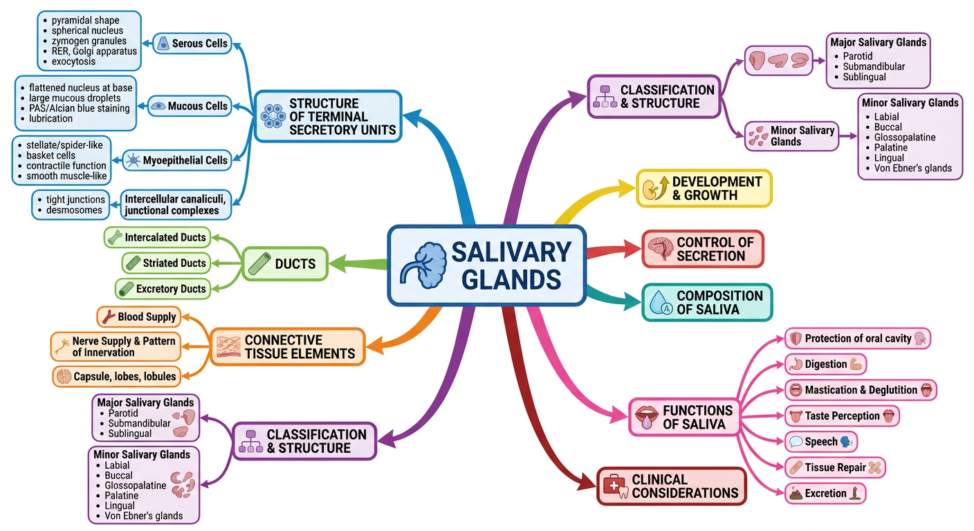
- Central node - Salivary Glands
- 9 color-coded branches - each representing a major chapter section
- Structure of Terminal Secretory Units (blue) - Serous cells, Mucous cells, Myoepithelial cells, junctional complexes
- Ducts (green) - Intercalated, Striated, Excretory
- Connective Tissue (orange) - Blood supply, Nerve supply, Capsule
- Classification (purple) - Major glands (Parotid, Submandibular, Sublingual) and Minor glands
- Functions of Saliva (pink) - Protection, Digestion, Speech, Taste, Tissue repair, Excretion
- Development & Growth, Control of Secretion, Composition of Saliva, Clinical Considerations as additional branches