
Write a short note on triceps surae for Ms orthopedics theory examination
triceps surae muscles gastrocnemius soleus anatomy diagram
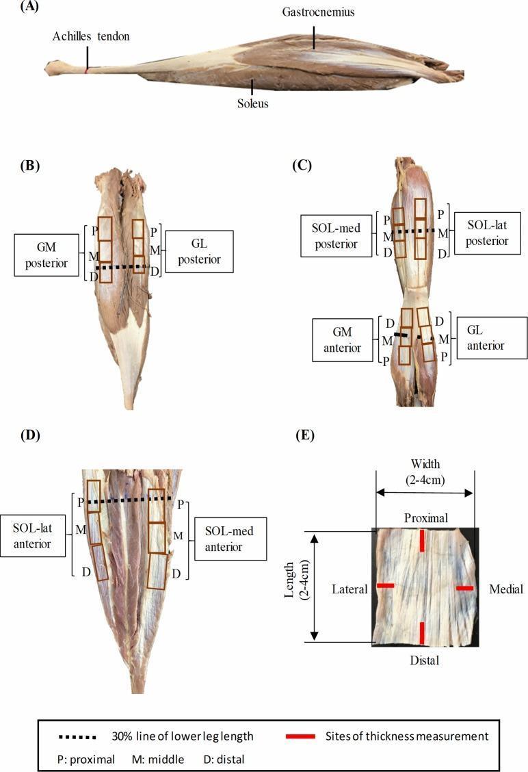
Educational anatomical diagram and specimen preparation of the human triceps surae muscle-tendon unit. (A) Lateral sagittal view demonstrating the spatial relationship between the superiorly positioned gastrocnemius, the underlying soleus, and the distal Achilles tendon. (B-D) Posterior and anterior views of dissected muscles (Gastrocnemius Medialis - GM, Gastrocnemius Lateralis - GL, and Soleus - SOL) illustrating the sites of aponeurosis harvesting. These views are annotated with rectangular grids indicating proximal (P), middle (M), and distal (D) sampling regions, alongside a dashed line representing 30% of the total lower leg length. (E) A high-magnification photograph of a typical rectangular aponeurosis specimen (2-4 cm), marked with specific sites for thickness measurement in both longitudinal and transverse orientations. This figure illustrates musculoskeletal anatomy, aponeurosis morphology, and standardized biomechanical testing preparation for the posterior compartment of the lower leg.
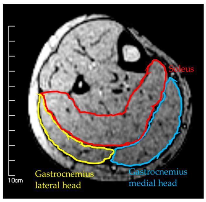
This diagnostic image is an axial T1-weighted MRI cross-section of the human lower leg, demonstrating the musculoskeletal anatomy of the posterior compartment. The primary focus is the triceps surae muscle group, with color-coded overlays identifying specific muscle boundaries for cross-sectional area (CSA) measurement. The Soleus (SOL) is delineated in red, showing a broad, crescent-shaped morphology that occupies the deep portion of the posterior compartment. Situated superficially to the soleus are the two heads of the gastrocnemius: the Gastrocnemius Medial Head (GM), outlined in blue, and the Gastrocnemius Lateral Head (GL), outlined in yellow. The image clearly shows the fascial planes separating these muscles and their relative positions to the tibia and fibula bones. This visual serves as an educational tool for identifying lower limb anatomy and understanding diagnostic imaging techniques used to quantify muscle mass, volume, and atrophy in clinical research or sports medicine.
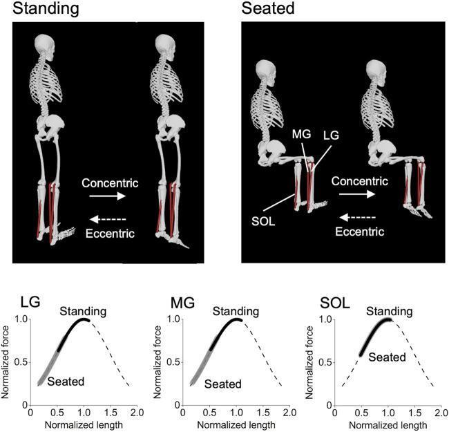
This musculoskeletal biomechanics diagram illustrates the physiological effects of standing versus seated calf-raise exercises on the triceps surae muscle group. The top section displays anatomical skeletal models performing the exercises: a 'Standing' model with knees at 0° extension and a 'Seated' model with knees flexed at 90°. Arrows indicate the concentric (plantarflexion) and eccentric (dorsiflexion) phases, with labels identifying the lateral gastrocnemius (LG), medial gastrocnemius (MG), and soleus (SOL) muscles. The bottom section presents three force-length curves correlating normalized force to normalized muscle length for the LG, MG, and SOL. The curves demonstrate that for the biarticular LG and MG, the standing position shifts the muscle operating range toward longer, more optimal muscle lengths compared to the seated position. In contrast, the monoarticular SOL shows nearly identical operating ranges in both positions. This content is intended for exercise physiology and sports medicine education to explain how joint positioning influences muscle recruitment and hypertrophic potential.
SHORT NOTE: TRICEPS SURAE
Introduction
- Gastrocnemius - medial head
- Gastrocnemius - lateral head
- Soleus
Anatomy
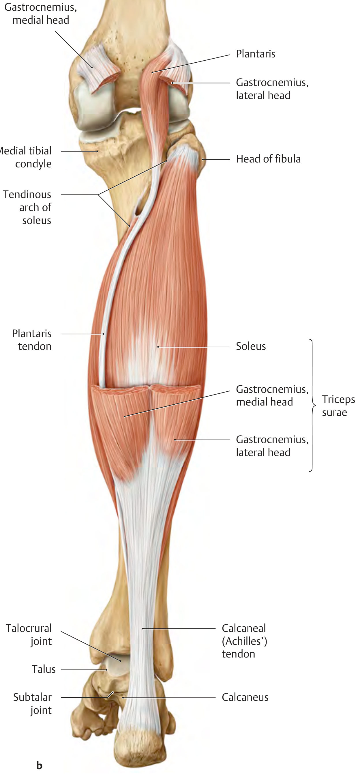
Origin
| Component | Origin |
|---|---|
| Soleus | Posterior surface of the head and neck of fibula; attached to the soleal line of tibia via a tendinous arch |
| Gastrocnemius, medial head | Medial epicondyle of the femur |
| Gastrocnemius, lateral head | Lateral epicondyle of the femur |
Insertion
- The calcaneal tendon averages 20-25 cm in length, with a mean cross-sectional area of ~70-80 mm² and breaking strength of 60-100 N/mm² - capable of bearing nearly 1 tonne in a healthy state.
Nerve Supply
Blood Supply
Actions
| Joint | Action |
|---|---|
| Talocrural (ankle) joint | Plantar flexion (all three heads) |
| Knee joint | Flexion (gastrocnemius only - biarticular muscle) |
- The gastrocnemius is a biarticular muscle crossing both the knee and ankle; the soleus is monoarticular crossing only the ankle.
- Together they are the primary plantarflexors and are active during the push-off phase of gait.
- The triceps surae (with tibialis anterior) also stabilizes the upper ankle joint in the sagittal plane.
Relations
- The triceps surae lies in the superficial posterior compartment of the leg.
- The deep posterior compartment (tibialis posterior, flexor digitorum longus, flexor hallucis longus) lies deep to it.
- The common peroneal (fibular) nerve winds around the neck of the fibula; the tibial nerve and posterior tibial vessels run between the superficial and deep compartments.
Applied Anatomy
1. Achilles Tendon Rupture
- Most common site is 2-6 cm proximal to the calcaneal insertion - the zone of poorest blood supply.
- Typically seen in sportsmen after repetitive microtrauma causing degeneration and loss of tensile strength.
- Clinical features: sudden whip-like snapping sound, loss of active plantarflexion, positive Simmonds (Thompson) test.
- Residual plantarflexion is still possible via the deep flexors.
2. Calcaneus Gait
- Weakness of the triceps surae (gastrocnemius-soleus complex) causes calcaneus gait - increased ankle dorsiflexion during heel strike due to inability to plantarflex adequately.
3. Contracture
- Tight gastrocnemius contributes to equinus deformity (e.g., in cerebral palsy, post-polio).
- Silfverskiold test differentiates isolated gastrocnemius tightness (equinus corrects with knee flexion) from combined gastrocnemius + soleus tightness (equinus persists regardless of knee position).
4. Muscle-splitting Approaches
- The interval between the two heads of gastrocnemius is used in approaches to the posterior knee and proximal tibia.
Summary Box
| Feature | Detail |
|---|---|
| Components | Gastrocnemius (2 heads) + Soleus |
| Origin | Femoral epicondyles (gastrocnemius); Fibula head/neck + Soleal line of tibia (soleus) |
| Insertion | Calcaneal tuberosity via Achilles tendon |
| Nerve | Tibial nerve S1, S2 |
| Action | Plantarflexion (all); Knee flexion (gastrocnemius) |
| Clinical relevance | Achilles rupture, equinus, calcaneus gait |