
can you clarify the distinct roles of a diagnostic Medial Branch Block (MBBB) versus a therapeutic intra-articular facet joint injection?" (Diagnostic value of dual MBBB, placebo effect management).
"medial branch block" AND "facet joint" AND diagnostic
"medial branch block" AND "placebo" AND "false positive"
"medial branch block" AND "radiofrequency ablation" AND "dual block"
Diagnostic Medial Branch Block (MBB) vs. Therapeutic Intra-Articular Facet Joint Injection: Distinct Roles
1. Anatomical Basis
2. Diagnostic MBB - What It Is and Why It Is Preferred
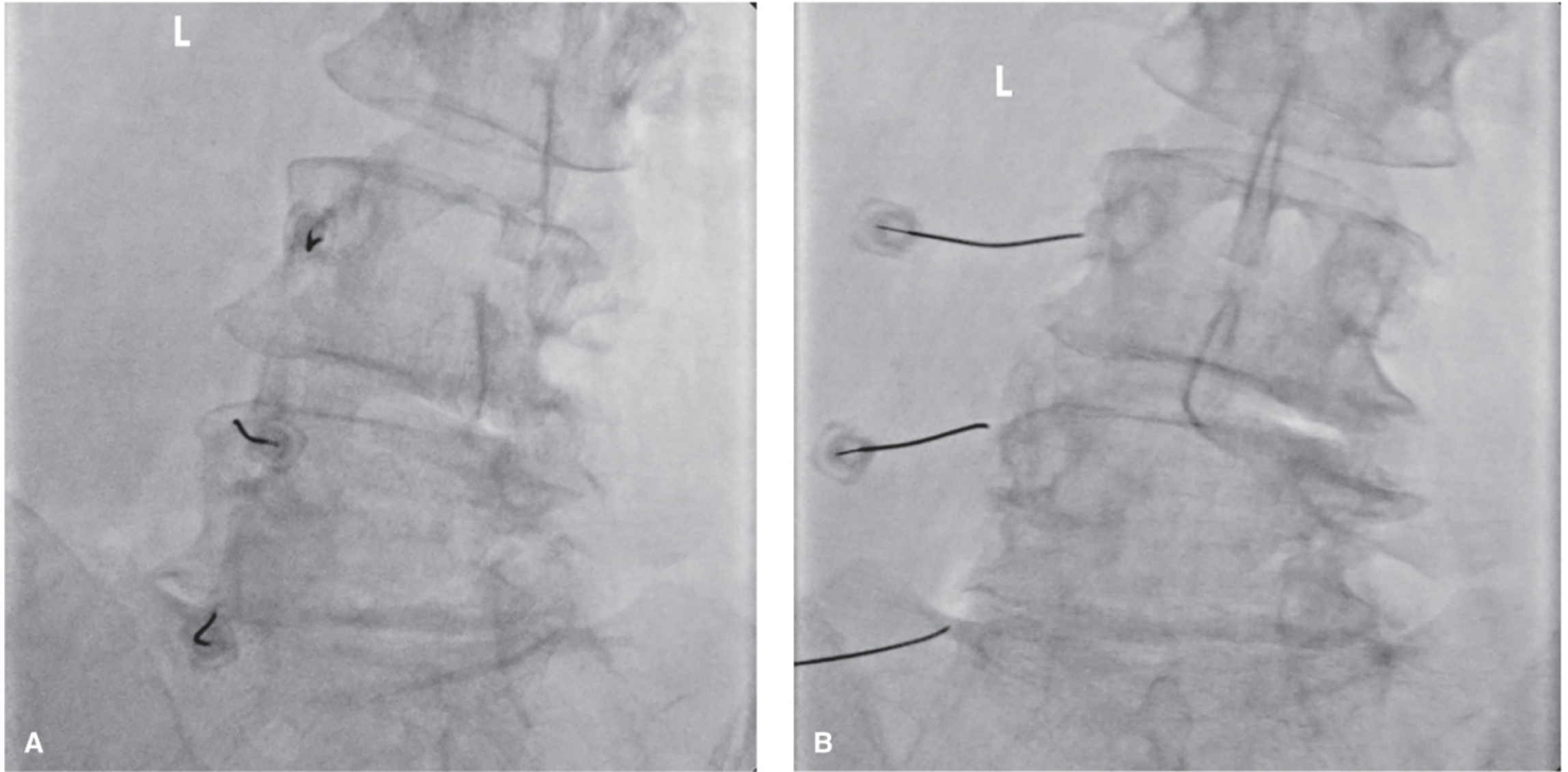
- Has better "face validity" - it directly tests the same nerve that RFA will later target
- High failure rate of intra-articular injections (the joint capsule is often degenerate, tight, or osteoarthritic and will not accept the full volume)
- Studies demonstrate better RFA outcomes when MBBs are used to select candidates, compared to intra-articular screening
- Per Barash's Clinical Anesthesia: "Guidelines recommend MBBs rather than intra-articular facet injections as the predictive tool before lumbar or cervical RFA based on better face validity, high failure rate for intra-articular injections, and studies demonstrating better outcomes when MBBs are used to identify candidates for RFA."
3. The Dual (Comparative/Controlled) MBB Protocol - Placebo Effect Management
- Placebo effect - the procedural experience, expectation, and attention bias
- Systemic absorption of local anesthetic
- Needle trauma causing temporary relief unrelated to facet pathology
- Regression to the mean (patients seek help when pain peaks; any intervention at that point may seem effective)
| Block | Agent | Expected Duration |
|---|---|---|
| Block 1 | Lidocaine (short-acting) | ~2-3 hours |
| Block 2 | Bupivacaine (long-acting) | ~4-6 hours |
- True positive (concordant): Patient gets short relief with lidocaine and long relief with bupivacaine - the duration of relief matches the expected pharmacokinetic duration of each agent. This is a "concordant positive" response and is the most reliable predictor of RFA benefit.
- False positive: Patient reports equal duration of relief from both agents (suggesting placebo or non-specific response), or reports pain relief far exceeding the drug's pharmacodynamic window.
- True negative: No significant relief from either block.
4. Therapeutic Intra-Articular Facet Joint Injection
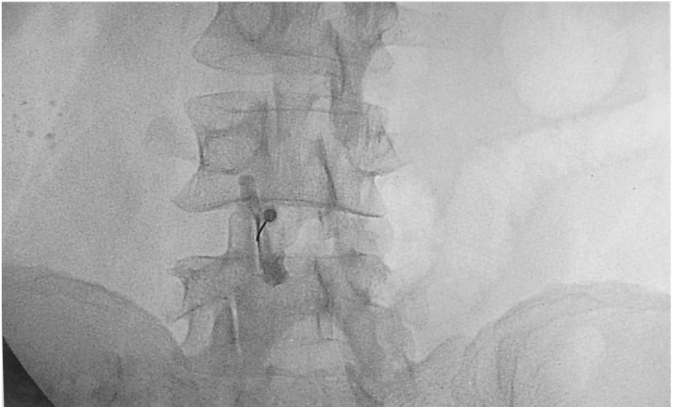
- Acute inflammatory facet arthropathy (e.g., synovitis following trauma or a flare of spondyloarthropathy)
- Short-to-medium term pain relief as a bridge while other treatments are implemented
- Where MRI shows active joint effusion or bone marrow edema adjacent to a facet joint
- Does not reliably distinguish the facet joint as the pain generator (therapeutic effect does not equal diagnostic confirmation)
- The joint capsule is frequently too degenerate, fibrosed, or space-limited to accept full injectate volume - failed entry is common in advanced facet OA
- Evidence for sustained efficacy is weak - both Firestein & Kelley's Rheumatology and Barash's Clinical Anesthesia note that long-term effectiveness is unproven
- Cannot be used to select patients for RFA
5. Side-by-Side Comparison
| Feature | Diagnostic MBB | Therapeutic Intra-Articular Injection |
|---|---|---|
| Target | Medial branch nerve | Joint cavity (synovium/capsule) |
| Injectate | Local anesthetic only (small volume, 0.5-1 mL) | Corticosteroid ± LA (1-2 mL) |
| Primary goal | Confirm facet as pain generator | Reduce intra-articular inflammation |
| Gateway to RFA? | Yes - specifically required | No (poor predictor of RFA outcome) |
| Dual block needed? | Yes, to exclude placebo/false positives | Not applicable |
| Therapeutic benefit expected? | Transient (diagnostic window only) | Yes, weeks to months potentially |
| Technical failure rate | Low (nerve is accessible) | Moderate-high (degenerate capsule) |
| Evidence base | Moderate (supported as RFA prerequisite) | Weak for long-term benefit |
6. Clinical Pathway Summary
Suspected facet joint pain (axial LBP, worse with extension/rotation,
no radiculopathy, facet arthrosis on imaging)
↓
Dual comparative MBB (lidocaine day 1, bupivacaine day 2)
↓
Concordant positive response (≥50-80% relief, duration-matched)?
YES → Proceed to RFA (lumbar/cervical medial branch neurotomy)
NO → Facet joint unlikely the primary pain generator; re-investigate
↓
Intra-articular injection may be considered adjunctively:
- For acute inflammatory flare
- As bridge therapy pending RFA
- Where RFA is not indicated/available
7. Recent Evidence Note
what are the critical imaging modalities or risk considerations you keep in mind when performing these injections near the spine?” (Fluoroscopy vs. Ultrasound guidance, avoiding vascular complications, patient selection).
"spinal injection" AND "vascular complication" AND fluoroscopy
"epidural steroid injection" AND "vascular" AND "complication"
"fluoroscopy" AND "ultrasound" AND "medial branch block" OR "facet injection"
PMID: 38296041
fluoroscopy guided spine injection contrast spread
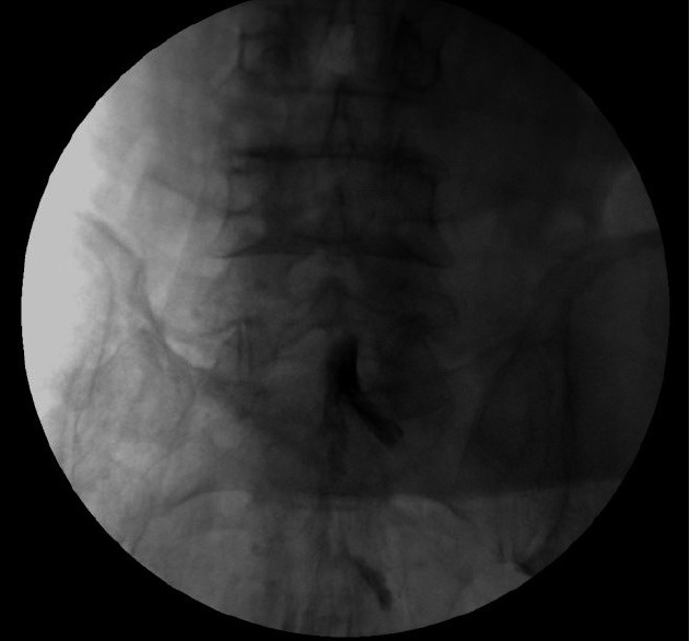
**Imaging Modality:** Fluoroscopy (Anteroposterior view) **Anatomical Region:** Lumbar spine and sacrum (L4-S1 region). **Procedure/Observation:** This image captures a fluoroscopically guided spinal injection, likely an epidural or subarachnoid administration. A radiopaque contrast medium (injectate) is visible as a focal, hyperdense (dark in this inverted-style display) accumulation near the midline at the lumbosacral junction. **Characteristic Visual Features:** - **Distribution:** The contrast agent exhibits a gravity-dependent settling pattern. Rather than a linear spread along nerve roots or symmetric cephalocaudal flow, the injectate pools preferentially toward the dependent side of the spinal canal. - **Positioning:** The anatomical orientation and flow pattern are consistent with the patient being in a lateral decubitus position during the procedure. - **Landmarks:** Lumbar vertebral bodies, intervertebral spaces, and the sacral promontory are visible, providing spatial orientation for the needle placement and subsequent spread of medication. **Clinical Significance:** This image demonstrates the baricity and gravitational influence on the distribution of injectate within the spinal compartments, a critical factor in determining the dermatomal level and lateralization of anesthetic or corticosteroid effect.
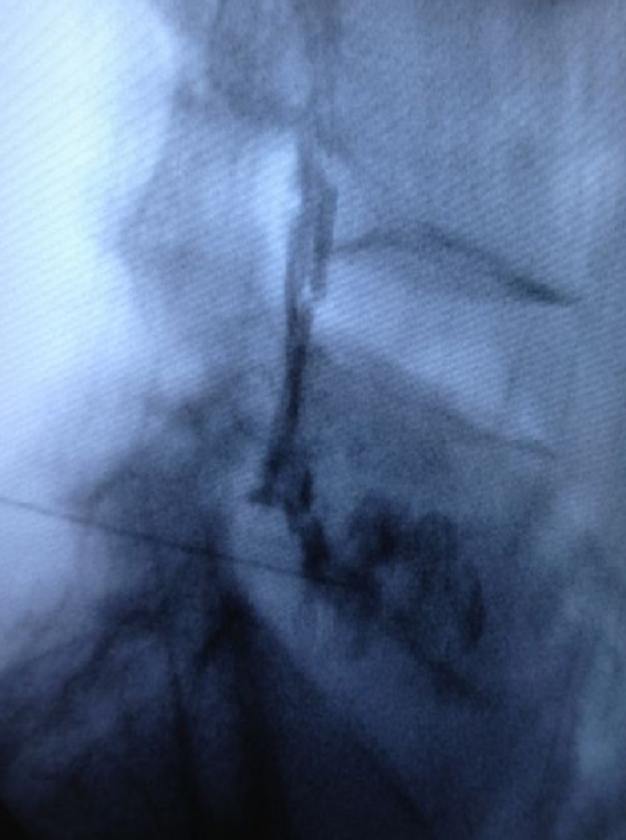
**Imaging Modality:** Fluoroscopy (Lateral projection) **Anatomical Region:** Lumbar spine, specifically the lumbosacral junction. **Procedure:** Transforaminal epidural injection/block. **Observation and Key Features:** The image displays a lateral fluoroscopic view of the lower lumbar vertebrae. A spinal needle is visualized with its tip positioned within the neural foramen. Radiopaque contrast medium has been injected, demonstrating a characteristic flow pattern. The contrast outlines the nerve root (radiculogram) and extends into the ventral epidural space, confirming intra-articular and epidural distribution. **Visual Characteristics:** - **Needle Placement:** The needle trajectory is directed toward the upper posterior aspect of the neuroforamen ("safe triangle" zone). - **Contrast Pattern:** Linear, vertical opacification consistent with epidural spread, alongside tracking along the exiting spinal nerve root. - **Bony Landmarks:** The vertebral bodies, intervertebral disc spaces, and the sacral promontory are visible, though partially obscured by soft tissue density and contrast. **Clinical Utility:** This image serves as intraoperative documentation for a guided transforaminal block, utilized in the management of radiculopathy. The lateral view is essential to verify the needle depth and confirm that the contrast is spreading ventrally into the epidural space rather than posteriorly or intravascularly.
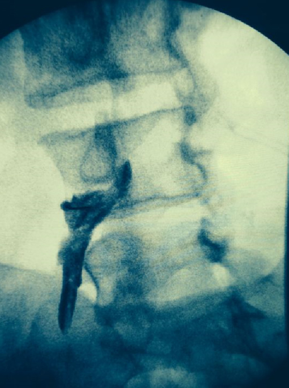
**Imaging Modality:** Fluoroscopy (Spot film) **Anatomical Region:** Lumbar spine, oblique view. **Procedure/Intervention:** Fluoroscopically guided transforaminal epidural injection (transforaminal nerve block). **Characteristic Visual Features:** - **Needle Placement:** A radiopaque spinal needle is visualized with its tip positioned within the neural foramen, typically at the "6 o'clock" position of the pedicle in an oblique projection. - **Contrast Pattern:** A radiopaque contrast medium (epidurogram) is seen delineating the targeted nerve root sheath. The flow pattern demonstrates characteristic spread along the spinal nerve within the neuroforamen and extending into the lateral epidural space. - **Landmarks:** Distinct visualization of the vertebral bodies, pedicles ("scotty dog" appearance), and the intervertebral foramen. **Clinical Context:** This image demonstrates a diagnostic and therapeutic interventional procedure used to treat radicular pain. The contrast spread confirms the correct anatomical localization of the needle tip and ensures the medication will reach the ventral epidural space while avoiding intravascular or intrathecal injection. **Key Diagnostic Features:** Linear contrast opacification along the course of the lumbar nerve root exiting the foramen.
Imaging Guidance and Risk Considerations for Spinal Facet/MBB Injections
1. The Injections in Anatomical Context
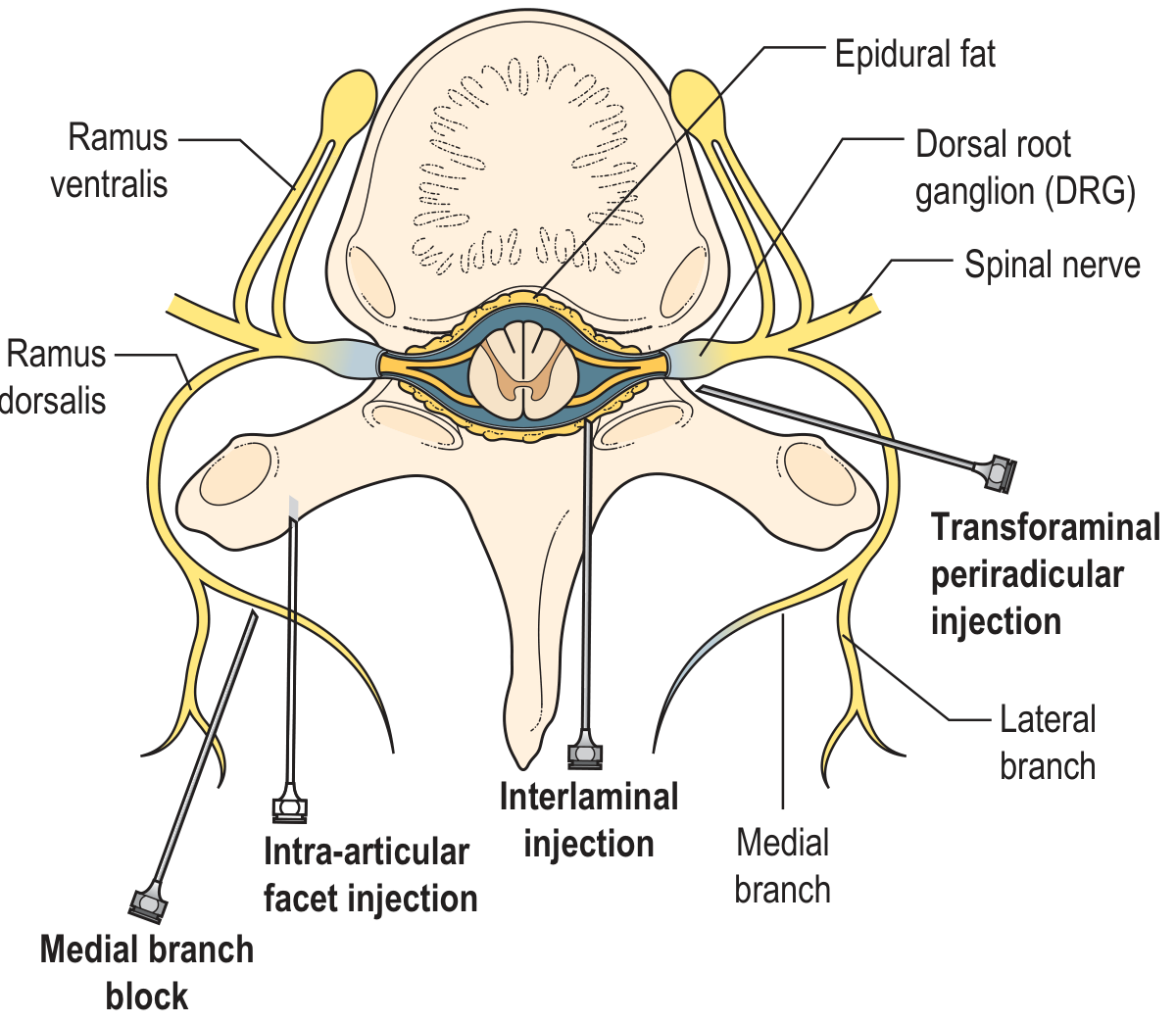
2. Imaging Modality Selection
Fluoroscopy - the Reference Standard
- Real-time visualization of needle tip position as it advances to the target landmark (transverse process-SAP junction for lumbar MBB; oblique "scotty dog" view for the joint space)
- Contrast confirmation - a small test injection of radiopaque iodinated contrast before any drug is given demonstrates whether the needle is intra-articular, periarticular, intravascular, or intrathecal. The spread pattern is immediately interpretable
- Digital Subtraction Angiography (DSA) mode - the most important safety upgrade. Real-time DSA subtracts background bone from each frame, making blood vessel opacification immediately visible even with tiny volumes of contrast. Per Grainger & Allison: "the latest guidelines on spine interventional procedures underline the utility of real-time high-quality fluoroscopy, ideally with digital subtraction angiography, to effectively exclude intra-arterial injections"
- Particulate steroid use requires mandatory contrast + live fluoroscopy (or DSA) before injection
- Radiation dose is the main drawback - occupational exposure to the operator and procedural dose to the patient (especially with repeated injections)
Ultrasound
- Pain outcomes with ultrasound were not significantly different from fluoroscopy or CT guidance
- Correct needle placement confirmed by fluoroscopy/CT ranged from 88% to 100% for ultrasound-guided attempts
- Procedural time was longer with ultrasound
- Conclusion: ultrasound cannot yet replace fluoroscopy/CT for dorsal and medial branch blocks in lumbar facet syndrome
CT Guidance
- Post-surgical anatomy with hardware obscuring fluoroscopic views
- Cervical or upper thoracic levels where fluoroscopy windows are narrow
- Anomalous anatomy (scoliosis, transitional vertebrae, congenital fusions)
3. Vascular Complications - the Most Feared Risks
The Artery of Adamkiewicz and Catastrophic Spinal Cord Infarction
Particulate vs. Non-Particulate Steroids - a Critical Safety Decision
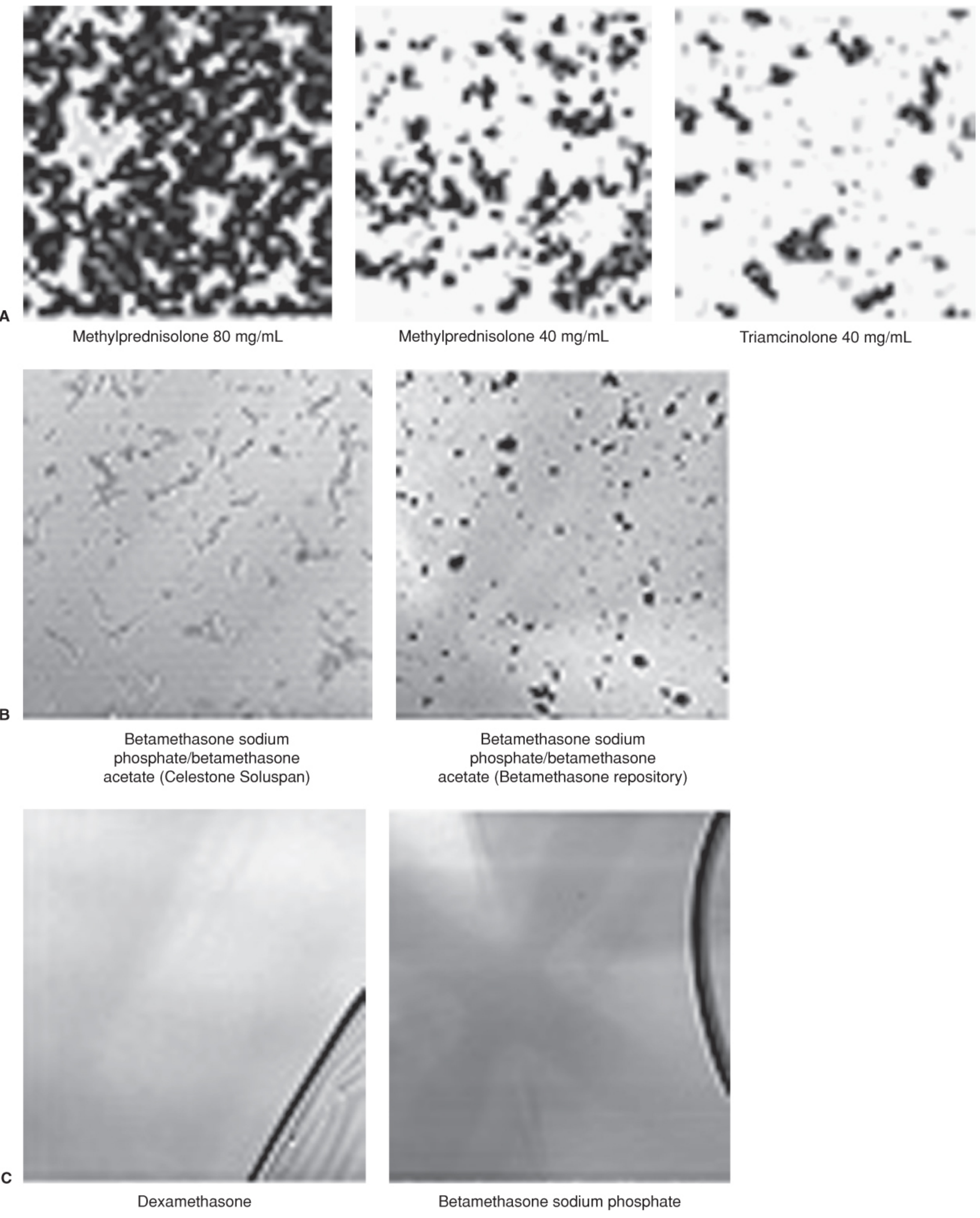
| Steroid | Particulate? | Risk Level for Transforaminal |
|---|---|---|
| Methylprednisolone (Depo-Medrol) | Yes - large amorphous aggregates | High - avoid above L3 |
| Triamcinolone | Yes - amorphous particles | High - avoid above L3 |
| Betamethasone (Celestone Soluspan) | Yes - rod-like crystals | Moderate/high |
| Dexamethasone | No - pure solution | Preferred for cervical and upper lumbar TF injections |
- Interlaminar ESIs require image guidance with AP, lateral, or contralateral oblique views plus contrast
- Cervical interlaminar ESI should not be performed above C6-C7
- Transforaminal ESIs require contrast injection under real-time fluoroscopy and/or DSA before steroid injection
- Particulate steroids must not be used in cervical TF ESIs
- For lumbar TF ESIs, particulate steroids may still be used at lower lumbar levels (L4-S1) in specific situations - but non-particulate (dexamethasone) is becoming the default
Intravascular Injection - Detection Protocol
- Systemic local anesthetic toxicity (LAST) - cardiovascular collapse, seizures
- Inadvertent epidural vein injection causing rapid intravascular spread
- Subdural or intrathecal injection causing high/total spinal
- Aspiration before any injection (necessary but not sufficient - small vessels may not aspirate)
- Test dose of contrast under live fluoroscopy - intravascular spread shows as a vascular blush or linear vessel filling, not the expected perineuronal or joint-cavity spread
- DSA mode - the gold standard; detects vascular opacification even with 0.1-0.2 mL contrast
- Slow, incremental injection - allows early detection of systemic symptoms before full dose is delivered
- Patient communication - the awake patient reporting sudden tinnitus, metallic taste, dizziness, or palpitations is an early warning signal
4. Other Significant Complications
| Complication | Mechanism | Frequency | Prevention |
|---|---|---|---|
| Dural puncture / post-dural puncture headache | Unintentional thecal sac entry | ~2.5% (lumbar interlaminar) | Careful technique, lateral fluoroscopy view |
| Epidural hematoma | Venous plexus hemorrhage (especially in anticoagulated patients) | Rare (<0.1%) | Anticoagulation management (see below) |
| Epidural/spinal abscess | Infection tracking along needle tract | Very rare | Aseptic technique; avoid in active infection/immunosuppression |
| Arachnoiditis | Intrathecal steroid with preservative (ethylene glycol) | Case reports | Avoid intrathecal steroid injection; use preservative-free formulations |
| Nerve root injury | Direct trauma | Rare | Avoid injection into nerve proper (paresthesia = needle too central) |
| Spinal cord ischemia | Particulate embolism into radiculomedullary artery | Very rare but catastrophic | DSA, non-particulate steroid, avoid high lumbar/cervical TF |
| Facial flushing / hyperglycemia | Systemic steroid absorption | Common, transient | Counsel diabetic patients; monitor glucose |
5. Patient Selection - Risk Stratification
Anticoagulation and Bleeding Risk
- Low risk (can proceed with routine precautions): Aspirin alone, NSAIDs, superficial MBB
- Moderate risk (requires cessation and bridging discussion): Clopidogrel (stop 5-7 days), warfarin (hold, target INR <1.5 for epidural), direct oral anticoagulants (DOACs - hold 24-72 hrs based on drug and renal function)
- High risk (avoid or delay): Active anticoagulation with therapeutic INR, thrombocytopenia (<50,000), coagulopathy
Active Infection
- Systemic bacteremia/sepsis
- Local skin infection overlying the injection site
- Epidural abscess or spinal infection
- Immunocompromised states (uncontrolled diabetes, chemotherapy, high-dose systemic steroids, HIV) - skin flora may seed the injection site; use additional antiseptic precautions and lower threshold to abort
Allergy to Contrast or Local Anesthetic
- Iodinated contrast allergy: premedicate with corticosteroid + antihistamine protocol, or consider non-contrast CT guidance or ultrasound
- Amide local anesthetic allergy (rare): skin test, use ester alternative (chloroprocaine), or consult allergist
Other Relative Contraindications
- Pregnancy - fluoroscopy radiation to fetus (use ultrasound guidance or defer unless necessary)
- Uncontrolled diabetes - steroid-induced hyperglycemia can be severe; monitor blood glucose for 48-72 hrs post-injection
- Previous failed injections at same level - review technique and targeting before repeating; consider alternative diagnosis
- Spinal instability or recent fusion hardware - altered anatomy changes spread patterns; CT guidance preferred
6. Summary Decision Framework
Before any spinal/facet injection:
1. IMAGING MODALITY
- Lumbar MBB / facet injection: Fluoroscopy (preferred) | Ultrasound (acceptable, longer procedure)
- Cervical or upper thoracic: Fluoroscopy with DSA mode mandatory
- Post-surgical / altered anatomy: CT guidance
- Obese patient: Fluoroscopy preferred over ultrasound
2. CONTRAST PROTOCOL
- Always inject test contrast under LIVE fluoroscopy before steroid/LA
- Use DSA mode for cervical and upper lumbar transforaminal work
- Confirm spread pattern: intra-articular / perineuronal vs. vascular blush
3. STEROID SELECTION
- Cervical TF injection: Dexamethasone ONLY (non-particulate)
- Lumbar TF above L3: Dexamethasone strongly preferred
- Lumbar TF L4-S1 / intra-articular facet: Methylprednisolone or triamcinolone acceptable
- Frequency: ≥2 weeks between injections; max 3-4/year
4. PATIENT CHECKLIST
☐ Anticoagulants held appropriately?
☐ Active infection excluded?
☐ Contrast allergy screened?
☐ Diabetes - glucose monitoring plan in place?
☐ Pregnancy excluded?
☐ Consent covers catastrophic risks (spinal cord ischemia, hematoma, abscess)?
what are the critical imaging modalities or risk considerations you keep in mind when performing these injections near the spine?” (Fluoroscopy vs. Ultrasound guidance, avoiding vascular complications, patient selection).
Quick Reference: Imaging Guidance & Risk Considerations for Spinal Facet/MBB Injections
Imaging Modality Comparison
| Modality | Best For | Key Advantage | Key Limitation |
|---|---|---|---|
| Fluoroscopy | All MBBs, facet injections - standard | Real-time + contrast confirmation; DSA mode detects intravascular injection | Radiation exposure |
| Fluoroscopy + DSA | Cervical and upper lumbar TF injections - mandatory | Subtracts bone; shows even 0.1 mL vascular opacification in real time | Requires DSA-capable C-arm |
| Ultrasound | Lumbar MBBs where fluoroscopy unavailable; radiation-sensitive patients | No radiation; Doppler shows adjacent vessels | Longer procedure time; cannot confirm intra-articular placement; inferior in obese patients |
| CT guidance | Post-surgical anatomy, hardware, upper thoracic, anomalous spine | Highest anatomical precision | Highest radiation dose; no real-time injection monitoring without CT fluoro |
Vascular Complications - the Non-Negotiables
- Dominant anterior spinal artery supply; typically enters at T9-L1 on the left, but variants reach L5
- Travels through the neural foramen alongside a nerve root - exactly where transforaminal needles are placed
- Accidental intra-arterial injection of particulate steroid → microembolism → spinal cord infarction → paraplegia (catastrophic, documented in case reports)
- Methylprednisolone, triamcinolone, betamethasone = particulate → can aggregate and embolize
- Dexamethasone = non-particulate (pure solution) → no neurological complications reported to date
- Rule: Cervical TF injections = dexamethasone only. Upper lumbar TF (above L3) = dexamethasone strongly preferred. L4-S1 level facet/TF = particulate agents are still used but dexamethasone is becoming the default
- Aspirate (necessary but not sufficient - small vessels may not bleed back)
- Inject test contrast under live fluoroscopy - intravascular = vascular blush or linear vessel fill; correct placement = perineuronal/joint spread
- DSA mode for cervical and upper lumbar TF - gold standard for excluding intra-arterial injection
- Slow, incremental injection with the patient awake - tinnitus, metallic taste, palpitations, or paresthesia = stop immediately
- Never inject before contrast confirmation
Other Complications at a Glance
| Complication | Frequency | Key Prevention |
|---|---|---|
| Dural puncture / post-dural puncture headache | ~2.5% lumbar interlaminar | Lateral fluoroscopy view; cautious advancement |
| Epidural hematoma | Rare (<0.1%) | Hold anticoagulants appropriately |
| Epidural/spinal abscess | Very rare | Aseptic technique; exclude active infection |
| Arachnoiditis | Case reports | Never inject preserved steroids intrathecally |
| Spinal cord ischemia | Very rare, catastrophic | DSA + non-particulate steroid at high-risk levels |
| Steroid-induced hyperglycemia | Common, transient | Counsel diabetic patients; glucose monitoring 48-72 hrs |
| Facial flushing, adrenal suppression | Common transient | Limit frequency to 3-4 injections/year; ≥2 weeks between |
Patient Selection Checklist
- Aspirin alone: proceed with routine precautions for MBB/facet injection
- Clopidogrel: stop 5-7 days before
- Warfarin: hold; target INR <1.5 for epidural procedures
- DOACs (apixaban, rivaroxaban, etc.): hold 24-72 hrs based on drug and renal function
- Risk is procedure-specific - peripheral MBBs carry lower hematoma risk than epidural injections
- Active systemic bacteremia or sepsis
- Local skin infection at the injection site
- Known epidural abscess or spinal infection
- Uncontrolled coagulopathy
- Immunocompromised (uncontrolled diabetes, chemotherapy, high-dose steroids, HIV)
- Iodinated contrast allergy: premedicate or use CT/ultrasound
- Pregnancy: avoid fluoroscopy; use ultrasound or defer
- Post-surgical/hardware anatomy: switch to CT guidance
Practical Safety Hierarchy
BEFORE THE NEEDLE GOES IN:
Hold anticoagulants → Check INR/platelets → Screen for infection
→ Consent for catastrophic risks → Choose correct steroid
AT THE NEEDLE TIP:
Fluoroscopy ON → Confirm landmark → Inject contrast under live view
→ DSA if cervical or upper lumbar TF → Confirm spread pattern
→ Only then inject drug, slowly, in increments
STEROID SELECTION RULE:
Cervical TF or above L3 → Dexamethasone ONLY
L4-S1 facet / intra-articular → Particulate acceptable; dexamethasone preferred
Max 3-4 injections/year; ≥2 weeks between sessions
what are the primary anatomical sources of low back pain we should look out for, and how do we differentiate them clinically? (discogenic vs. facet vs. myofascial).
"low back pain" AND "differential diagnosis" AND discogenic AND facet
PMID: 39063567
low back pain anatomy disc facet joint nerve root sources
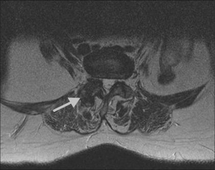
This diagnostic image is an axial T2-weighted MRI of the lumbar spine at the L2-3 level. The scan highlights the posterior elements of the spinal anatomy, specifically focusing on the facet joints. An arrow indicates the right facet joint, which demonstrates advanced degenerative changes characteristic of facetal arthropathy. Visible features include irregular joint morphology, bone remodeling, and altered signal intensity compared to normal spinal structures. The vertebral body is visible anteriorly, while the posterior elements (laminae and spinous process) enclose the spinal canal. The surrounding paraspinal musculature and soft tissues are shown in cross-section. This imaging is clinically significant for diagnosing sources of facet-mediated low back pain and planning interventional procedures such as facet joint injections or medial branch blocks. It serves as an educational example of spinal osteoarthritis and age-related degenerative disc disease manifestations in radiological studies.
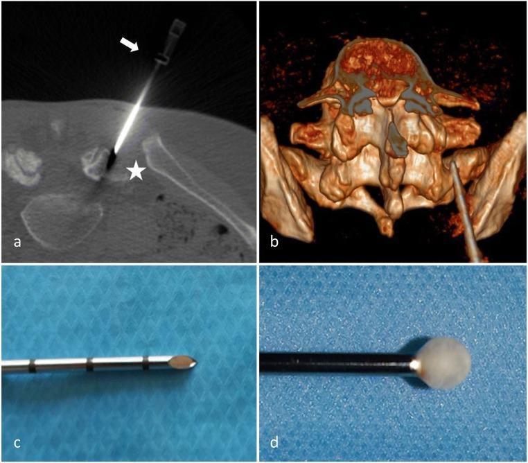
This four-panel image illustrates facet joint cryoneurolysis at the right L5-S1 level using CT guidance. Panel (a) is an axial CT image showing a cryoprobe (white arrow) precisely positioned against the ala of the sacrum (white star), a landmark for targeting the medial branch of the dorsal rami. Panel (b) provides a 3D CT reconstruction of the lumbosacral junction, depicting the posterior approach of the cryoprobe relative to the spinal anatomy. Panels (c) and (d) are close-up clinical photographs of the cryoprobe tip. Panel (c) shows the 12G metallic cryoprobe with visible depth markings and a sharp, conical tip. Panel (d) demonstrates the Joule-Thompson effect, showing a spherical, opaque 'ice-ball' formation at the tip during the freezing cycle. This procedure is used for chronic low back pain management by ablating nociceptive nerve fibers in the facet joint capsule. The visual content serves as an educational guide for interventional radiologists and pain management specialists regarding needle trajectory and the physical mechanism of cryoablation.
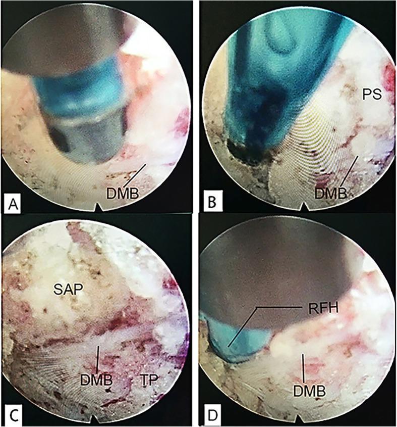
This set of four endoscopic clinical photographs (labeled a–d) illustrates the procedural steps for identifying and treating the dorsal medial branch (DMB) of the spinal nerve during endoscopic radiofrequency ablation for chronic low back pain. Panel A shows the initial exposure of the DMB. Panel B highlights the nerve as it is embedded within the thick periosteum (PS). Panel C demonstrates the critical anatomical landmarks at the junction of the superior articular process (SAP) and the transverse process (TP) base, where the DMB is typically situated. The SAP appears as a mottled, reddish-white bony surface, while the TP shows a lighter, striated texture. Panel D depicts the application of a radiofrequency head (RFH) to the DMB for neurotomy. These images serve as an educational guide for spinal surgeons and interventional pain specialists to understand the endoscopic anatomy and target localization required for facet joint denervation.
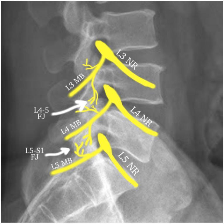
This lateral lumbar spine X-ray contains an anatomical overlay diagram illustrating the dual innervation of lumbar facet joints (zygapophyseal joints). The visual focuses on the L3, L4, and L5 nerve levels. The L4-L5 facet joint (L4-5 FJ) is shown receiving innervation from the L3 and L4 medial branches (MB), which originate from their respective nerve roots (NR). The L5-S1 facet joint (L5-S1 FJ) is shown being innervated by the L4 medial branch and the L5 dorsal ramus/medial branch. Yellow graphic overlays map the pathway of these nerves as they exit the neural foramina and descend over the transverse processes to supply the joints. The image serves as an educational guide for interventional pain management, specifically for identifying targets in medial branch blocks or radiofrequency ablation (RFA) to treat chronic low back pain originating from facet arthropathy.
Primary Anatomical Sources of Low Back Pain and Clinical Differentiation
The Diagnostic Challenge
- Intervertebral disc (discogenic / radicular)
- Facet (zygapophyseal) joints
- Myofascial / paraspinal muscle
- Sacroiliac (SI) joint
- Spinal stenosis (neurogenic claudication)
- Non-mechanical / referred ("red flag") sources
Anatomy of Innervation - Why Pain Patterns Overlap
1. Discogenic Pain (Anterior Column Source)
- Age 40-60 years, chronic relapsing pattern
- Axial low back pain, worse with sitting and forward flexion (intradiscal pressure increases in flexion and with prolonged sitting)
- Pain that eases with standing and walking (unloads the disc partially)
- "Centralization" phenomenon on repeated movement assessment - pain moves from periphery toward the spine with repeated extension (McKenzie pattern) - strongly suggests discogenic source
- Pain provoked by Valsalva maneuver (coughing, sneezing) if annular fissure is present
- No neurological deficit unless there is concurrent radiculopathy from disc herniation
- Shooting, lancinating pain radiating below the knee in a dermatomal distribution (true sciatica)
- L4 root: Pain down medial shin to medial foot; weakness of tibialis anterior (foot drop); reduced knee jerk
- L5 root: Pain down lateral shin to dorsum of foot and great toe; weakness of EHL (great toe extension)
- S1 root: Pain down posterior thigh/calf to lateral foot and little toe; weakness of peronei and gastrocnemius; reduced ankle jerk
- Positive straight leg raise (SLR) test at <60° with reproduction of dermatomal leg pain (not just back pain) is the most sensitive sign of lumbar nerve root compression - Bradley & Daroff's Neurology notes this is a key differentiator from facet pain where SLR is negative
- MRI: disc height loss, T2 "black disc" dehydration, Modic changes (type I = active endplate edema/inflammation; type II = fatty replacement; type III = sclerosis)
- High-intensity zone (HIZ): Focal posterior annular T2 hyperintensity suggesting annular tear - but HIZ is also found in asymptomatic individuals
- Modic type I changes are the strongest MRI correlate of clinically symptomatic discogenic pain
- Provocative discography remains the definitive test for pure discogenic pain (concordant reproduction of "usual" pain with injection), but it is controversial - false-positive rates are significant and the procedure may accelerate disc degeneration
2. Facet (Zygapophyseal) Joint Pain (Posterior Column Source)
- Axial low back pain, worse with extension, hyperextension, and ipsilateral rotation
- Pain relieved by forward flexion (opens the facet joint space)
- Referred pain into the buttock and posterior thigh, typically not below the knee - this is the key differentiator from true radiculopathy
- Pain with prolonged standing and walking (both load the posterior elements)
- Morning stiffness is common
- Physical examination: paraspinal tenderness over the affected joint level; pain reproduced with extension-rotation to the symptomatic side; SLR test is negative; no neurological deficit
3. Myofascial Pain (Paraspinal Muscle Source)
- Quadratus lumborum - deep to erector spinae; refers pain to the iliac crest and greater trochanter
- Gluteus medius/minimus - refers pain down the lateral thigh/leg, mimicking L5 radiculopathy
- Paraspinal/erector spinae group - diffuse axial pain with ipsilateral radiation
- Piriformis - refers pain down the posterior thigh, mimicking sciatica (piriformis syndrome)
- Acute onset following a specific mechanical event (lifting, twisting, prolonged posture) or more insidiously with repetitive microtrauma
- Diffuse, aching, non-dermatomal pain - poorly localized, no specific movement direction reliably aggravates or relieves it
- Palpable taut band with a discrete, exquisitely tender spot (trigger point) that reproduces the patient's referred pain pattern on pressure
- Local twitch response - a brisk visible muscle contraction when the trigger point is snapped or needled - highly specific for true trigger point
- SLR negative, no neurological deficit, no dermatomal distribution
- Restricted range of motion without a clear directional pattern
4. Sacroiliac (SI) Joint Pain
- Unilateral pain at and below the PSIS (posterior superior iliac spine) - the "Fortin finger sign" (patient points with one finger to the PSIS area)
- Pain radiating to the groin, anterior thigh, or posterior thigh, occasionally below the knee
- Walking upstairs or single-leg standing typically aggravates pain (sacroiliac shear loading)
- Tenderness directly over the SI joint sulcus
- Multiple provocative tests increase specificity when clustered:
- FABER/Patrick test (hip flexion-abduction-external rotation) - stresses the SI joint
- Gaenslen's test - contralateral hip hyperflexion with ipsilateral hip extended
- Thigh thrust / posterior shear test - most specific single test
- Per Bradley & Daroff's Neurology: three positive provocative tests combined with SI joint tenderness adequately diagnose SI dysfunction
5. Spinal Stenosis (Neurogenic Claudication)
- Bilateral leg pain, aching or cramping, brought on by walking or standing - neurogenic claudication
- Pain relieved by sitting, forward flexion, or lying in fetal position (flexion opens the canal)
- Patients adopt a stooped posture when walking ("shopping cart sign")
- Pain resolves over minutes with rest - distinguishing it from vascular claudication (which resolves in seconds with standing still and does not require sitting/flexion)
- SLR may be normal (unlike acute disc herniation)
- Variable neurological findings that fluctuate with posture
6. Red Flag Sources - Do Not Miss
| Red Flag Source | Alarm Features |
|---|---|
| Malignancy (primary or metastatic) | Age >50, prior cancer history, unexplained weight loss, night pain that wakes from sleep, pain not relieved by rest |
| Vertebral fracture | Osteoporosis risk factors, steroid use, trauma, sudden onset after minor mechanism |
| Infection (discitis, epidural abscess) | Fever, IV drug use, recent procedure, immunosuppression, ESR/CRP elevated |
| Cauda equina syndrome | Bowel/bladder dysfunction, saddle anesthesia, bilateral leg weakness - surgical emergency |
| Referred visceral pain | Aortic aneurysm (pulsatile mass, vascular risk factors), renal colic (flank-to-groin radiation, urinalysis), pancreatic/retroperitoneal pathology |
Master Differentiation Table
| Feature | Discogenic | Facet | Myofascial | SI Joint | Spinal Stenosis |
|---|---|---|---|---|---|
| Pain location | Central/axial | Axial ± buttock | Diffuse axial ± referred | Unilateral PSIS | Bilateral legs |
| Below knee radiation | Yes (with radiculopathy) | Rarely | Possible (non-dermatomal) | Occasionally | Yes (neurogenic claudication) |
| Aggravated by | Flexion, sitting, Valsalva | Extension, rotation, standing | Palpation of trigger point, sustained posture | Stairs, single-leg stance, FABER | Walking, standing, extension |
| Relieved by | Standing, extension | Flexion | Stretch, heat | Rest | Sitting, flexion ("shopping cart") |
| SLR test | Positive (if radiculopathy) | Negative | Negative | Negative | May be negative |
| Neurological signs | Possible (dermatomal) | None | None | None | Variable, positional |
| Palpation finding | Paraspinal tenderness | Facet joint tenderness | Taut band + trigger point | SI sulcus tenderness | Paraspinal ± diffuse |
| Diagnostic test | MRI + discography | Dual comparative MBB | Clinical (trigger point exam) | Cluster of provocation tests + SI injection | MRI canal dimensions |
| MRI correlation | Moderate (Modic I > HIZ) | Poor | None | Moderate (sacroiliitis) | Good |
| Key age group | 40-60 | 40-70+ | Any age | 20-50 (often female) | >60 |
A Structured Clinical Approach
Step 1: EXCLUDE RED FLAGS
Night pain, constitutional symptoms, bowel/bladder change,
neurological progression → urgent imaging / referral
Step 2: NEUROLOGICAL SCREEN
Dermatomal pain below knee? → SLR test
Positive SLR + dermatomal deficit → RADICULOPATHY (disc herniation or foraminal stenosis)
Bilateral leg claudication + walking-limited → STENOSIS
Step 3: MOVEMENT PATTERN ANALYSIS
Pain worse in FLEXION + sitting → DISCOGENIC
Pain worse in EXTENSION + rotation → FACET
No clear directional pattern + trigger points → MYOFASCIAL
Pain at PSIS + provocation cluster → SI JOINT
Step 4: CONFIRMATORY TESTING
Discogenic → MRI (Modic I changes); provocative discography if pre-surgical
Facet → Dual comparative MBB (lidocaine/bupivacaine)
Myofascial → Clinical (trigger point injection response)
SI joint → Cluster of 3+ provocative tests; fluoroscopic SI joint injection
Step 5: RECOGNIZE OVERLAP
Most patients over 50 have multiple simultaneous sources.
Treat the dominant source first; reassess the residual pain.