
Dexa scan
DEXA scan bone mineral density dual energy x-ray absorptiometry
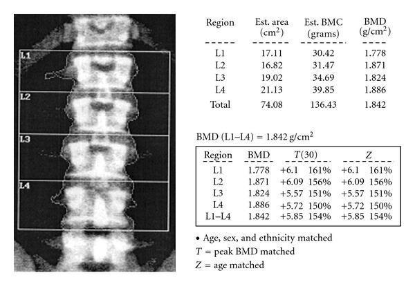
This diagnostic image and data summary illustrate a Dual-energy X-ray Absorptiometry (DEXA) scan of the lumbar spine (L1-L4) used for bone mineral density (BMD) assessment. The visual component features a posterior-anterior (PA) view of the lumbar vertebrae, where each vertebral body (L1, L2, L3, L4) is segmented and delineated by rectangular regions of interest (ROI). Accompanying tables provide quantitative data including estimated area (cm²), Bone Mineral Content (BMC in grams), and BMD (g/cm²). The total BMD for L1-L4 is reported as 1.842 g/cm², with significantly elevated T-scores and Z-scores reaching +5.85. This clinical finding indicates abnormally high bone density, which is essential for diagnosing conditions like osteopetrosis or sclerosing bone dysplasias in endocrinology and orthopedics. The educational focus is on the interpretation of DEXA scan outputs and the recognition of supra-normal bone density values.
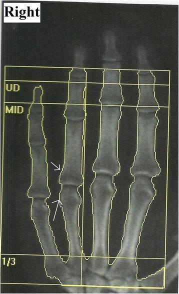
This monochrome diagnostic image is a Dual-Energy X-ray Absorptiometry (DEXA) scan of the right hand, presented in a posterior-anterior orientation. The scan captures the distal metacarpals and the phalanges of the second through fifth digits. Superimposed on the anatomy is a yellow analysis grid with horizontal markers labeled 'UD' (ultra-distal), 'MID' (middle), and '1/3' (distal shaft), which are standard regions of interest for bone mineral density quantification. White arrows indicate juxta-articular osteopenia at the proximal and middle phalanges of the second digit, characterized by increased radiolucency and reduced bone density near the joint margins. This visual finding is characteristic of regional osteoporosis, often associated with Complex Regional Pain Syndrome (CRPS) Type I. The image serves as a clinical example of using DEXA imaging to diagnose and monitor metabolic bone changes in peripheral extremities following localized trauma or neurological insult.
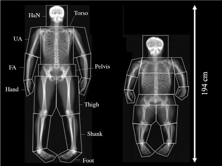
This diagnostic image shows a side-by-side comparison of two whole-body Dual-energy X-ray Absorptiometry (DEXA) scans, highlighting body composition and skeletal morphology in a control subject (left) and an individual with achondroplasia (right). Both scans utilize a standardized 15-segment analysis model, with boundaries demarcated by geometric shapes (squares, rhomboids, and pentagons) along the transverse axis of the joints. Segments are labeled as Head and Neck (HaN), Torso, Pelvis, Upper Arm (UA), Forearm (FA), Hand, Thigh, Shank, and Foot. The achondroplasia scan demonstrates characteristic disproportionate dwarfism, including rhizomelic shortening of the long bones (UA and Thigh), a macrocephalic head appearance relative to body size, and a significantly reduced overall height compared to the control. The segmental analysis is used in clinical research to quantify differences in bone mineral content (BMC), bone mineral density (BMD), and soft tissue distribution between the two phenotypes. The image serves as a clinical illustration of skeletal dysplasia and the application of DEXA for detailed anthropometric and volumetric bone mineral density assessment.
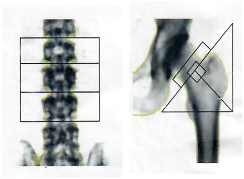
This diagnostic image displays dual-energy X-ray absorptiometry (DEXA) scans used for Bone Mineral Density (BMD) assessment. The visual is divided into two clinical views: an anteroposterior (AP) view of the lumbar spine and a proximal femur scan. In the lumbar spine image, horizontal rectangular Regions of Interest (ROIs) are demarcated over the L2, L3, and L4 vertebrae to calculate specific density values. The right image shows the proximal hip, featuring geometric overlays that isolate the femoral neck, Ward's triangle, and the greater trochanter for analysis. These scans are critical in diagnosing metabolic bone diseases such as osteopenia, osteoporosis, or osteomalacia, which in this clinical context relates to drug-induced renal impairment and hypophosphatemia. The grayscale variations within the skeletal structures represent the relative mineral density, where higher-density cortical bone appears darker compared to more lucent trabecular areas.
DEXA Scan (Dual-Energy X-ray Absorptiometry)
What Is It?
Sites Measured
- Lumbar spine (L1-L4)
- Proximal femur (femoral neck, Ward's triangle, greater trochanter)
- Distal forearm/radius - particularly important in hyperparathyroidism, where cortical bone loss predominates - Cummings Otolaryngology
T-Score and Z-Score Interpretation
| T-Score | Interpretation |
|---|---|
| Above -1.0 | Normal |
| -1.0 to -2.5 | Osteopenia (low bone mass) |
| -2.5 or below | Osteoporosis |
| -2.5 or below + fragility fracture | Severe osteoporosis |
- T-score: Compares BMD to a young healthy adult reference population
- Z-score: Compares BMD to age-matched and sex-matched reference population (used in premenopausal women, children, and younger men)
Indications - Who Should Be Tested?
- Women ≥65 years and men ≥70 years, regardless of risk factors
- Younger postmenopausal women, women in menopausal transition, and men aged 50-69 with clinical risk factors for fracture
- Adults who have had a fracture after age 50
- Adults with conditions or medications associated with bone loss (e.g., rheumatoid arthritis, glucocorticoids ≥5 mg prednisone/day for ≥3 months)
- Monitoring response: repeat BMD testing 1-2 years after initiating osteoporosis therapy, then every 2 years - Harrison's Principles of Internal Medicine 22E
Clinical Uses Beyond Osteoporosis
-
Hyperparathyroidism: DEXA is part of the NIH criteria for surgery in asymptomatic primary hyperparathyroidism - a T-score worse than -2.5 at any site (lumbar spine, total hip, femoral neck, or distal radius) is a surgical indication - Cummings Otolaryngology
-
Glucocorticoid therapy monitoring: Baseline and serial BMD testing is recommended for patients on long-term corticosteroids - Andrews' Diseases of the Skin
-
GI/Liver disease: Patients with IBD and liver disease are at risk for metabolic bone disease and benefit from DEXA screening - Sleisenger & Fordtran's Gastrointestinal and Liver Disease
-
Body composition: Whole-body DEXA can quantify fat mass, lean mass, and bone mineral content by body segment
DEXA vs. Quantitative CT (QCT)
Sample DEXA Images
Key Limitations
- Does not measure bone quality (microarchitecture), only quantity
- Results can be falsely elevated by osteoarthritis, aortic calcification, or vertebral fractures (in AP lumbar views)
- Radiation dose is very low (~1-10 µSv), far below diagnostic CT
- Positioning and software variations between machines can affect comparability
Treatment Implications
- Bisphosphonates (e.g., alendronate, risedronate) - effective for 3-5 years of fracture risk reduction
- Calcium and vitamin D supplementation
- Fall prevention - balance/gait training, ambulatory aids, environmental modification
- Exercise to improve bone quality
- Textbook of Family Medicine 9e
More detailed explanation of dexa
DEXA scan lumbar spine femoral neck T-score report osteoporosis
DEXA Scan - Detailed Explanation
1. Physical Principle - How It Works
"The intensity of a photon beam is altered by the thickness, density, and chemical composition of an object in its path."
- Pfenninger and Fowler's Procedures for Primary Care
- Bone mineral content (BMC) in grams
- Bone mineral density (BMD) = BMC / scanned area (g/cm²)
- Lean body mass vs. fat mass in whole-body scans
2. What It Measures
| Output | Unit | Clinical Use |
|---|---|---|
| BMD (areal) | g/cm² | Osteoporosis diagnosis |
| BMC | g | Pediatric bone assessment |
| T-score | SD from young adult mean | Osteoporosis/fracture risk |
| Z-score | SD from age-matched mean | Secondary bone loss |
| % Body fat | % | Obesity/body composition |
| Lean mass | kg | Sarcopenia, nutrition |
3. Sites Measured
| Site | Why Important |
|---|---|
| Lumbar spine (L1-L4) | Rich in trabecular bone; very sensitive to early bone loss; most responsive to treatment |
| Femoral neck | Best predicts hip fracture risk; used in FRAX calculator |
| Total hip | Combines femoral neck + trochanter + intertrochanteric region |
| Distal radius/forearm (1/3 site) | Important in hyperparathyroidism (cortical bone loss) and when spine/hip cannot be measured |
| Whole body | Body composition; pediatric use |
4. T-Score and Z-Score - Full Interpretation
T-Score (used in postmenopausal women and men ≥50 years)
| T-Score | Classification |
|---|---|
| > -1.0 | Normal |
| -1.0 to -2.5 | Osteopenia (low bone mass) |
| ≤ -2.5 | Osteoporosis |
| ≤ -2.5 + fragility fracture | Severe osteoporosis |
- Source: WHO criteria, confirmed by Firestein & Kelley's Textbook of Rheumatology
Z-Score (used in premenopausal women and men <50 years)
5. FRAX - Fracture Risk Calculator
- Age, sex, weight, height
- Prior fragility fracture
- Parental hip fracture history
- Current glucocorticoid use
- Secondary osteoporosis causes
- Alcohol intake (>2 drinks/day)
- Current smoking
- Femoral neck T-score (optional but improves accuracy)
- 10-year probability of hip fracture
- 10-year probability of major osteoporotic fracture (hip + spine + forearm + humerus)
- 10-year hip fracture risk ≥3%, OR
- 10-year major osteoporotic fracture risk ≥20%
6. Types of Osteoporosis Detected
| Type | Key Features |
|---|---|
| Type I (Postmenopausal) | Primarily trabecular bone; vertebral + distal radius fractures |
| Type II (Age-related, >70 yrs) | Both trabecular and cortical bone; hip + pelvic fractures; related to poor calcium absorption |
| Secondary osteoporosis | Due to an underlying cause (see below) |
7. Secondary Causes to Exclude Before Diagnosing Primary Osteoporosis
- CBC (malignancy, malabsorption)
- Comprehensive metabolic panel (calcium, phosphorus, creatinine, liver enzymes, albumin)
- 25(OH) Vitamin D
- Alkaline phosphatase
- 24-hour urinary calcium
- Additional: TSH, PTH, serum protein electrophoresis as needed
- Vitamin D deficiency
- Hyperparathyroidism (primary or secondary)
- Hyperthyroidism
- Cushing syndrome
- Hematologic disorders / malignancy
- Celiac disease / malabsorption
- Chronic glucocorticoid use
- Hypogonadism / premature menopause
8. Screening Indications (Who Gets a DEXA?)
- Women ≥65 years and men ≥70 years regardless of risk factors
- Postmenopausal women and men 50-69 with ≥1 risk factor
- Adults with fragility fracture after age 50
- Patients on glucocorticoids ≥5 mg prednisone/day for ≥3 months
- Conditions: rheumatoid arthritis, hyperparathyroidism, malabsorption, HIV
- Estrogen-deficient women where BMD would influence treatment decision
- Monitoring: Repeat 1-2 years after starting therapy, then every 2 years
9. Risk Factors for Osteoporosis (Detected by DEXA)
- White race, female sex, northern European descent
- Sedentary lifestyle, thinness
- Smoking, heavy alcohol use
- Diet low in calcium and vitamin D
- Phenytoin use (impairs vitamin D metabolism)
- History of breastfeeding
- Positive family history of osteoporosis
- Premature menopause
10. Radiation Dose
- Absolutely contraindicated in first trimester pregnancy
- Relatively contraindicated in second and third trimesters
11. Treatment Thresholds and Pharmacotherapy
When to Treat (US guidelines - Goldman-Cecil):
- Hip or vertebral fracture (any T-score)
- T-score ≤ -2.5 at femoral neck or spine
- Low bone mass (T-score -1.0 to -2.5) + 10-year hip fracture risk ≥5% OR major fracture risk ≥20%
Drug Classes:
| Drug | Mechanism | Key Notes |
|---|---|---|
| Bisphosphonates (alendronate, risedronate, ibandronate, zoledronic acid) | Inhibit osteoclast function/survival | First-line; taken fasting, must stay upright 30-60 min; contraindicated in GFR <35; benefit persists after 5 years of use |
| Denosumab | RANK-L inhibitor; reduces osteoclast formation | Injected every 6 months; stopping leads to rapid bone loss - must transition to bisphosphonate |
| Teriparatide | Recombinant PTH 1-34; anabolic (builds bone) | Only anabolic agent approved for corticosteroid-induced osteoporosis; followed by bisphosphonate |
| Romosozumab | Sclerostin inhibitor; dual anabolic/antiresorptive | Compared to teriparatide in postmenopausal osteoporosis |
| Calcium + Vitamin D | Substrate for mineralization | Calcium 1200 mg/day + Vitamin D 800 IU/day for glucocorticoid-induced bone loss |
- Sources: Goldman-Cecil Medicine, Firestein & Kelley's Rheumatology, Dermatology 2-Volume Set 5e
12. Limitations of DEXA
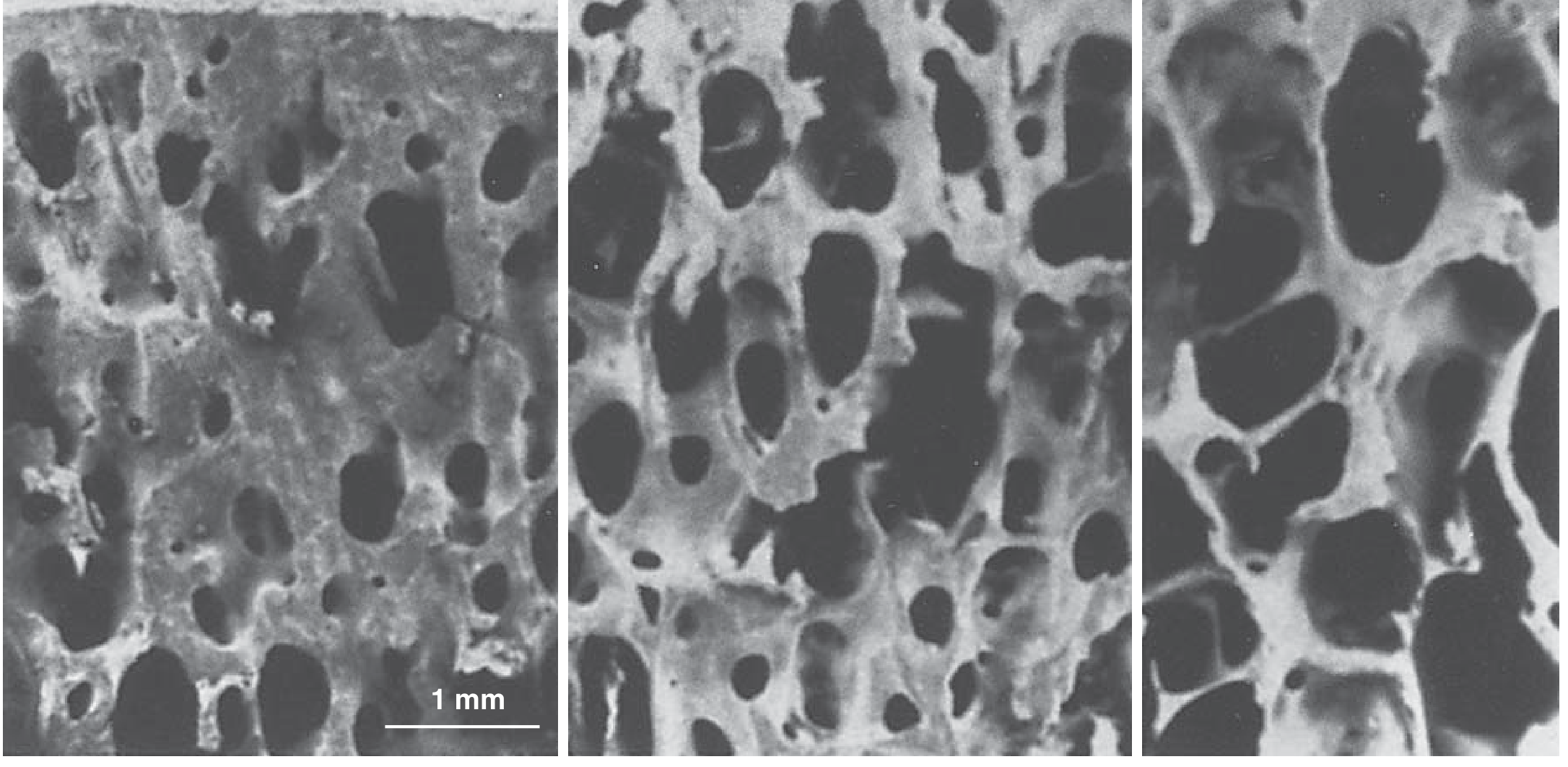
- Measures areal (2D) bone density (g/cm²), not volumetric - larger bones appear denser even if not truly so
- Cannot assess bone microarchitecture or quality - only quantity
- False elevation of BMD by: osteoarthritis, aortic calcification, vertebral fractures, scoliosis (in AP lumbar views)
- Results not easily comparable between different manufacturer scanners
- Precision errors with poor repositioning between follow-up scans
- Plain radiographs cannot detect bone loss until >30% is lost - making DEXA the only reliable early detection tool - Miller's Review of Orthopaedics
Newer Alternatives:
- Quantitative CT (QCT): Measures true volumetric BMD; separates trabecular from cortical; more precise but higher radiation
- HR-pQCT and microMRI: Assess bone microarchitecture; research tools
- Biomechanical CT (BCT): FDA-approved for opportunistic BMD assessment via routine CT; not recommended for treatment monitoring
- Ultrasound: No radiation; cannot diagnose osteoporosis reliably; not preferred over DXA
Compound palmar ganglion
Compound Palmar Ganglion
Definition
"This is nothing but chronic inflammation of the common sheath of the flexor tendons leading to swelling of this sheath (ulnar bursa) above and below the flexor retinaculum."
- S. Das: A Manual on Clinical Surgery, 13th Ed.
Aetiology (Causes)
| Cause | Context |
|---|---|
| Tuberculosis (tuberculous tenosynovitis of the ulnar bursa) | Predominant cause in developing countries (India, Southeast Asia) |
| Rheumatoid arthritis | Predominant cause in Western countries |
- S. Das Manual on Clinical Surgery / Bailey & Love's Short Practice of Surgery 28th Ed.
Anatomy - Why the Hourglass Shape?
- Proximally: about 2.5 cm above the flexor retinaculum (into the distal forearm)
- Distally: into the palm
Clinical Features
Symptoms
- Painless or mildly aching swelling at the wrist/lower forearm and palm - often the presenting complaint
- Restricted finger movements due to tendon involvement
- Thenar and hypothenar muscle wasting (from disuse or nerve compression)
- Paraesthesia in the median nerve distribution (thumb, index, middle, radial half of ring finger) - due to median nerve compression within the carpal tunnel
Signs (on Examination)
-
Inspection: Hourglass-shaped swelling - one lobe above the wrist (distal forearm) and one lobe below (palm), with a visible waist at the level of the flexor retinaculum
-
Fluctuation: The swelling is fluctuant (fluid-filled). Cross-fluctuation can be elicited between the two lobes above and below the flexor retinaculum - this is the pathognomonic sign.
-
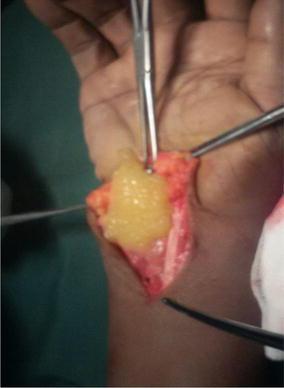
Melon-seed bodies (rice bodies): With careful palpation, small loose bodies within the bursa can be felt moving - these are fibrin deposits that have become rounded and organized within the synovial fluid. They are felt as mobile, small nodular structures."With careful palpation one can feel movements of the melon-seed bodies within the bursa."
- S. Das Manual on Clinical Surgery
-
Non-tender or mildly tender (in contrast to acute septic tenosynovitis, which is very tender)
Classic Clinical Image

Intraoperative Finding - Rice Bodies
Pathology
- Fibrin particles
- Melon-seed bodies (organized fibrin deposits, also called "rice bodies")
- Tuberculous granulomas (caseating) if TB etiology
Differential Diagnosis
| Condition | Key Distinction |
|---|---|
| Simple ganglion | Unilocular; tense, hard swelling; no cross-fluctuation; gelatinous fluid; dorsal wrist more common |
| Effusion of wrist joint | Swelling limited to joint extent; cross-fluctuation elicited anteriorly AND posteriorly (not just above/below retinaculum) |
| Lipoma | Non-fluctuant; lobulated |
| Acute suppurative tenosynovitis | Acutely painful; red; Kanavel's signs present; fever |
Complications
- Carpal tunnel syndrome: Median nerve compression within the carpal tunnel due to the expanding synovial swelling. The patient develops:
- Tingling/numbness in thumb, index, middle fingers (median nerve territory)
- Thenar wasting
- Weakness of thumb opposition
- Bailey & Love's Surgery 28th Ed. describes this as "occasionally causing symptoms of carpal tunnel syndrome"
Diagnosis
- Clinical - the cross-fluctuation sign above and below the retinaculum is characteristic
- Biopsy of the synovium - confirms the underlying etiology (caseating granulomas = TB; pannus = RA)
- Ziehl-Neelsen staining / TB culture if TB is suspected
- MRI/Ultrasound - can confirm bilocular fluid-filled sheath, assess extent, and detect rice bodies
Treatment
Surgical
- Thorough excision of the thickened synovium from both lobes of the sheath
- Removal of all rice bodies/melon-seed bodies
- Division of the flexor retinaculum if carpal tunnel syndrome is present (simultaneous carpal tunnel release)
Medical
- Antitubercular therapy (ATT) if TB is the cause: standard 4-drug regimen (Isoniazid + Rifampicin + Pyrazinamide + Ethambutol) for 6-9 months - surgery is combined with ATT, not a substitute
- DMARDs (methotrexate, etc.) if rheumatoid arthritis is the underlying cause
- Synovectomy alone without ATT risks recurrence in TB
Summary Table
| Feature | Detail |
|---|---|
| Definition | Chronic TB (or RA) tenosynovitis of ulnar bursa/common flexor sheath |
| Shape | Hourglass / dumbbell - bilocular above and below flexor retinaculum |
| Pathognomonic sign | Cross-fluctuation between the two lobes |
| Contents | Inflammatory fluid + rice bodies (melon-seed bodies) |
| Complication | Carpal tunnel syndrome (median nerve compression) |
| Diagnosis | Clinical + synovial biopsy |
| Treatment | Synovectomy + ATT (TB) or DMARDs (RA) |