
Only digit and metatarsal head is removed in which amputation and describe various foot amputation
Reading File
Quick Answer: The amputation where only the digit and metatarsal head is removed is called a Ray amputation. Foot amputations range from toe-only (phalangeal) to transmetatarsal, Lisfranc (tarsometatarsal), Chopart (midtarsal), and Syme's (ankle disarticulation).
Searching sources...
Finding Sources
Finding Sources
Reading File
Finding Sources
Reading File
Reading File
Finding Sources
Finding Sources
Reading File
Reading File
Reading File
Reading File
Reading File
Finding Sources
Reading File
Reading File
I now have all the information needed for a comprehensive answer. Let me also fetch the key anatomical diagram that shows all foot amputation levels.
Loading Image
Here is a complete, textbook-based answer.
Foot Amputations
Direct Answer to Your Question
Ray amputation is the procedure in which only the digit (toe/phalanges) and its corresponding metatarsal head are removed. It is performed when the disease process (infection, osteomyelitis, gangrene) is more proximal in the toe and involves the web space or metatarsal head - beyond what a simple digital amputation can address.
- Current Surgical Therapy 14e defines it precisely: "A ray amputation is a more involved procedure resulting in removal of the entire phalanx. It is performed when the disease process is more proximal in the toe, with involvement of the web space or metatarsal head."
- Sabiston Textbook of Surgery adds: "A ray amputation may be used when there is a need to resect the MTP joint or the distal metatarsal bone has become involved with infection."
Levels of Foot Amputation (Distal to Proximal)
The diagram below shows all levels clearly:
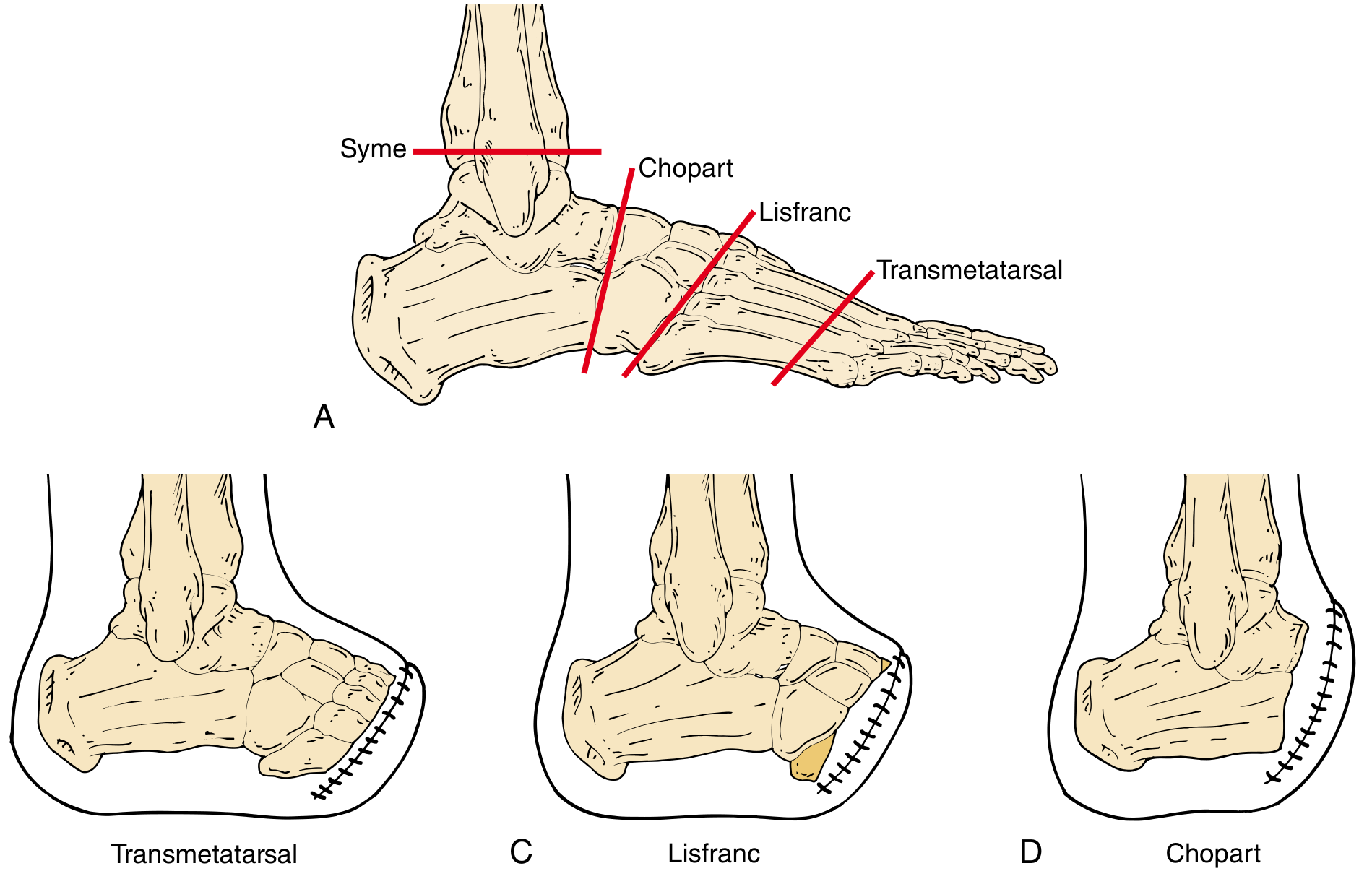
1. Digital (Toe) Amputation
Level: Distal or through the proximal phalanx
Indication: Gangrene, infected neuropathic ulceration, or osteomyelitis confined distal to the base of the proximal phalanx.
- Great toe amputation is the least well-tolerated because the great toe provides propulsion during ambulation.
- The most tolerable digital amputation involves the 3rd digit.
- A "fishmouth" circular incision is used at the base of the toe; the phalanx is divided with bone shears or a power saw, and the cut end is smoothed with a rongeur.
- Interphalangeal joint disarticulation is avoided (avascular cartilage impairs healing).
- Postoperative care: non-weight-bearing or heel-only weight bearing in a rigid soled shoe.
2. Ray Amputation
Level: Digit + metatarsal head (and metatarsal shaft to a healthy margin)
Indication: Disease involving the web space or metatarsal head; proximal phalanx involvement necessitating MTP joint resection.
Technique:
- A racquet-shaped incision - circular component around the base of the digit, with a longitudinal limb extending proximally over the metatarsal.
- Metatarsal is transected proximal to the head at a healthy margin.
- Digital arteries and nerves are carefully identified during medial/lateral dissection around the metatarsal.
- Wound may be left open if gross infection is present.
Special considerations:
- First ray amputation significantly affects gait stability (propulsion) - requires proper footwear and physical therapy.
- Fifth ray amputation: incision must be extended over the lateral border of the foot.
- If the 2nd ray is amputated, consider a Lapidus-type procedure to stabilize the foot.
- Removing multiple central rays may allow approximation of medial and lateral borders ("closing" the foot).
(Source: Campbell's Operative Orthopaedics 15e; Mulholland & Greenfield's Surgery 7e)
3. Transmetatarsal Amputation (TMA)
Level: Through the shafts of all five metatarsals (junction of middle and distal thirds)
Indication: Multiple toe infections up to the MTP joint level or extensive forefoot tissue loss involving several toes; when leaving one or two toes would be non-functional.
Technique:
- Long plantar flap + short dorsal flap ("fish-mouth" design).
- Dorsal incision at mid-metatarsal shaft level; metatarsals are sectioned in a beveled fashion (dorsal-distal to plantar-proximal) approximately 5 mm proximal to the skin incision.
- All plantar skin proximal to the MTP joints must be preserved.
- Tendons are divided under tension to allow retraction; alternatively, flexor and extensor tendons may be sutured to each other (myoplasty).
- Plantar flap is rotated dorsally for closure.
Key adjunct: Percutaneous Achilles tendon lengthening (or gastrocnemius recession) is nearly always required to prevent plantarflexion contracture, as the shortened foot lever arm limits dorsiflexion and increases terminal stump pressure during walking.
4. Lisfranc Amputation (Tarsometatarsal Disarticulation)
Level: Tarsometatarsal (TMT) joints
Indication: Insufficient soft tissue coverage for a TMA; infection or necrosis extending into the proximal forefoot.
Technique:
- Disarticulation of 1st, 3rd, 4th, and 5th tarsometatarsal joints; the 2nd metatarsal is transected ~2 cm distal to the medial cuneiform (to preserve the proximal transverse arch and base of 2nd metatarsal).
- Long plantar flap used for coverage.
- Peroneus brevis insertion at the base of the 5th metatarsal is preserved to avoid equinovarus deformity.
- Tibialis anterior and peroneus longus insertions at the medial cuneiform are meticulously preserved (or reattached) to prevent contracture.
- Achilles tendon is released; plantar fascia is approximated to dorsal periosteum.
- Plaster cast applied with talus in slight dorsiflexion.
Complication: Equinovarus deformity (due to loss of dorsiflexor attachments) - may require ankle arthrodesis or revision.
5. Chopart Amputation (Midtarsal / Transverse Tarsal Disarticulation)
Level: Calcaneocuboid - talonavicular joints (transverse tarsal joints)
Indication: More extensive foot disease not amenable to Lisfranc; offers less limb shortening than Syme's; preserves talus and calcaneus.
Technique:
- Begin with a posterior incision and tenectomy of the Achilles tendon (excise 2 cm) BEFORE the main procedure - this is essential to prevent progressive equinus deformity.
- "Fish-mouth" incision with longer plantar flap; medial and lateral incisions begin at the transverse tarsal joints.
- Anterior tibial and long extensor tendons are resected distally and prepared for transfer.
- One or more ankle dorsiflexors must be transferred (to balance the foot stump).
- Patient placed in slight dorsiflexion rigid dressing for 6 weeks.
Complications:
- High risk of equinus deformity (loss of all dorsiflexor attachments + unopposed Achilles pull) - can prevent ambulation and cause stump breakdown.
- Limited weight-bearing surface leads to continued pressure ulceration.
- May require tibiotalocalcaneal arthrodesis for stabilization.
(Campbell's Operative Orthopaedics 15e)
6. Syme's Amputation (Ankle Disarticulation)
Level: Ankle joint (tibiotalar), with removal of all foot bones; heel pad is retained.
Indication: Severe foot trauma (original primary indication); designed to preserve maximum limb length while keeping growth plates intact; allows ambulation without a prosthesis.
Relative contraindications: Neurotrophic ulcers, ischemic vascular disease (poor heel pad vascularity).
Key features:
- Preserves the heel fat pad (which serves as the end-bearing weight surface).
- Results in a longer residual limb than BKA - better rehabilitation potential.
- Compared with Chopart, provides a more stable end-bearing stump but greater limb shortening.
Boyd amputation is a variant: all foot bones except the calcaneus are removed, and the calcaneus is fused to the distal tibia - preserving an even longer limb.
Summary Table
| Amputation | Level | Bones Removed | Key Feature |
|---|---|---|---|
| Digital | Through proximal phalanx | Distal/mid phalanx | For distal gangrene/osteomyelitis |
| Ray | Digit + metatarsal head/shaft | Phalanges + metatarsal head | Racquet incision; for web space/MTP disease |
| Transmetatarsal (TMA) | Mid-metatarsal shafts | All 5 metatarsal heads + toes | Needs Achilles lengthening; long plantar flap |
| Lisfranc | Tarsometatarsal joints | All metatarsals + toes | Preserve 2nd met base; prevent equinovarus |
| Chopart | Transverse tarsal joints | All metatarsals + toes + navicular + cuboid | Achilles tenectomy first; equinus is major risk |
| Syme's | Ankle disarticulation | Entire foot | Heel pad preserved; no prosthesis needed |
Sources:
- Mulholland and Greenfield's Surgery 7e - Digital and Ray Amputations section
- Current Surgical Therapy 14e - Minor Amputations / Amputation Types
- Sabiston Textbook of Surgery - Toe and Ray Amputation, Lisfranc and Chopart sections
- Campbell's Operative Orthopaedics 15e - Midfoot Amputations, Techniques 17.6-17.8
This is a shared conversation. Sign in to Orris to start your own chat.