
Define diabetes mellitus. Write criteria for diagnosis of a new case of diabetes mellitus. Socine → Enumerate complications of diabetes and describe any one an detail. Animation
diabetes complications
American Diabetes Association Standards of Care 2026 diagnosis diabetes diagnostic criteria
https://diabetesjournals.org/care/article-pdf/49/Supplement_…
color fundus photograph diabetic retinopathy microaneurysms hard exudates neovascularization
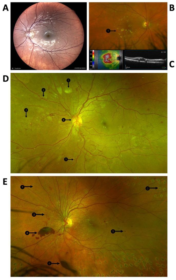
A multi-panel medical image illustrating the clinical manifestations and progression of diabetic retinopathy using fundus photography and Optical Coherence Tomography (OCT). Panel A displays a color fundus photograph of the posterior pole showing microaneurysms and dot hemorrhages indicative of diabetic maculopathy. Panel B highlights clinically significant macular edema (CSME) with visible hard exudates (B1). Panel C provides corresponding OCT imaging, including a retinal thickness heat map and a cross-sectional scan showing intraretinal fluid. Panels D and E are widefield fundus photographs demonstrating advanced proliferative diabetic retinopathy (PDR). Key pathological features identified with numbered arrows include cotton wool spots (D1), venous beading (D2), intraretinal microvascular abnormalities (IRMA) (D3/E1), neovascularization at the disc (D4), and venous loops (D5). Panel E further illustrates end-stage complications and treatment, showing laser photocoagulation scars (E2), retinal neovascularization (E3), pre-retinal hemorrhage (E4), blot hemorrhage (E5), and vitreous hemorrhage (E6). This compilation serves as an educational tool for identifying the transition from non-proliferative to proliferative diabetic disease.
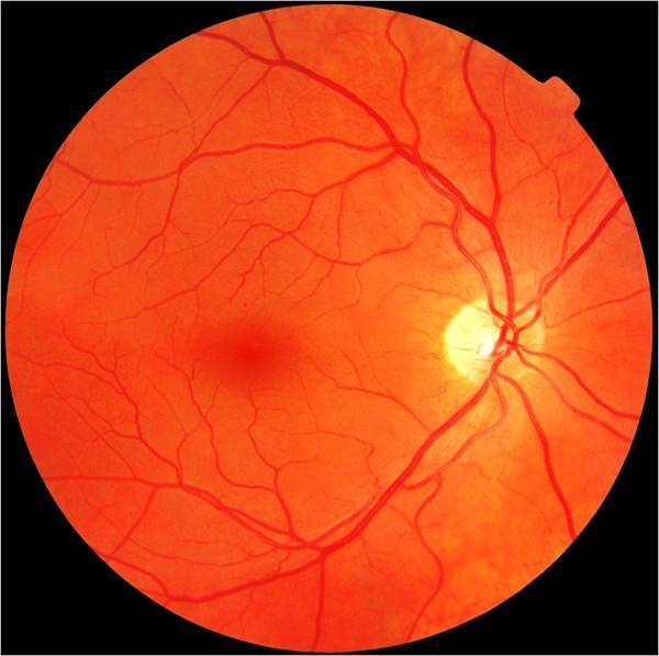
This clinical diagnostic image is a color fundus photograph of the human retina, specifically demonstrating mild non-proliferative diabetic retinopathy (NPDR). The image captures the posterior pole, showing the optic disc as a well-defined, yellowish-white circular structure located on the nasal side (right side of the frame). From the optic disc, retinal arteries and veins emerge and branch across the orange-hued retinal background toward the periphery. Centrally and slightly temporal to the disc, the macula is visible as a darker, less vascularized region. Pathological features characteristic of early-stage diabetic retinopathy are present, including isolated microaneurysms that appear as small, discrete red dots. There is also a subtle focal red lesion in the temporal region, which may represent a small blot hemorrhage. The overall vascular architecture remains relatively preserved without significant neovascularization, hard exudates, or cotton wool spots, aligning with a 'mild' classification on the Diabetic Retinopathy Disease Severity Scale. This image serves as an educational example for identifying early microvascular changes in diabetic patients during routine screening.
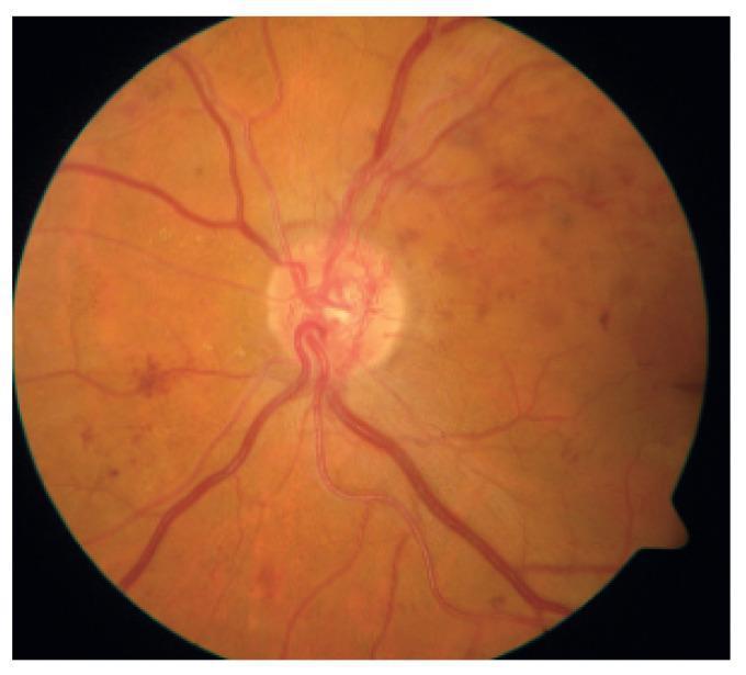
This color fundus photograph displays the posterior pole of a human eye with hallmarks of Proliferative Diabetic Retinopathy (PDR). The optic disc is centrally located and serves as the origin for the major retinal vascular arcades. A key diagnostic feature is the presence of neovascularization of the disc (NVD), characterized by a fine, tangled, and tortuous network of abnormal new blood vessels proliferating on and immediately adjacent to the optic nerve head. The surrounding retina shows multi-focal pathology, including scattered dot-and-blot intraretinal hemorrhages and smaller microaneurysms, particularly prominent in the temporal and nasal quadrants. Faint, yellowish deposits suggestive of hard exudates are visible in the peripapillary region. The vascular caliber appears irregular, and the overall fundus background exhibits a slightly hazy appearance, possibly due to vitreous changes or early preretinal hemorrhage. This image serves as a clinical example of advanced diabetic microvascular disease requiring urgent ophthalmic intervention.
Definition
Diagnosis of a new case of diabetes mellitus
| Test | Diagnostic value |
|---|---|
| Fasting plasma glucose (FPG) after at least 8 hours fast | ≥126 mg/dL (≥7.0 mmol/L) |
| 2-hour plasma glucose during 75-g oral glucose tolerance test | ≥200 mg/dL (≥11.1 mmol/L) |
| HbA1c using a standardized assay | ≥6.5% (48 mmol/mol) |
| Random plasma glucose with classic symptoms of hyperglycemia or hyperglycemic crisis | ≥200 mg/dL (≥11.1 mmol/L) |
- FPG: 100-125 mg/dL
- 2-hour OGTT: 140-199 mg/dL
- HbA1c: 5.7%-6.4%
Complications of diabetes mellitus
1. Acute metabolic complications
- Hypoglycemia - usually due to insulin or oral antidiabetic therapy.
- Diabetic ketoacidosis (DKA) - more common in type 1 DM.
- Hyperosmolar hyperglycemic state (HHS) - more common in type 2 DM.
- Lactic acidosis - may occur with severe illness, hypoxia, renal failure, or rarely metformin accumulation.
2. Chronic complications
A. Microvascular complications
- Diabetic retinopathy
- Diabetic nephropathy
- Diabetic neuropathy
- Peripheral symmetric polyneuropathy
- Autonomic neuropathy
- Mononeuropathy
B. Macrovascular complications
- Coronary artery disease and myocardial infarction
- Cerebrovascular disease and stroke
- Peripheral arterial disease causing claudication, ischemic ulcers, and gangrene
C. Other complications
- Diabetic foot: ulcer, infection, Charcot joint, gangrene, amputation
- Recurrent infections: skin, urinary tract, candidiasis, tuberculosis
- Cataract and glaucoma
- Gastroparesis, diarrhea, constipation
- Erectile dysfunction and bladder dysfunction
- Poor wound healing
Diabetic retinopathy in detail
Definition
Risk factors
- Long duration of diabetes
- Poor glycemic control and high HbA1c
- Hypertension
- Dyslipidemia
- Diabetic kidney disease
- Pregnancy
- Smoking
Pathogenesis
- Loss of retinal capillary pericytes and basement membrane thickening.
- Capillary leakage, leading to retinal edema and hard exudates.
- Capillary occlusion and retinal ischemia.
- Retinal ischemia stimulates VEGF release.
- VEGF causes abnormal new-vessel formation, termed neovascularization.
- Fragile new vessels bleed easily, causing vitreous hemorrhage, fibrosis, traction retinal detachment, and blindness.
Classification
1. Non-proliferative diabetic retinopathy (NPDR)
- Microaneurysms, the earliest clinical lesion
- Dot and blot retinal hemorrhages
- Hard exudates
- Cotton-wool spots
- Venous beading
- Intraretinal microvascular abnormalities (IRMA)
- Macular edema
2. Proliferative diabetic retinopathy (PDR)
- New vessels on the disc, NVD
- New vessels elsewhere in the retina, NVE
- Preretinal or vitreous hemorrhage
- Fibrovascular proliferation
- Tractional retinal detachment
- Neovascular glaucoma
3. Diabetic macular edema
Clinical features
- Blurring or distortion of vision
- Difficulty reading
- Floaters, especially with vitreous hemorrhage
- Sudden painless loss of vision
- Reduced central vision in macular edema
Screening
- Type 1 DM: first dilated retinal examination within 5 years after onset, then annually.
- Type 2 DM: retinal examination at the time of diagnosis, then annually.
- More frequent review is needed when retinopathy is present or progressing.
- Pregnancy requires eye assessment before conception or early in the first trimester and follow-up during pregnancy.
Prevention and treatment
- Optimize glycemic control to slow onset and progression.
- Control blood pressure and treat dyslipidemia.
- Anti-VEGF intravitreal injections are preferred for center-involving macular edema and are also used in proliferative disease.
- Panretinal laser photocoagulation is used for proliferative diabetic retinopathy.
- Vitrectomy is indicated for non-clearing vitreous hemorrhage or tractional retinal detachment.