
/teacchme erythropoiesis
I'll start by reading the medical library skill, then pull comprehensive content on erythropoiesis.
Reading File
Now let me search the library for erythropoiesis content in parallel:
Finding Sources
Finding Sources
Excellent results. Now let me read the key blocks in parallel - the histology section (most comprehensive on development), the biochemistry section, and the respiratory medicine section:
Reading File
Reading File
Reading File
Now let me read the core histology erythropoiesis section and kidney section, plus fetch the key diagram image:
Reading File
Reading File
Loading Image
Now let me read the detailed histology section on erythropoiesis stages and the kinetics section:
Finding Sources
Reading File
Let me find the actual erythropoiesis development section in the histology textbook:
Finding Sources
Finding Sources
Now let me read the Henry's normoblastic maturation section and the Guyton physiology section in parallel:
Reading File
Reading File
Excellent! Now let me get the Guyton EPO and hemoglobin formation sections, and also fetch the normoblast images:
Reading File
Reading File
Loading Image
I now have rich, comprehensive content from multiple authoritative textbooks. Let me also fetch the hemoglobin synthesis diagram and the Henry's normoblast image for visual completeness:
Loading Image
Reading File
I now have comprehensive content from multiple authoritative sources. Let me compile the full teaching response.
Erythropoiesis - A Complete Teaching Guide
Sources: Guyton & Hall Medical Physiology | Henry's Clinical Diagnosis & Management | Basic Medical Biochemistry | Histology: A Text and Atlas | Brenner & Rector's The Kidney
1. What Is Erythropoiesis?
Erythropoiesis is the process by which red blood cells (RBCs) are generated from hematopoietic stem cells in the bone marrow. It is the body's principal mechanism for maintaining adequate oxygen delivery to tissues.
Key numbers to remember:
- ~250 billion RBCs are replaced daily
- 2.5 - 3.0 million erythrocytes are produced every second
- ~1% of circulating RBCs is replaced each day
- Each mature RBC lives approximately 120 days
2. The Stem Cell Hierarchy (Progenitor Stages)
Production of RBCs follows a strictly ordered pathway of commitment and differentiation:
Hematopoietic Stem Cell (HSC)
        ↓
    CFU-GEMM  (Colony-Forming Unit - Granulocyte, Erythroid, Monocyte, Megakaryocyte)
        ↓
    BFU-EMeg → BFU-E  (Burst-Forming Unit - Erythroid)
        ↓
    CFU-E  (Colony-Forming Unit - Erythroid)   ← EPO acts here primarily
        ↓
  PRONORMOBLAST  (first morphologically recognizable cell)
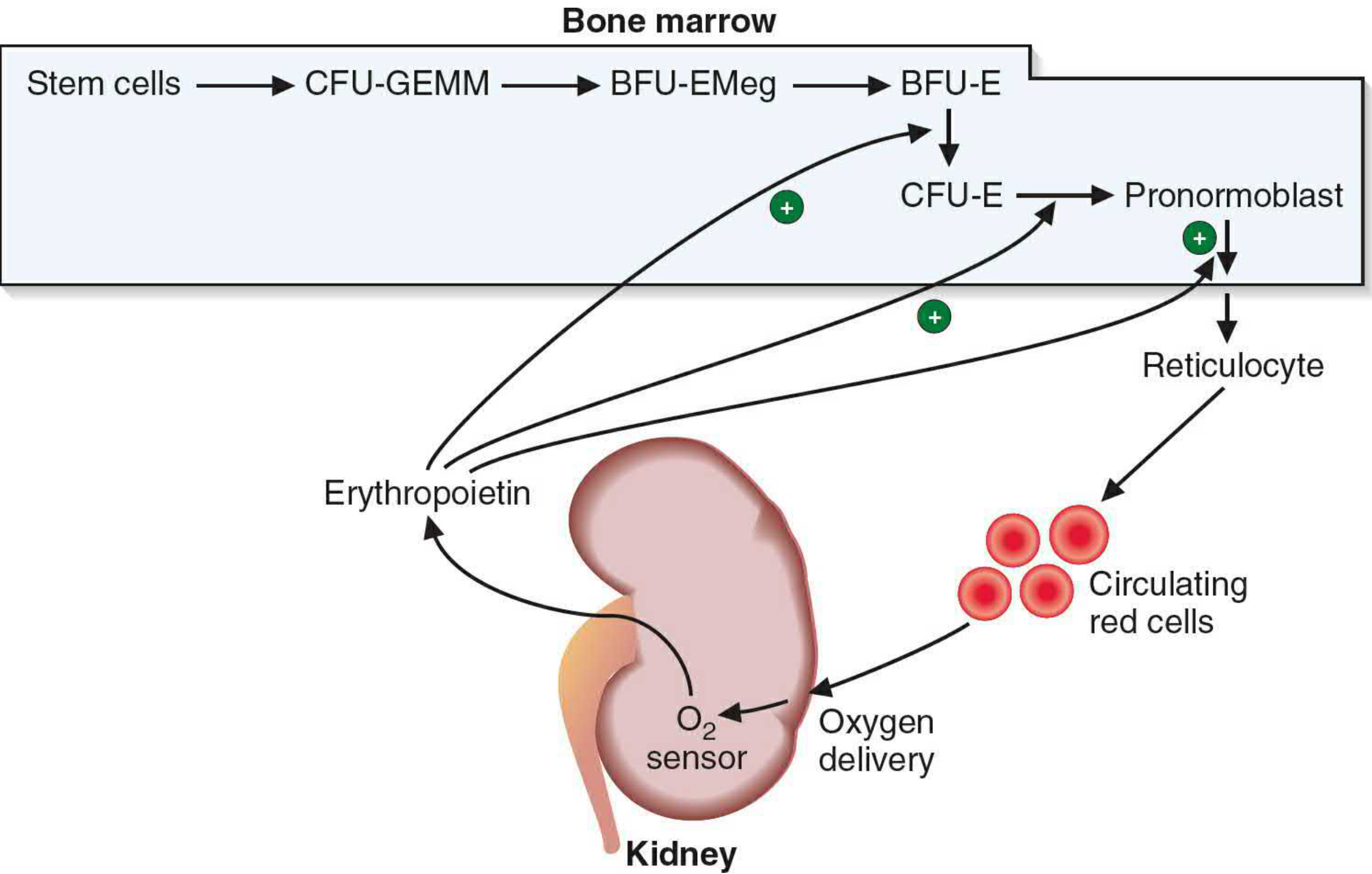
Erythropoietin (EPO) acts at multiple points - mainly on BFU-E, CFU-E, and the pronormoblast - to drive commitment and acceleration of maturation. - Basic Medical Biochemistry, p. 1536
3. Normoblastic Maturation (Morphological Stages)
The pronormoblast passes through 4 mitotic divisions over ~3 days, yielding up to 16 reticulocytes per pronormoblast:
Stage 1: Pronormoblast (Rubriblast)
- Largest erythroid precursor (~20 µm diameter)
- Fine, uniform chromatin; prominent nuclear membrane; 1+ nucleoli
- Cytoplasm is basophilic (rich in RNA), no granules
- Undergoes mitosis → 2 basophilic normoblasts
- Henry's, p. 658
Stage 2: Basophilic Normoblast (Prorubricyte)
- Slightly smaller; chromatin more coarsely clumped
- Parachromatin (non-chromatin nuclear material) stains pink - "spoke-wheel" pattern
- Cytoplasm is deeply basophilic - packed with polysomes actively making RNA
- Cell borders often irregular with pseudopodia
Stage 3: Polychromatophilic Normoblast (Rubricyte)
- Nucleus occupies ~half the cell area; chromatin moderately condensed
- Cytoplasm now shows polychromasia - mixed red (hemoglobin) + blue (RNA) → gray color
- This is the stage where hemoglobin synthesis becomes visible
- Undergoes 1-2 more mitotic divisions
- RNA content begins to decline as Hb accumulates
Stage 4: Orthochromatic Normoblast (Metarubricyte)
- Nucleus is pyknotic (small, dense) - mitosis is NO longer possible
- Cytoplasm is predominantly pink-gray (abundant hemoglobin, few polysomes)
- Nucleus is extruded via cytoplasmic contractions → forms reticulocyte
Stage 5: Reticulocyte
- Anucleate but retains ribosomes, mRNA, mitochondria, Golgi remnants
- Stains polychromatophilic on Romanowsky stains (residual RNA)
- Remains in bone marrow 2-3 days, then circulates 1-2 days
- Matures in the spleen where RNA and organelles are lost
- Normal reticulocyte count: slightly less than 1% of circulating RBCs
Stage 6: Mature Erythrocyte
- Biconcave disc, ~8 µm diameter
- No nucleus, no organelles
- Circulates for ~120 days
4. The EPO Axis - Regulation of Erythropoiesis
The regulation of RBC mass is a classic negative feedback loop centered on tissue oxygen delivery:
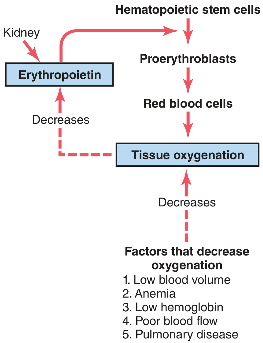
Figure: When tissue oxygenation decreases, the kidney releases EPO, which drives proerythroblast production and accelerates maturation. - Guyton & Hall Medical Physiology, p. 443
EPO Facts:
| Feature | Detail |
|---|---|
| Chemical nature | Glycoprotein, MW ~30.4 kDa (or ~34,000 per Guyton) |
| Primary source | Kidney - ~90% (cortex/outer medulla fibroblast-like interstitial cells + pericytes) |
| Secondary source | Liver - ~10% |
| Molecular trigger | Hypoxia → HIF-1 (Hypoxia-Inducible Factor-1) binds hypoxia response element on EPO gene → transcription of EPO mRNA |
| Time course | EPO rises within minutes to hours of hypoxia; new RBCs appear in circulation ~5 days later |
Factors that trigger EPO release:
- Anemia (reduced RBC mass)
- High altitude (low PO₂)
- Pulmonary disease / cardiac failure
- Norepinephrine, epinephrine, prostaglandins (secondary stimulants)
- Cobalt (in mining regions, inhibits prolyl hydroxylases that degrade HIF)
EPO actions on bone marrow:
- Stimulates proerythroblast production from hematopoietic stem cells
- Accelerates passage through erythroblastic maturation stages
- Promotes early release of reticulocytes from marrow
Clinical note: Bilateral nephrectomy or chronic kidney disease destroys the EPO source, leaving only hepatic EPO (~10%), which produces only 1/3 to 1/2 the RBCs needed - hence the anemia of chronic kidney disease. - Guyton & Hall, p. 443
5. Hemoglobin Synthesis
Hb synthesis begins at the polychromatophilic erythroblast stage and continues through reticulocyte maturation:
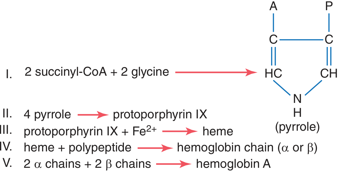
The 5 steps:
| Step | Reaction |
|---|---|
| I | 2 succinyl-CoA + 2 glycine → pyrrole |
| II | 4 pyrroles → protoporphyrin IX |
| III | protoporphyrin IX + Fe²⁺ → heme |
| IV | heme + polypeptide → hemoglobin chain (α or β) |
| V | 2 α chains + 2 β chains → Hemoglobin A (HbA, MW 64,458) |
Each hemoglobin molecule carries 4 heme groups → can bind 4 O₂ molecules (8 oxygen atoms).
RNA dynamics during maturation:
- Pronormoblast and basophilic normoblast: highest RNA content
- RNA declines progressively through polychromatophilic → orthochromatic normoblast
- Reticulocyte: no nucleus, no new RNA synthesis, but existing RNA persists 1-2 days enabling continued Hb synthesis
- Henry's Clinical Diagnosis, p. 658
6. Erythroblastic Islands
In the bone marrow, developing erythroid cells cluster around central macrophages forming erythroblastic islands:
- The macrophage supplies iron via transferrin to developing normoblasts
- It phagocytoses the extruded nucleus from orthochromatic normoblasts
- Iron is transferred from plasma transferrin → pronormoblast onwards
- Islands are disrupted when marrow aspirate is spread on slides; fragments of macrophage cytoplasm may remain attached to normoblasts (visible on Prussian blue stain)
- Henry's Clinical Diagnosis, p. 658
7. Kinetics Summary
| Parameter | Value |
|---|---|
| Divisions per pronormoblast | 3-4 mitotic divisions |
| Time from pronormoblast to reticulocyte | ~3 days |
| Reticulocytes per pronormoblast | Up to 16 |
| Time in marrow as reticulocyte | 2-3 days |
| Time in circulation as reticulocyte | ~1 day |
| RBC lifespan | ~120 days |
| Normal reticulocyte % | < 1% of circulating RBCs |
8. Effective vs. Ineffective Erythropoiesis
| Term | Meaning |
|---|---|
| Total erythropoiesis | Total production of hemoglobin/RBCs |
| Effective erythropoiesis | Production of RBCs that actually reach circulation |
| Ineffective erythropoiesis | Precursors that die in the marrow before release (e.g., thalassemia, B12/folate deficiency, myelodysplasia) |
In β-thalassemia, unpaired α-globin chains form aggregates that destroy red cell precursors in the marrow - a classic cause of high ineffective erythropoiesis and resultant iron overload from increased iron absorption. - Robbins Pathology
9. Clinical Connections
| Condition | Erythropoiesis Effect |
|---|---|
| Chronic kidney disease | Low EPO → normocytic anemia |
| High altitude | Hypoxia → ↑EPO → polycythemia |
| β-thalassemia | Ineffective erythropoiesis, bone marrow expansion, extramedullary hematopoiesis |
| Iron deficiency | Impairs heme synthesis → hypochromic, microcytic RBCs |
| B12/folate deficiency | Impairs DNA synthesis → megaloblastic anemia (large, dysfunctional cells) |
| Polycythemia vera | Autonomous overproduction, EPO-independent |
| MDS | Ineffective erythropoiesis → anemia despite a full marrow |
Sources
- Guyton and Hall Textbook of Medical Physiology, p. 442-445
- Henry's Clinical Diagnosis and Management by Laboratory Methods, p. 658-659
- Basic Medical Biochemistry - A Clinical Approach, 6e, p. 1536
- Histology: A Text and Atlas With Correlated Cell and Molecular Biology, pp. 667-729
- Brenner and Rector's The Kidney, p. 2465
This is a shared conversation. Sign in to Orris to start your own chat.