
Blood supply of pancreas diagram , relation diagram and pancreas easy diagram for exam drawing all easy diagrams
arterial blood supply of pancreas diagram
relations of the pancreas posterior abdominal wall
pancreas parts head neck body tail pancreatic duct
ls -la /tmp/workspace/ ; mkdir -p /tmp/workspace/pancreas-diagrams
mkdir -p /tmp/workspace/pancreas-diagrams/src /tmp/workspace/pancreas-diagrams/output && cat > /tmp/workspace/pancreas-diagrams/src/draw.py <<'EOF' import matplotlib matplotlib.use("Agg") import matplotlib.pyplot as plt from matplotlib.patches import Polygon, Ellipse, FancyArrowPatch, Circle, PathPatch from matplotlib.path import Path import numpy as np OUT="/tmp/workspace/pancreas-diagrams/output/" YEL="#f7e08a"; RED="#d62828"; BLUE="#1d4ed8"; GRN="#2a9d4a"; PINK="#f6c1c7" def setup(title, w=10, h=7): fig, ax = plt.subplots(figsize=(w,h)) ax.set_xlim(0,100); ax.set_ylim(0,70); ax.axis("off") ax.set_title(title, fontsize=16, fontweight="bold", pad=10) return fig, ax def lab(ax, text, xy, xytext, ha="left", color="k", fs=11): ax.annotate(text, xy=xy, xytext=xytext, ha=ha, va="center", fontsize=fs, color=color, arrowprops=dict(arrowstyle="-", color=color, lw=1.1)) def pancreas_poly(ax, alpha=1): # head (low left), neck, body (rising to right), tail (top right) pts=[(26,18),(36,14),(42,20),(44,27),(52,31),(64,36),(76,44),(86,50),(89,54),(84,56),(74,52),(62,46),(50,42),(40,40),(31,36),(26,28)] p=Polygon(pts, closed=True, fc=YEL, ec="#8a6d00", lw=2, alpha=alpha, joinstyle="round") ax.add_patch(p); return p # ---------- Diagram 1: Parts & ducts ---------- fig,ax=setup("Fig 1. Pancreas: parts and ducts (anterior view)") # duodenum C-loop th=np.linspace(np.pi/2,-np.pi/2,60) cx,cy=34,24 xs=cx-12*np.cos(th)*-1; # simple C: draw as thick arc open to the right ang=np.linspace(np.radians(80),np.radians(280),80) ax.plot(cx+19*np.cos(ang)*-1*-1*-1 if False else cx+ 20*np.cos(ang), cy+ 15*np.sin(ang), color="#e8a0a8", lw=22, solid_capstyle="round", zorder=1) ax.plot(cx+20*np.cos(ang), cy+15*np.sin(ang), color=PINK, lw=18, solid_capstyle="round", zorder=1) pancreas_poly(ax) # ducts duct=[(31,22),(36,24),(44,30),(54,35),(66,41),(78,48),(86,53)] ax.plot(*zip(*duct), color="#1b6ca8", lw=3, zorder=5) # accessory duct ax.plot([40,36,33],[29,26,22], color="#7aa6c2", lw=2, zorder=5) ax.plot([32,33,34],[22,21,20], color="#1b6ca8", lw=3, zorder=5) # CBD ax.plot([20,22,28,31],[56,40,28,22], color=GRN, lw=4, zorder=5) ax.add_patch(Circle((31,21),1.4,fc="k",zorder=6)) # uncinate ax.add_patch(Polygon([(26,18),(33,17),(40,22),(33,23)],fc="#e9c94a",ec="#8a6d00",lw=1,zorder=3)) lab(ax,"HEAD (in C of duodenum)",(30,24),(4,6)) lab(ax,"Uncinate process",(34,19),(30,6),) lab(ax,"NECK (1.5-2 cm)\nin front of portal vein / SMV",(42,29),(10,64)) lab(ax,"BODY (triangular on section)",(60,40),(40,64)) lab(ax,"TAIL (in lienorenal ligament,\nreaches splenic hilum)",(87,53),(62,64),fs=10.5) lab(ax,"Main duct (of Wirsung)",(68,42),(72,26),color="#1b6ca8") lab(ax,"Accessory duct (of Santorini)",(38,28),(52,16),color="#4a7fa0") lab(ax,"Common bile duct",(21,48),(2,44),color=GRN) lab(ax,"Ampulla of Vater\n(major duodenal papilla, D2)",(31,21),(52,5)) lab(ax,"Duodenum (C-loop)",(14,22),(0,14),color="#b5545f") fig.savefig(OUT+"fig1_pancreas_parts.png",dpi=160,bbox_inches="tight",facecolor="white"); plt.close(fig) # ---------- Diagram 2: Arterial supply ---------- fig,ax=setup("Fig 2. Arterial supply of the pancreas (schematic)",10.5,8) ax.set_ylim(0,80) # pancreas pts=[(24,20),(34,16),(40,22),(42,28),(52,32),(64,37),(76,44),(86,50),(89,54),(84,56),(74,52),(62,47),(50,43),(40,41),(30,37),(24,30)] ax.add_patch(Polygon(pts,fc=YEL,ec="#8a6d00",lw=2)) def art(xy_list,color=RED,lw=3,ls="-"): ax.plot(*zip(*xy_list),color=color,lw=lw,ls=ls,solid_capstyle="round") # aorta ax.plot([48,48],[2,76],color="#e07070",lw=10,solid_capstyle="butt",alpha=.5) ax.text(48,0.2,"Aorta",ha="center",fontsize=10,color="#a02020") # celiac trunk art([(48,66),(48,60)]) ax.text(50,70,"COELIAC TRUNK (T12)",fontsize=11,fontweight="bold",color=RED) # splenic: runs along upper border to the right art([(48,60),(56,62),(66,58),(76,56),(86,58)]) ax.text(60,64,"Splenic artery",fontsize=11,color=RED) # CHA to left art([(48,60),(40,62),(33,60)]) ax.text(34,64,"Common hepatic a.",fontsize=11,color=RED,ha="center") # GDA art([(33,60),(30,52),(30,42),(30,34)]) ax.text(21,52,"Gastroduodenal a.",fontsize=11,color=RED,ha="center") ax.annotate("To liver\n(proper hepatic a.)",xy=(33,60),xytext=(28,72),fontsize=9,ha="center",arrowprops=dict(arrowstyle="->")) # Superior pancreaticoduodenal anterior/posterior art([(30,34),(27,28),(26,22)],lw=2.5) art([(30,34),(33,28),(33,22)],lw=2.5) ax.text(11,26,"Anterior &\nposterior SUPERIOR\npancreatico-\nduodenal aa.",fontsize=10,color=RED,ha="center") art([(30,40),(27,43)],lw=2) # Right gastroepiploic art([(30,34),(24,34),(18,34)],lw=2,ls="--") ax.text(1,37,"Rt gastro-\nepiploic a.",fontsize=9,color=RED) # SMA art([(48,48),(48,36),(48,22),(48,12)]) ax.text(50,10,"SUPERIOR MESENTERIC a. (L1)",fontsize=11,fontweight="bold",color=RED) # Inferior pancreaticoduodenal art([(48,20),(40,18),(33,19),(29,20)],lw=2.5) ax.text(27,12,"INFERIOR pancreatico-\nduodenal a.",fontsize=10,color=RED,ha="center") ax.annotate("",xy=(33,23),xytext=(33,19),arrowprops=dict(arrowstyle="<->",color="purple",lw=1.5)) ax.text(36,24.8,"anastomosis",fontsize=9,color="purple") # Splenic branches to body/tail for x,y,name in [(60,57.5,""),(70,56.5,"")]: pass art([(58,61),(58,52)],lw=2) ax.text(58.5,50.4,"Dorsal pancreatic a.",fontsize=9.5,color=RED) art([(66,58),(66,50)],lw=2) art([(74,56),(74,50)],lw=2) ax.annotate("Pancreatica magna\n(great pancreatic a.)",xy=(66,50),xytext=(80,40),fontsize=10,color=RED,arrowprops=dict(arrowstyle="-",color=RED)) ax.annotate("Caudal pancreatic a.\n(to tail)",xy=(80,55),xytext=(84,66),fontsize=10,color=RED,arrowprops=dict(arrowstyle="-",color=RED)) art([(58,52),(52,44),(44,38)],lw=2,color="#d6609a") ax.text(60,32,"Inferior pancreatic a.\n(from dorsal pancreatic;\njoins SMA branches)",fontsize=9.5,color="#b03070") # Spleen ax.add_patch(Ellipse((93,62),8,14,fc="#c9b6e4",ec="#6b4fa0")) ax.text(93,62,"Spleen",rotation=90,ha="center",va="center",fontsize=9) ax.text(4,3,"Head + uncinate: gastroduodenal (superior) + SMA (inferior) arcades\nNeck, body, tail: splenic artery branches",fontsize=10.5,bbox=dict(fc="#fff7cc",ec="#8a6d00")) ax.text(41,38,"HEAD",fontsize=9,alpha=.6); ax.text(70,45,"BODY",fontsize=9,alpha=.6,rotation=30) fig.savefig(OUT+"fig2_arterial_supply.png",dpi=160,bbox_inches="tight",facecolor="white"); plt.close(fig) # ---------- Diagram 3: Relations (posterior view of pancreas bed) ---------- fig,ax=setup("Fig 3. Relations of the pancreas (structures behind it, L1-L2)",10.5,7.5) pts=[(26,26),(36,22),(42,28),(44,34),(52,38),(64,43),(76,50),(86,56),(89,60),(84,62),(74,58),(62,52),(50,48),(40,46),(31,42),(26,34)] ax.add_patch(Polygon(pts,fc=YEL,ec="#8a6d00",lw=2,alpha=.9)) ax.text(33,31,"HEAD",fontsize=11,fontweight="bold") ax.text(60,46,"BODY",fontsize=11,fontweight="bold",rotation=25) ax.text(80,57,"TAIL",fontsize=11,fontweight="bold",rotation=35) # Duodenum C ang=np.linspace(np.radians(80),np.radians(280),80) ax.plot(34+20*np.cos(ang),32+14*np.sin(ang),color=PINK,lw=14,zorder=0) ax.text(9,32,"Duodenum\n(D1-D3)",fontsize=10,ha="center",color="#b5545f") # IVC, aorta behind head/neck ax.add_patch(Ellipse((36,12),9,6,fc="#9db7f5",ec=BLUE,lw=1.5)); ax.text(36,12,"IVC",ha="center",va="center",fontsize=10) ax.add_patch(Circle((53,11),3.4,fc="#f08080",ec=RED,lw=1.5)); ax.text(53,11,"Ao",ha="center",va="center",fontsize=10) ax.plot([53,53],[14.4,22],color=RED,lw=2.5); ax.text(54.5,19,"SMA",fontsize=10,color=RED) ax.plot([41,57],[18,18],color=BLUE,lw=3); ax.text(44,14.3,"Left renal vein",fontsize=9.5,color=BLUE) # SMV / portal confluence ax.add_patch(Ellipse((46,31),4,5,fc="#9db7f5",ec=BLUE,lw=1.5)); ax.annotate("Portal vein / SMV\nconfluence (behind neck)",xy=(46,30),xytext=(48,3),fontsize=10,color=BLUE,arrowprops=dict(arrowstyle="-",color=BLUE)) # splenic vein ax.plot([48,58,70,82,90],[36,40,46,53,58],color=BLUE,lw=3) ax.text(70,38,"Splenic vein\n(runs behind body)",fontsize=10,color=BLUE) # CBD in groove behind head ax.plot([30,30,31],[46,38,30],color=GRN,lw=4); ax.text(22,48,"CBD (groove on\nposterior head)",fontsize=10,color=GRN) # left kidney, adrenal ax.add_patch(Ellipse((78,28),9,16,fc="#d9a066",ec="#8a5a20")); ax.text(78,28,"Left\nkidney",ha="center",va="center",fontsize=9) ax.add_patch(Polygon([(70,38),(76,38),(73,44)],fc="#e8c547",ec="#8a6d00")); ax.text(60,28,"Left supra-\nrenal gland",fontsize=9.5) ax.annotate("",xy=(72,38),xytext=(68,31),arrowprops=dict(arrowstyle="-")) ax.text(95,67,"Hilum of spleen\n(tail, lienorenal lig.)",fontsize=9.5,ha="right") ax.text(2,64,"Posterior relations: IVC, aorta, SMA origin, left renal vein, CBD, portal/SMV/splenic veins,\nleft kidney, left suprarenal, crus of diaphragm",fontsize=9.5,bbox=dict(fc="#eef6ff",ec=BLUE)) fig.savefig(OUT+"fig3_posterior_relations.png",dpi=160,bbox_inches="tight",facecolor="white"); plt.close(fig) # ---------- Diagram 4: Anterior relations & sagittal ---------- fig,ax=setup("Fig 4. Sagittal section at L1-L2: relations of body of pancreas (easy exam sketch)",10,7) ax.set_xlim(0,100); ax.set_ylim(0,70) # vertical structures from front (left) to back (right) ax.add_patch(Polygon([(2,12),(18,12),(18,58),(2,58)],fc="#f6c1c7",ec="#b5545f",lw=1.5)) ax.text(10,35,"STOMACH\n(posterior\nwall)",ha="center",va="center",fontsize=10) ax.add_patch(Ellipse((26,38),8,4,fc="#fff",ec="#999",ls="--")); ax.text(26,45,"Lesser sac",fontsize=9.5,ha="center",style="italic") # pancreas body (oval) ax.add_patch(Polygon([(32,30),(42,34),(48,40),(44,46),(34,44),(30,38)],fc=YEL,ec="#8a6d00",lw=2)) ax.text(39,38,"Pancreas\nbody",ha="center",va="center",fontsize=10,fontweight="bold") # splenic vein ax.add_patch(Ellipse((49,33),5,3,fc="#9db7f5",ec=BLUE)); ax.text(49,29,"Splenic v.",fontsize=9,color=BLUE,ha="center") # SMA ax.add_patch(Circle((53,26),1.8,fc="#f08080",ec=RED)); ax.text(56,26,"SMA",fontsize=9.5,color=RED) # left renal vein ax.plot([52,66],[20,20],color=BLUE,lw=3); ax.text(56,17.5,"Left renal v.",fontsize=9,color=BLUE) # aorta & vertebra ax.add_patch(Circle((62,30),4,fc="#f08080",ec=RED)); ax.text(62,30,"Ao",ha="center",va="center",fontsize=10) ax.add_patch(Polygon([(72,8),(88,8),(88,60),(72,60)],fc="#eee",ec="#777")); ax.text(80,35,"Vertebra\nL1-L2",ha="center",va="center",fontsize=10) # transverse mesocolon root ax.plot([36,36],[30,22],color="#555",lw=2); ax.annotate("Root of transverse\nmesocolon (along\nlower border)",xy=(36,26),xytext=(18,6),fontsize=9.5,arrowprops=dict(arrowstyle="-")) ax.text(1,66,"FRONT",fontsize=10,fontweight="bold"); ax.text(92,66,"BACK",fontsize=10,fontweight="bold") ax.text(18,62,"Anterior surface: covered by peritoneum of lesser sac;\nstomach in front. Tuber omentale at the neck-body junction touches lesser omentum.",fontsize=9) ax.annotate("Duodenojejunal flexure\n(below body)",xy=(46,22),xytext=(30,12),fontsize=9.5,arrowprops=dict(arrowstyle="-")) fig.savefig(OUT+"fig4_sagittal_relations.png",dpi=160,bbox_inches="tight",facecolor="white"); plt.close(fig) EOF cd /tmp/workspace/pancreas-diagrams/src && python draw.py && ls ../output
Quick answer
fig1 pancreas parts
Image · PNG
fig2 arterial supply
Image · PNG
fig3 posterior relations
Image · PNG
fig4 sagittal relations
Image · PNG
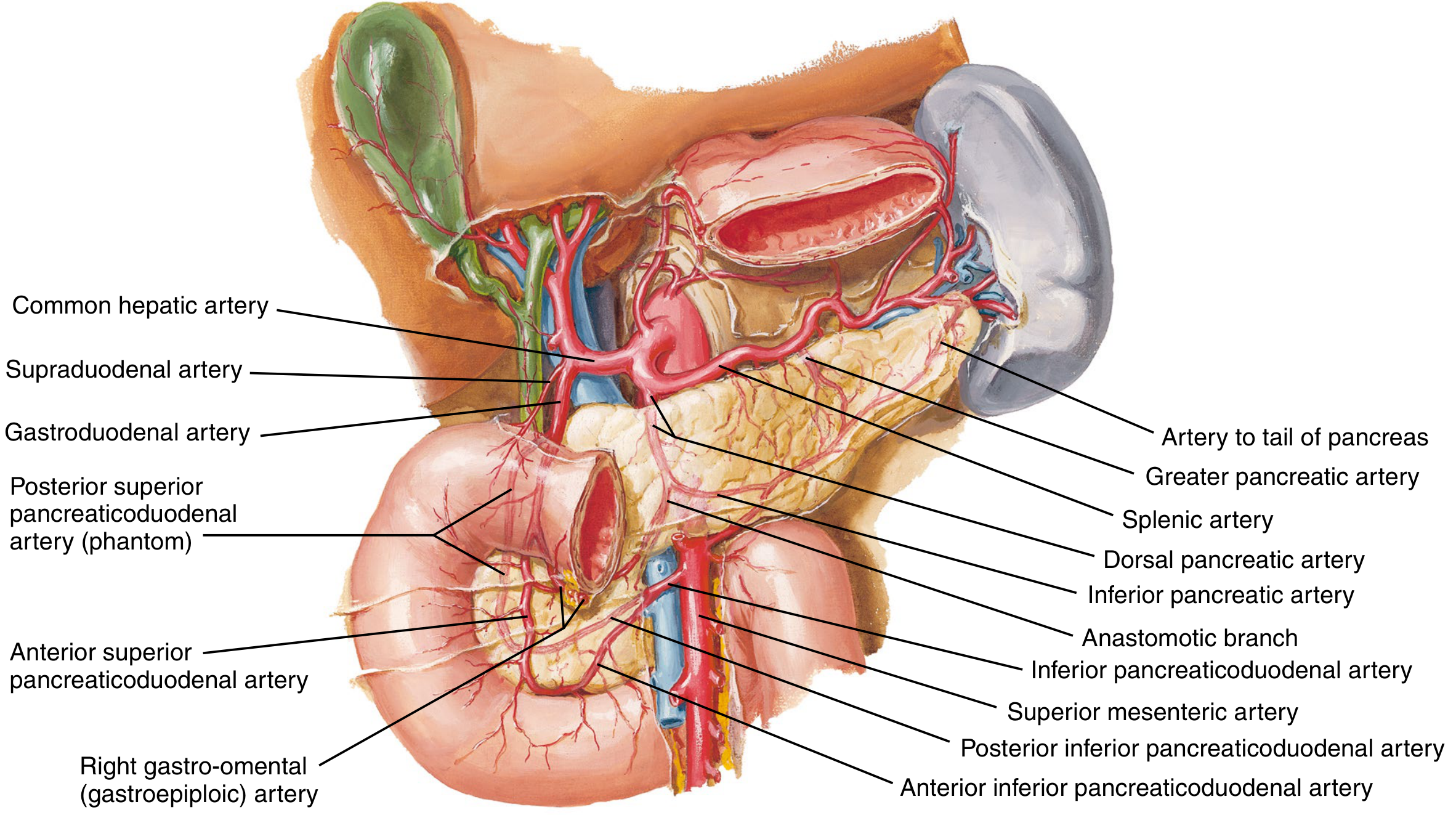
Diagram 1: Pancreas parts (easy sketch)
- Draw a "C" shape for the duodenum, open to the left.
- Fill the C with a rounded head. Add a small hook at its lower left, the uncinate process.
- Draw a short narrowing in front of the portal vein/SMV, the neck.
- Extend a long, slightly upward-sloping body to the left.
- End in a tail that touches the spleen.
- Draw the main pancreatic duct (Wirsung) as a line down the middle, from tail to head.
- Join it with the common bile duct at the ampulla of Vater, which opens at the major duodenal papilla in D2.
- Add the accessory duct (Santorini) as a small branch opening about 2 cm above the main papilla.
Diagram 2: Arterial supply (flow-chart style)
AORTA
 |-- COELIAC TRUNK (T12)
 |     |-- Splenic artery (along upper border of body and tail)
 |     |     |-- Dorsal pancreatic a. -> inferior pancreatic a.
 |     |     |-- Pancreatica magna (great pancreatic a.)
 |     |     |-- Caudal pancreatic a. (tail)
 |     |-- Common hepatic a.
 |           |-- Gastroduodenal a.
 |                 |-- Superior pancreaticoduodenal a. (anterior + posterior)
 |
 |-- SUPERIOR MESENTERIC ARTERY (L1)
       |-- Inferior pancreaticoduodenal a. (anterior + posterior)
- Head and uncinate process: these are supplied by two arcades, one from the gastroduodenal artery (above) and one from the SMA (below). The two arcades anastomose. This links the coeliac and SMA territories.
- Neck, body and tail: the splenic artery supplies these.
- Veins: these follow the arteries.
- The head drains through the pancreaticoduodenal veins into the SMV or portal vein.
- The body and tail drain through the splenic vein.
- The splenic vein joins the SMV behind the neck to form the portal vein.
- Lymph: the pancreaticosplenic nodes along the splenic artery, the pyloric nodes and the nodes around the SMA are the main groups.
Diagram 3: Relations
- IVC, with the right renal vein
- Aorta, with the SMA arising from it
- Left renal vein, passing between the aorta and the SMA
- Left suprarenal gland
- Left kidney
- A portal vein/SMV confluence behind the neck.
- The splenic vein running along the back of the body and tail.
- The CBD in a groove on the posterior head.
| Part | Anterior | Posterior | Other |
|---|---|---|---|
| Head | Root of transverse mesocolon, transverse colon, pylorus region | IVC, right renal vein, right crus, CBD | Right: D2. Left: SMV. Below: D3 |
| Neck | Pylorus | Portal vein formation (SMV + splenic vein) | Gastroduodenal artery in front-right |
| Body | Lesser sac, then stomach | Aorta, SMA, left crus, left suprarenal, left kidney, left renal vein, splenic vein | Below: duodenojejunal flexure and jejunal loops |
| Tail | Lies in the lienorenal ligament | Left kidney | Reaches the splenic hilum with the splenic vessels |
Quick exam tips
- Use yellow for the pancreas, red for arteries, blue for veins and green for the bile duct. Colours score well in diagrams.
- Label with straight lines and write the labels on the right side only.
- Draw in this order: duodenal C, pancreas, ducts, then vessels.
Fast brooo
fig1 pancreas parts
Image · PNG
fig2 arterial supply
Image · PNG
fig3 posterior relations
Image · PNG
fig4 sagittal relations
Image · PNG
1. Easy pancreas diagram (parts and ducts)
 Liver/CBD                                   TAIL -> splenic hilum
     |                                     /
  ___|____          NECK     BODY    ___/
 / D2    \__  HEAD  \______/ ~~~~~~~~
|  C-loop  |  (uncinate     main duct (Wirsung)
 \__D3____/    process)
   Ampulla of Vater (D2)  <- CBD + main duct join
- Draw the duodenal C-loop.
- Draw the head inside the C.
- Draw the neck, then the body sloping up to the left.
- Draw the tail ending at the spleen.
- Draw the main duct running along the whole gland.
- Add the accessory duct (Santorini) above the head.
- Add the CBD joining the main duct at the ampulla.
2. Blood supply diagram
                 AORTA
          ________|________
         |                 |
   COELIAC TRUNK (T12)   SMA (L1)
     |           |           |
 Common      Splenic a.   Inferior pancreatico-
 hepatic a.      |         duodenal a. (ant + post)
     |           |              |
 Gastroduodenal  |              |
     |           |              |
 Superior pancreatico-          |
 duodenal (ant + post) <--anastomose-->
  = HEAD + UNCINATE

 Splenic a. gives: dorsal pancreatic -> inferior (transverse) pancreatic a.
                   pancreatica magna
                   caudal pancreatic a.
  = NECK + BODY + TAIL
- Head and uncinate: the superior pancreaticoduodenal arteries (anterior and posterior) come from the gastroduodenal artery, which comes from the common hepatic artery and the coeliac trunk. The inferior pancreaticoduodenal arteries come from the SMA. The two sets form an arcade between the coeliac and SMA territories.
- Neck, body and tail: these are supplied by the splenic artery, which runs along the upper border. Its branches are the dorsal pancreatic artery, the pancreatica magna and the caudal pancreatic artery. The inferior (transverse) pancreatic artery arises from the dorsal pancreatic artery.
3. Relations diagram
           FRONT (anterior)
   Stomach / lesser sac / transverse mesocolon root
              |
   [HEAD]--[NECK]--[BODY]--------[TAIL]
     |        |        |             |
  IVC      Portal v./   Aorta, SMA,   Splenic hilum,
  R renal  SMV +        L crus, L     lienorenal lig.
  vein,    splenic v.   suprarenal,
  CBD      confluence   L kidney,
                        L renal vein,
                        splenic vein
           BACK (posterior)
- Right: D2 (descending duodenum).
- Above and below: D1 and D3.
- Anterior: the root of the transverse mesocolon, with the transverse colon and the pylorus above.
- Posterior: IVC, right renal vein, right crus and the CBD in a groove.
- Left: the SMV and SMA (the uncinate process lies behind them).
- Anterior: pylorus.
- Posterior: the SMV and splenic vein joining to form the portal vein.
- Anterior surface: the lesser sac and the stomach behind it.
- Posterior surface: aorta, SMA origin, left crus, left suprarenal gland, left kidney and left renal vein. The splenic vein runs along it.
- Inferior surface: the duodenojejunal flexure and jejunal loops.
- It lies in the lienorenal ligament with the splenic vessels, at the hilum of the spleen.
Exam tips
- Draw the pancreas in yellow, arteries in red and veins in blue, with labels on one side.
- Always show the coeliac trunk and SMA coming off the aorta, with the splenic artery along the top border.
- Mention the "watershed" at the gastroduodenal and SMA arcade junction. This is why the head and duodenum are removed together in a Whipple procedure.