
Write short note on ca breat including its classification and their explanation
Reading File
Now let me search the medical library for breast cancer classification content in parallel.
Finding Sources
Finding Sources
Excellent findings. Now let me read the most relevant sections - Sabiston's classification box, Robbins' classification, and Schwartz's invasive classification - all in parallel.
Reading File
Reading File
Reading File
Now let me read more from Schwartz (molecular/biomarker classification) and Robbins (deeper on molecular subtypes), plus the Bailey & Love molecular classification table, all in parallel.
Reading File
Reading File
Reading File
Now let me get the Robbins molecular subtype detail and fetch the key classification diagram image, plus get more detail on individual cancer types from Schwartz.
Reading File
Reading File
Loading Image
Excellent - I have very rich content from multiple authoritative textbooks. Let me read the remaining invasive cancer types from Schwartz and get Robbins molecular subtypes detail.
Reading File
Finding Sources
I now have excellent, comprehensive content from multiple authoritative textbooks. Let me compile the full short note.
Carcinoma of the Breast - Short Note
Introduction
Carcinoma of the breast (Ca breast) is the most common malignant tumor in women worldwide. In the United States, it is the second leading cause of cancer-related deaths in females. Its etiology is multifactorial - key risk factors include female sex, increasing age, prolonged estrogen exposure (early menarche, late menopause, nulliparity, late first pregnancy), germline mutations (BRCA1, BRCA2), strong family history, high breast density, prior chest irradiation, and postmenopausal hormone therapy. - Robbins Pathologic Basis of Disease, p. 972; Schwartz's Principles of Surgery, p. 591
Classification of Breast Cancer
Breast cancer is classified by three complementary systems: histological (morphology), clinical (biomarker expression), and molecular (gene expression profiling).
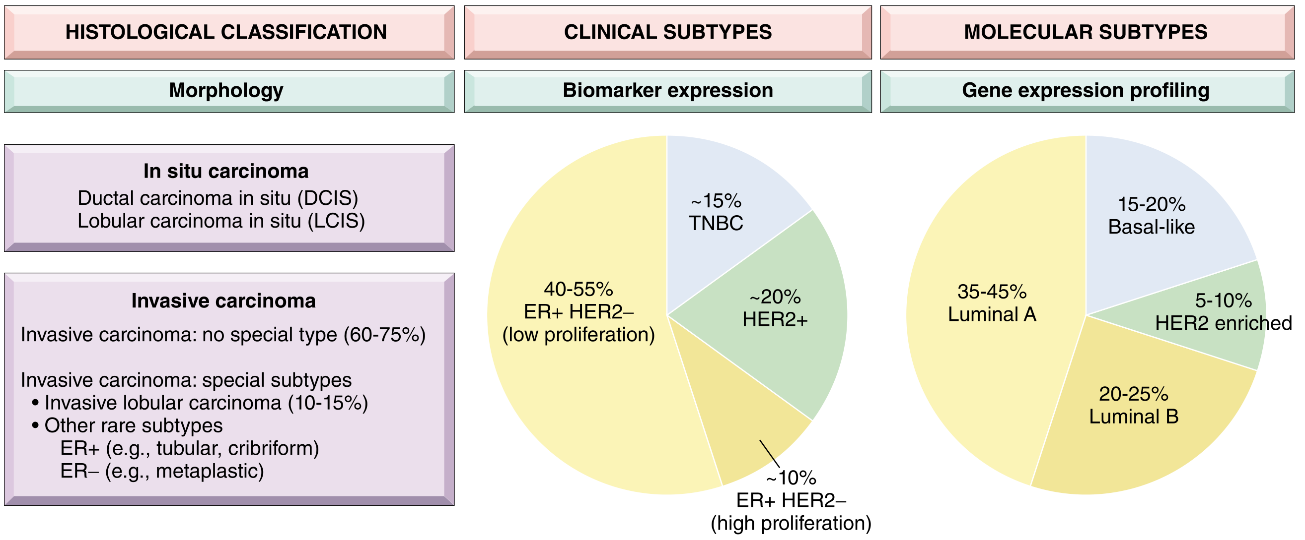
A. Histological Classification
I. Noninvasive (In Situ) Carcinomas (15-20%)
These are cancers confined within the basement membrane.
1. Ductal Carcinoma In Situ (DCIS)
Also called intraductal carcinoma. Subtypes include:
- Papillary and cribriform DCIS - generally lower-grade; slower progression to invasive cancer
- Solid and comedo DCIS - generally higher-grade; central necrosis produces calcifications visible on mammography as pleomorphic, linear microcalcifications
As cells within the duct undergo central necrosis, the debris calcifies and produces the characteristic segmental calcifications on mammography. If untreated, DCIS transforms into invasive cancer recapitulating the morphology of the in situ cells. - Sabiston Textbook of Surgery, p. 2313
2. Lobular Carcinoma In Situ (LCIS)
Neoplastic cells distend the acini but preserve the lobular architecture. Considered a risk marker rather than an obligate precursor. Cells are small with compact, bland nuclei.
II. Invasive Carcinomas (~80-85%)
1. Invasive Ductal Carcinoma - No Special Type (NST) - 50-80%
The most common breast cancer, also called scirrhous or simplex carcinoma. It presents as a solitary, firm mass with poorly defined margins, most often in peri- or postmenopausal women in their 5th-6th decade. Cut surface shows a central stellate configuration with chalky white or yellow streaks. Axillary lymph node metastases occur in up to 25% of screen-detected and 60% of symptomatic cases. About 75% express estrogen receptor. - Schwartz's Principles of Surgery, p. 592-593
2. Invasive Lobular Carcinoma - 5-15%
Second most common type. Histologically shows small cells with rounded nuclei arranged in a "single-file" (Indian file) pattern within a fibrous stroma. It is frequently multifocal, multicentric, and bilateral. Over 90% express estrogen receptor. Can be difficult to detect mammographically due to its insidious, diffuse growth pattern. Intracytoplasmic mucin may displace the nucleus to form a signet-ring cell appearance. - Schwartz's Principles of Surgery, p. 594
3. Medullary Carcinoma - ~4%
Characterized by three features: (a) dense lymphoreticular infiltrate of lymphocytes and plasma cells, (b) large pleomorphic nuclei with active mitosis, and (c) sheet-like growth with minimal ductal differentiation. It is soft and hemorrhagic grossly. A frequent phenotype of BRCA1-associated breast cancer. Fewer than 10% show hormone receptor expression. Despite its aggressive histology, prognosis is better than IDC-NST due to the immune response. Bilaterality in 20% of cases. - Schwartz's Principles of Surgery, p. 593
4. Mucinous (Colloid) Carcinoma - ~2%
Defined by extracellular pools of mucin surrounding aggregates of low-grade cancer cells. Presents in older women as a bulky, glistening, gelatinous tumor. Over 90% express hormone receptors. Lymph node metastases occur in 33% of cases. Has a relatively favorable prognosis - 5-year survival ~73%. - Schwartz's Principles of Surgery, p. 593
5. Tubular Carcinoma - ~2%
A well-differentiated special type with haphazardly arranged small tubular elements on low-power microscopy. Diagnosed in up to 20% of mammographically detected cancers. About 94% express estrogen receptor. Axillary metastases occur in only ~10% of cases. Long-term survival approaches 100%. - Schwartz's Principles of Surgery, p. 594
6. Papillary Carcinoma - ~2%
Defined by papillae with fibrovascular stalks and multilayered epithelium. Presents in the 7th decade. Usually small (rarely >3 cm). About 87% express estrogen receptor. Low frequency of axillary lymph node metastases; prognosis similar to tubular and mucinous carcinoma. - Schwartz's Principles of Surgery, p. 593
7. Paget's Disease of the Nipple
Presents as a chronic eczematous eruption of the nipple that may progress to an ulcerated, weeping lesion. Pathognomonic finding: large, pale, vacuolated Paget cells in the rete pegs. Almost always associated with extensive underlying DCIS and sometimes with invasive cancer. CEA immunostaining is positive (vs. S-100 in melanoma). - Schwartz's Principles of Surgery, p. 592
8. Rare/Special Types (<5%)
- Adenoid cystic carcinoma (~1%) - excellent prognosis; rarely metastasizes to nodes
- Metaplastic carcinoma (~1%) - ER/PR/HER2-negative; aggressive
- Inflammatory carcinoma - not a histologic type but a clinical diagnosis; diffuse erythema and skin edema (peau d'orange) involving >1/3 of breast, caused by dermal lymphatic invasion; highly aggressive
III. Mixed Connective and Epithelial Tumors (<5%)
| Type | Notes |
|---|---|
| Phyllodes tumor (<1%) | Can be benign or malignant; leaf-like growth pattern |
| Carcinosarcoma (<1%) | Mixed epithelial and mesenchymal malignancy |
| Angiosarcoma (<1%) | Rare; may occur post-radiation to breast |
- Sabiston Textbook of Surgery, Box 68.2
B. Clinical (Biomarker-Based) Classification
Based on immunohistochemical expression of ER, PR, and HER2:
| Subtype | ER/PR | HER2 | Prevalence | Notes |
|---|---|---|---|---|
| ER+/HER2- (low proliferation) | Positive | Negative | 40-55% | Best prognosis; responds to endocrine therapy |
| ER+/HER2- (high proliferation) | Positive | Negative | ~10% | Higher Ki-67; more aggressive |
| HER2+ | Variable | Positive | ~20% | Targeted with trastuzumab |
| Triple Negative (TNBC) | Negative | Negative | ~15% | ER-, PR-, HER2-; worst prognosis; chemotherapy only |
C. Molecular (Gene Expression-Based) Classification
Based on gene expression profiling (PAM50 and similar panels), breast cancers are grouped into intrinsic subtypes:
| Subtype | ER/PR | HER2 | Ki-67 | Prevalence | Prognosis |
|---|---|---|---|---|---|
| Luminal A | Positive | Negative | Low | 35-45% | Best; responds well to endocrine therapy |
| Luminal B | Positive | Negative | High | 20-25% | Intermediate; higher recurrence risk |
| HER2-enriched | Negative | Positive | High | 5-10% | Poor without anti-HER2 therapy |
| Basal-like (TNBC) | Negative | Negative | Usually high | 15-20% | Worst; associated with BRCA1 mutations |
| Claudin-low | Negative | Negative | Variable | <5% | Stem-cell like features; poor prognosis |
- Bailey and Love's Short Practice of Surgery, Table 58.4; Sabiston Textbook of Surgery, Molecular Markers section
Key clinical point: Luminal A cancers arise predominantly in postmenopausal women of European descent. Triple-negative and HER2+ cancers are proportionally more common in younger women and women of African descent, accounting for the observed difference in breast cancer mortality across ethnic groups. - Robbins Pathologic Basis of Disease, p. 4406-4408
Summary
| Classification Axis | Major Categories |
|---|---|
| Invasiveness | In situ (DCIS, LCIS) vs. Invasive |
| Histological type | IDC-NST (most common), ILC, medullary, mucinous, tubular, papillary, Paget's |
| Biomarker | ER+/HER2-, HER2+, TNBC |
| Molecular subtype | Luminal A, Luminal B, HER2-enriched, Basal-like, Claudin-low |
Understanding all three classification axes is necessary for prognosis and to guide treatment decisions - endocrine therapy for ER+ tumors, anti-HER2 agents (trastuzumab, pertuzumab) for HER2+ tumors, and chemotherapy as the primary systemic option for TNBC.
This is a shared conversation. Sign in to Orris to start your own chat.