
Inguinal hernia
Reading File
Now let me search the medical library for inguinal hernia content in parallel:
Finding Sources
Finding Sources
Searching PubMed
"inguinal hernia"[MeSH Terms] AND repair[MeSH Terms]
Excellent sources found. Let me read the most comprehensive sections in parallel:
Reading File
Reading File
Reading File
Now let me read the main Schwartz's section on adult inguinal hernia and Bailey & Love for more clinical detail:
Reading File
Reading File
Reading File
Now let me get the Bailey & Love clinical section and the S Das section on clinical features and treatment:
Reading File
Reading File
Let me read surgical treatment from Bailey & Love and S Das:
Reading File
Reading File
Let me get the surgical treatment section and Hesselbach's triangle details:
Reading File
Reading File
Now let me get the surgical repair section from Bailey & Love and fetch a key anatomical image:
Reading File
Loading Image
I now have comprehensive information to produce a detailed, well-structured answer. Let me compile it.
Inguinal Hernia
An inguinal hernia is a protrusion of abdominal contents through a defect in the inguinal region. It is the most common hernia in both sexes, with men affected about 10 times more often than women. - Bailey and Love's Short Practice of Surgery 28th Edition, p. 1087
Anatomy of the Inguinal Canal
The inguinal canal is an oblique passage through the lower abdominal wall, approximately 4 cm long:
- Anterior wall: external oblique aponeurosis
- Posterior wall: transversalis fascia
- Roof: conjoint tendon (fused transversus abdominis and internal oblique)
- Floor: inguinal (Poupart's) ligament
The deep (internal) inguinal ring is a defect in the transversalis fascia, located midway between the anterior superior iliac spine and the pubic tubercle, approximately 2-3 cm above the femoral artery pulse. The inferior epigastric vessels lie just medial to it.
The superficial (external) inguinal ring is an inverted-V-shaped defect in the external oblique aponeurosis, just above the pubic tubercle.
Contents of the canal in males: testicular artery, pampiniform venous plexus, vas deferens, lymphatics, cremasteric muscle, ilioinguinal nerve, genital branch of genitofemoral nerve, and iliohypogastric nerve. In females, the round ligament passes through.
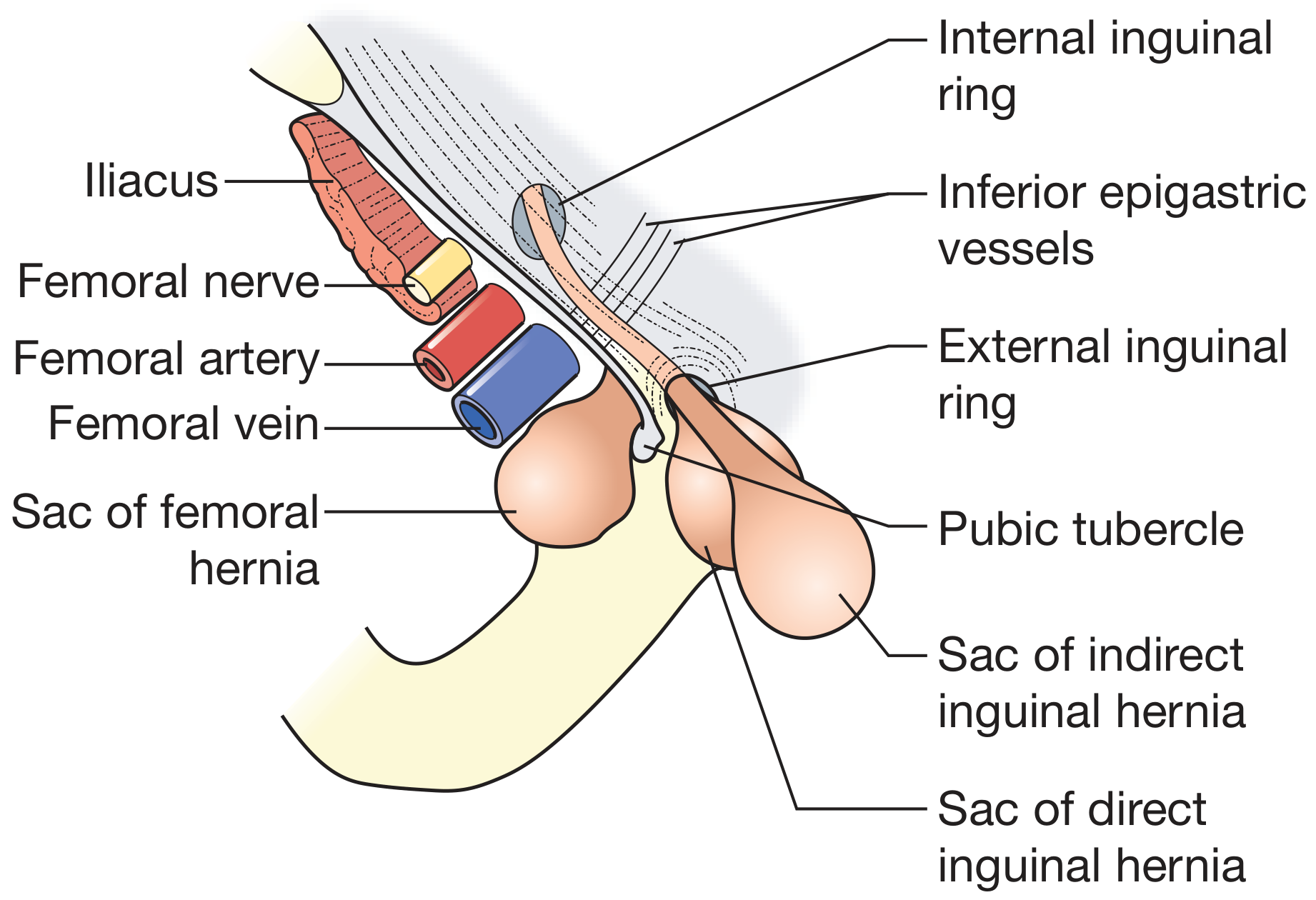
Figure: The close relationships of direct inguinal, indirect inguinal, and femoral hernias. Note the inferior epigastric vessels dividing the two types, and the separate femoral sac below the inguinal ligament. (Bailey and Love, 28th ed.)
Hesselbach's Triangle
The anatomical "floor" where direct hernias emerge. Boundaries:
- Medially: lateral border of rectus abdominis
- Laterally: inferior epigastric artery
- Inferiorly: inguinal ligament
This area is structurally weak because it consists only of transversalis fascia covered by external oblique aponeurosis - no muscular protection.
Classification
By Type
| Feature | Indirect (Oblique) | Direct (Medial) |
|---|---|---|
| Site of exit | Deep inguinal ring - lateral to inferior epigastric vessels | Hesselbach's triangle - medial to inferior epigastric vessels |
| Cause | Failure of obliteration of processus vaginalis (congenital) or acquired | Always acquired - muscle weakness |
| Frequency | >80% of inguinal hernias | ~20% |
| Age | Any age, including children | Predominantly >40 years |
| Descent to scrotum | Common (complete hernia) | Rare unless long-standing |
| Strangulation risk | Higher (narrow neck) | Lower (broad neck, wide opening) |
| Cough test | Controlled by pressure over deep ring | Appears medial to occluding finger |
S Das Manual on Clinical Surgery 13th ed., p. 608; Bailey and Love, p. 1088
By Extent (indirect hernias)
- Bubonocele - hernia does not exit the superficial ring
- Incomplete (funicular) - exits superficial ring but does not reach the scrotum
- Complete - descends to the bottom of the scrotum
By Clinical State
- Reducible - contents easily returned to abdomen
- Irreducible - contents cannot be returned (adhesions, large sac); no vascular compromise
- Obstructed/Incarcerated - irreducibility + intestinal obstruction; blood supply intact
- Strangulated - irreducibility + obstruction + loss of blood supply to contents (surgical emergency)
- Inflamed - rare; occurs when appendix, Meckel's diverticulum, or salpinx within sac becomes inflamed
Key distinction: A strangulated hernia is irreducible, has no cough impulse, and is extremely tense, tender, and often has overlying skin changes. An obstructed hernia shares irreducibility and obstruction but has no vascular compromise. - S Das, p. 609
Special Varieties
- Sliding hernia (hernia-en-glissade): Retroperitoneal bowel (caecum on right, sigmoid on left, or bladder) slides down forming the posterior wall of the sac. The bowel is not entirely within the sac. Common in older men, high risk of inadvertent bowel injury during repair.
- Pantaloon hernia: Coexisting direct and indirect hernia straddling the inferior epigastric vessels (like trouser legs).
- Richter's hernia: Only part of the bowel circumference is strangulated. Intestinal obstruction may be absent initially, making diagnosis deceptively easy to miss.
- Littre's hernia: Contains a Meckel's diverticulum.
- Maydl's hernia (W hernia / retrograde strangulation): Two loops in the sac with a third intervening loop in the abdomen. The loop outside the sac may appear viable while the loop inside the abdomen is already strangulated.
Embryology (Indirect/Congenital)
The processus vaginalis is a peritoneal tube that accompanies testicular descent. It normally obliterates before birth, leaving only the tunica vaginalis. Failure of obliteration leaves a patent communication = congenital indirect inguinal hernia. This is why premature infants have a very high incidence. All hernias in children are functionally of the indirect type. - Schwartz's Principles of Surgery 11th ed., p. 1771
Clinical Features
Symptoms:
- Groin bulge - appears on standing or coughing, reduces on lying down
- Dragging discomfort or pain, worse with activity
- Scrotal swelling if complete
Examination:
- Reduce the hernia (if not spontaneously reduced supine)
- Locate the deep ring (midpoint of inguinal ligament, 2-3 cm above femoral pulse)
- Apply pressure over deep ring and ask patient to cough:
- Hernia controlled = indirect
- Hernia appears medial to pressure point = direct
- Invagination test: Finger invaginated into the scrotum up the inguinal canal; feel the cough impulse. Impulse felt at the finger tip (directed obliquely upwards/laterally) = indirect. Impulse straight back against the pulp = direct.
- Transillumination: Negative (unlike hydrocele, which transilluminates)
- Cannot "get above" a scrotal hernia (differentiates from hydrocele)
A patient with a unilateral hernia has a 50% lifetime risk of a contralateral hernia. - Bailey and Love, p. 1089
Investigations
Usually clinical. Imaging is reserved for:
- Occult/uncertain diagnosis - ultrasound (first-line), MRI
- Irreducible or complicated hernia - CT abdomen
- Note: hernias may reduce spontaneously in the supine position during imaging, giving false negatives
Management
Conservative
- Watchful waiting is acceptable for asymptomatic or minimally symptomatic direct hernias in elderly patients who decline surgery.
- Trusses are not recommended - they do not prevent complications and may cause adhesions.
- Patients should be counselled to seek urgent review if the hernia becomes painful, irreducible, or tense.
Bailey and Love, p. 1089
Surgical
In children - Herniotomy: Simple ligation and excision of the sac at the internal ring. No floor repair needed as the inguinal floor is normal.
In adults - Hernioplasty (floor repair required):
Open Tension-Free Mesh Repair (Lichtenstein)
- Gold standard open technique
- Polypropylene mesh placed over the posterior wall of the inguinal canal, sutured to inguinal ligament inferiorly and conjoint tendon superiorly
- Very low recurrence rate (~1%), performed under local anaesthesia
- Named after Irving Lichtenstein (1989)
Open Suture Repairs (historical/still used)
- Bassini repair (1890): Conjoint tendon sutured to inguinal ligament behind the cord
- Shouldice repair: Multilayer running suture repair of the transversalis fascia; very low recurrence in dedicated centers
- Desarda repair: Physiological repair using a strip of external oblique aponeurosis
Laparoscopic/Minimally Invasive Repair
Two main techniques:
- TEP (Totally Extraperitoneal): Mesh placed in preperitoneal space without entering the peritoneal cavity; preferred approach in most laparoscopic centers
- TAPP (Transabdominal Preperitoneal): Peritoneal cavity entered, mesh placed in the preperitoneal space from inside
Indications for minimally invasive approach (strongly supported by evidence):
- Bilateral inguinal hernias (single procedure, two sides)
- Recurrent inguinal hernia after prior open repair
- Athletes/younger patients (less groin pain, faster return to activity)
Current Surgical Therapy 14th ed.; Bailey and Love, p. 1090
Robotic Repair
Increasing adoption; most robotic inguinal repairs were initially performed by urologists during prostatectomy, then adopted by general surgeons. Outcomes are comparable to laparoscopic. - Sabiston Textbook of Surgery
Complications
Of the Hernia
- Incarceration/strangulation (bowel or omentum)
- Intestinal obstruction
- Bowel gangrene and perforation
Of Surgery
- Recurrence (1-3% with mesh; higher with suture-only repair)
- Chronic groin pain (CPIIP - Chronic Post-Inguinal Hernioplasty Pain) - most significant long-term complication; up to 10-15% of patients
- Nerve injury: ilioinguinal, iliohypogastric, genitofemoral nerves - causes numbness or neuralgia
- Vas deferens injury - can cause infertility
- Testicular atrophy - from vascular injury to testicular vessels
- Mesh infection / seroma
- Hematoma
Pediatric Notes
- Male:female ratio 10:1; right side more common (due to later descent of right testis)
- Infants at highest risk for incarceration (narrow ring)
- Presentation: groin bulge noticed during diaper changes; firm irreducible bulge = incarceration
- Incarcerated hernia: attempt gentle reduction (may require sedation); schedule repair within 24 hours
- Strangulation = surgical emergency; may require bowel resection - Schwartz's, p. 1771
Differentiating Inguinal vs Femoral Hernia
| Feature | Inguinal | Femoral |
|---|---|---|
| Position relative to pubic tubercle | Medial and above inguinal ligament | Lateral and below inguinal ligament |
| Sex | Much more common in males | Relatively more common in females |
| Strangulation risk | Lower (indirect), very low (direct) | High (rigid femoral canal) |
| Cough impulse | In inguinal canal | At saphenous opening (~4 cm below/lateral to pubic tubercle) |
Even in women, the most common groin hernia is still inguinal, not femoral. - S Das, p. 611
Sources:
- Bailey and Love's Short Practice of Surgery, 28th ed., pp. 1087-1090
- S Das Manual on Clinical Surgery, 13th ed., pp. 608-611
- Schwartz's Principles of Surgery, 11th ed., pp. 1771-1772
- Gray's Anatomy for Students
- Current Surgical Therapy, 14th ed.
- Sabiston Textbook of Surgery
This is a shared conversation. Sign in to Orris to start your own chat.