

Explain
angiogenesis steps new blood vessel formation wound healing granulation tissue
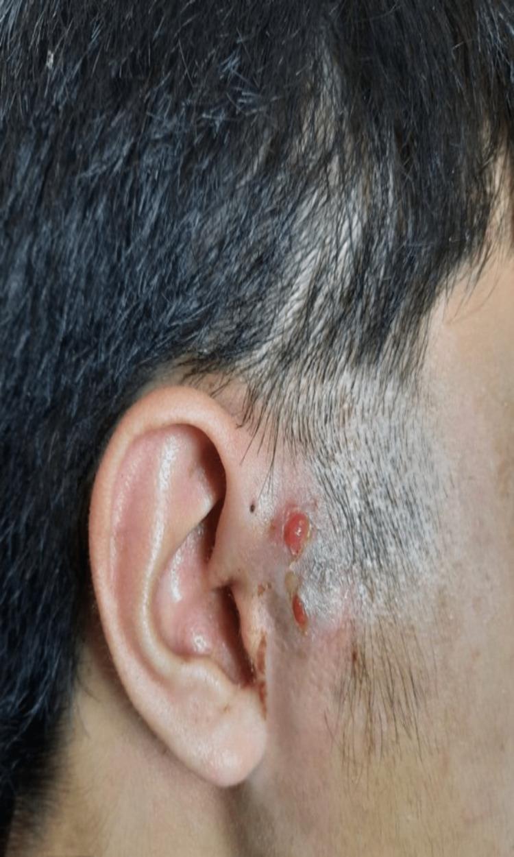
This clinical photograph shows a close-up of the right preauricular region and the adjacent pinna. The image displays a healing wound characterized by the formation of healthy, beefy red granulation tissue. The tissue appears raised, moist, and slightly lobulated (cobblestone appearance), indicative of active angiogenesis and the proliferative phase of wound healing. The granulation tissue is located near a preauricular sinus opening, which is visible as a small punctum. The surrounding perilesional skin exhibits signs of resolving inflammation, including mild erythema and localized hyperpigmentation. There is evidence of previous surgical intervention or drainage in the area, with some crusting and scabbing noted along the wound margins. The anatomical context and clinical presentation are consistent with a postoperative review of a preauricular sinus abscess, potentially secondary to tuberculous infection as indicated by the context. This image serves as a clinical example of secondary intention wound healing and common complications associated with congenital preauricular sinuses.
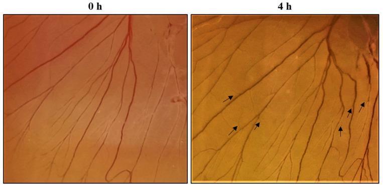
This clinical photograph displays a side-by-side comparison of an in vivo Chorioallantoic Membrane (CAM) assay used to evaluate the angiogenic potential of a SPEM (sericin and decellularized extracellular matrix) scaffold. The left image (0 h) represents the baseline vascular network, showing established reddish-brown blood vessels branching across the transparent membrane. The right image (4 h) illustrates the progression of angiogenesis after a four-hour treatment period. In this frame, black arrows highlight the rapid formation of new, finer capillary sprouts and secondary branching from the pre-existing vessels. The comparison demonstrates the scaffold's ability to stimulate neovascularization, a critical component of the proliferative phase of wound healing. This experimental model is highly relevant to regenerative medicine and the development of bioactive wound dressings that utilize growth factors to promote tissue regeneration and nutrient supply through enhanced blood vessel formation.
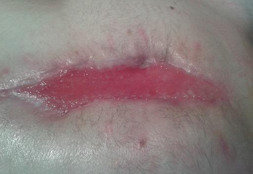
This clinical photograph displays a healing surgical wound on the anterior chest, following an omentoplasty (epiplooplasty) for the management of post-sternotomy mediastinitis. The wound bed is characterized by a central, horizontal, fusiform area of beefy-red granulation tissue, indicating active angiogenesis and secondary intention healing. The surface of the granulation tissue appears moist and glistening, with no evidence of purulent exudate or slough. The wound margins show signs of epithelialization, visible as a pale, whitish-pink rim of new tissue migrating inward. The surrounding periwound skin exhibits mild, diffuse erythema consistent with normal inflammatory response in the proliferative phase of healing. Fine terminal hair is visible on the surrounding thoracic skin. This image serves as a clinical example of using an omental flap to provide vascularized coverage for a complex sternal wound, facilitating eventual complete cicatrization.
Context: The Proliferative Phase of Wound Healing
(b) Fibroblasts Lay a Bed of Collagen
(c) Angiogenesis - The 7 Steps
Step 1 - Vasodilation (triggered by Nitric Oxide / NO)
- Injury signals cause local endothelial cells to produce nitric oxide (NO).
- NO is a potent vasodilator - it relaxes the smooth muscle of the vessel wall, widening the lumen.
- This increases blood flow to the area and sets the stage for the next steps.
Step 2 - Increased Vascular Permeability (induced by VEGF)
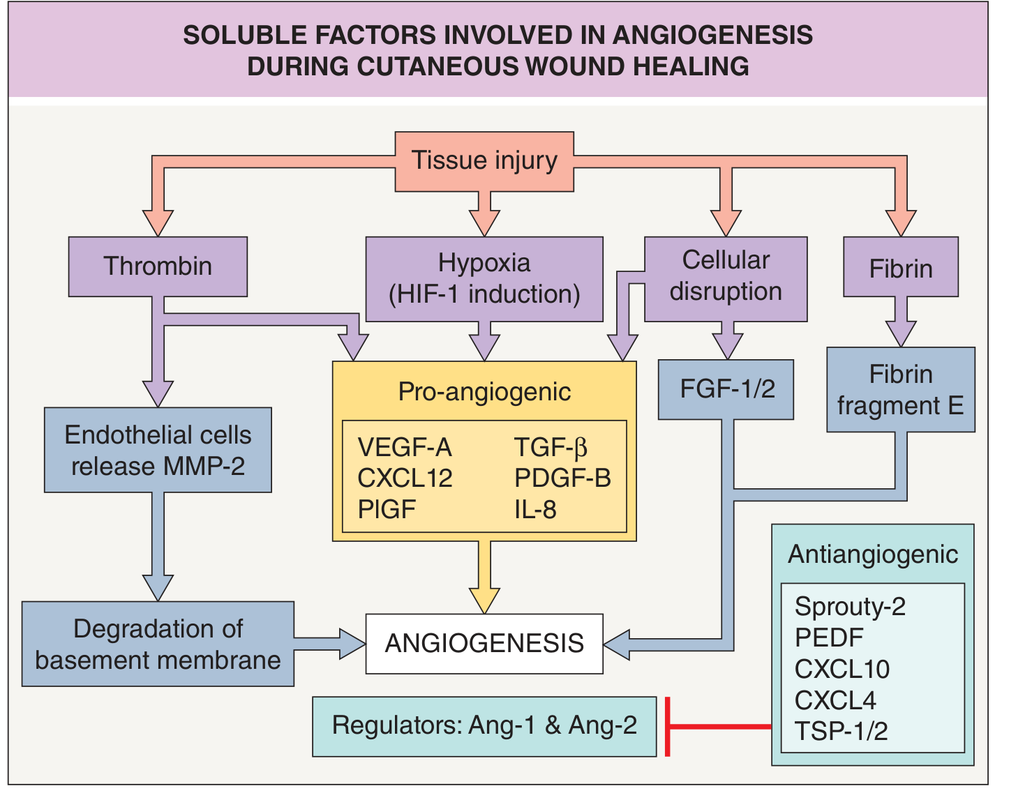
- VEGF (Vascular Endothelial Growth Factor) is the master regulator of angiogenesis. It is secreted primarily by hypoxic cells, macrophages, and platelets.
- VEGF makes the vessel wall "leaky," allowing proteins and fluid to escape into the surrounding tissue, creating a provisional extracellular matrix (ECM) scaffold for new vessel growth.
Step 3 - Separation of Pericytes from the Abluminal Surface
- Pericytes are the supportive cells that wrap around the outside (abluminal surface) of blood vessels, keeping them stable and quiescent.
- The diagram shows this clearly: the top image shows a quiescent vessel with pericytes (purple cells) tightly attached; the middle image shows them beginning to detach.
- Their detachment "destabilizes" the vessel, allowing it to become responsive to growth signals. This step is regulated by Ang-2 (angiopoietin-2), which loosens pericyte attachment.
Step 4 - Breakdown of the Basement Membrane
- The basement membrane is a thin, dense layer of ECM that surrounds the vessel and acts as a barrier.
- Enzymes called matrix metalloproteinases (MMPs) - especially MMP-2 (gelatinase A) - are released by endothelial cells in response to thrombin, and they digest this membrane.
- The bottom image in the diagram shows the basement membrane disrupted, creating a gap through which a vessel sprout can emerge.
Step 5 - Migration of Endothelial Cells toward the Injury
- The endothelial cells at the tip of the new sprout (called "tip cells") extend filopodia (finger-like projections) and migrate directionally toward the area of tissue injury, guided by the VEGF gradient (highest concentration near the hypoxic wound center).
Step 6 - Proliferation of Endothelial Cells (just behind the leading tip)
- While tip cells lead the way, the endothelial cells immediately behind them (called "stalk cells") proliferate rapidly to elongate the new sprout.
- This division happens just behind the leading front - think of tip cells as pathfinders and stalk cells as builders extending the tube.
Step 7 - Recruitment of Periendothelial Cells to Form a Mature Vessel
- Once the sprout reaches its target and connects with another sprout (anastomosis), pericytes and smooth muscle cells are recruited back to coat the new vessel.
- This is driven by PDGF-B (platelet-derived growth factor B) and Ang-1 (angiopoietin-1).
- The new vessel matures, stabilizes, and becomes functional.
End Product: Granulation Tissue
- Newly formed thin-walled capillaries (hence the red color)
- Proliferating fibroblasts
- Loose collagen (type III)
- Inflammatory cells (macrophages predominate)
Quick Summary Table
| Step | Event | Key Mediator |
|---|---|---|
| 1 | Vasodilation | NO |
| 2 | Increased permeability | VEGF |
| 3 | Pericyte detachment | Ang-2 |
| 4 | Basement membrane breakdown | MMP-2 |
| 5 | Endothelial cell migration | VEGF gradient |
| 6 | Endothelial cell proliferation | VEGF, FGF |
| 7 | Periendothelial cell recruitment | PDGF-B, Ang-1 |
| Result | Granulation tissue | - |