
Swine flu, clinical signs and symptoms, how to diagnose, management
H1N1 influenza chest x-ray pneumonia
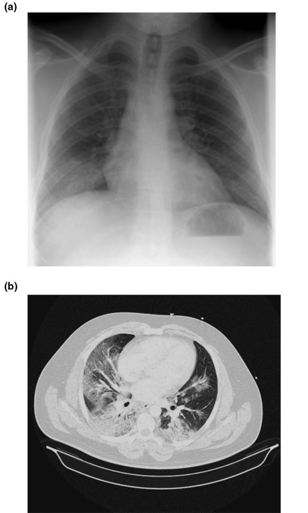
Educational comparison of radiological findings in viral pneumonitis (H1N1). Figure (a) is a posterior-anterior chest radiograph demonstrating bilateral, patchy alveolar opacities. These opacities are predominantly distributed in the lower lung zones (basal regions), with relative sparing of the upper lobes. Figure (b) is an axial thoracic computed tomography (CT) scan from the same clinical context. The CT image reveals diffuse, bilateral ground-glass opacities and areas of airspace consolidation throughout the lung parenchyma. The findings show a multilobar distribution, characteristic of severe viral pneumonia and acute lung injury. These images are intended for medical trainees to recognize the typical imaging patterns of viral-induced pulmonary infiltration, highlighting the transition from nonspecific findings on X-ray to more detailed parenchyma involvement visible on CT. Key educational concepts include pulmonary consolidation, ground-glass attenuation, and the radiological presentation of pandemic influenza A (H1N1)v.
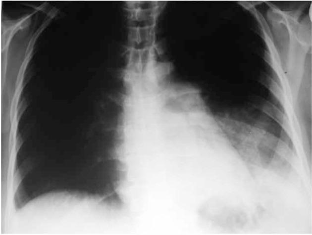
This diagnostic image is a posterior-anterior (PA) chest X-ray of a 30-year-old female patient diagnosed with H1N1 influenza. The primary abnormality is a significant homogenous opacity in the left lower and middle lung zones, characteristic of a pleural effusion. This finding is evidenced by the blunting of the left costophrenic angle and obscuration of the left hemidiaphragm silhouette. Additionally, there is prominence of the pulmonary segment along the left cardiac border, which may reflect vascular congestion or anatomical variation. The right lung field remains relatively clear with no definitive evidence of focal consolidation or infiltrates. The mediastinal contour appears stable, and the trachea is midline. This case illustrates a typical pulmonary complication of viral pneumonia, specifically H1N1, where pleural involvement can occur alongside parenchymal infection. It serves as an educational example for identifying secondary radiographic signs of respiratory viruses and differentiating pleural fluid from pulmonary consolidation.
"Influenza A" AND "H1N1" AND treatment
swine flu H1N1 symptoms diagnosis treatment CDC guidelines 2025
Swine Flu (Pandemic Influenza A H1N1)
Clinical Signs and Symptoms
- Influenza-like illness (ILI): fever, cough, sore throat, rhinorrhoea (runny/stuffy nose), headache, muscle pain (myalgia), and malaise
- No shortness of breath or dyspnoea
- Fatigue/weakness
- Gastrointestinal symptoms (diarrhoea and/or vomiting) can occur, especially in children, though without dehydration
- Shortness of breath, dyspnoea, tachypnoea, hypoxia, or radiological signs of pneumonia
- CNS involvement: encephalopathy, encephalitis, altered mental status, confusion, persistent seizures, severe weakness or paralysis
- Severe dehydration (decreased activity, dizziness, reduced urine output, lethargy)
- Secondary complications: renal failure, multi-organ failure, septic shock, rhabdomyolysis, myocarditis
- Exacerbation of underlying chronic disease (asthma, COPD, diabetes, cardiac/hepatic/renal disease)
- Persistent high fever beyond 3 days suggesting sustained viral replication or secondary bacterial infection
Diagnosis
-
Clinical/epidemiological grounds - When influenza is known to be circulating in a community, uncomplicated cases can be diagnosed clinically without lab confirmation.
-
Case definitions (per Park's textbook):
- Suspected: acute febrile respiratory illness with recent contact/travel/community exposure link
- Probable: positive influenza A but unsubtypable, or positive rapid antigen/immunofluorescence test
- Confirmed: laboratory-confirmed by RT-PCR, viral culture, or a four-fold rise in specific neutralizing antibodies
-
Laboratory tests (Sherris & Ryan's Medical Microbiology):
- RT-PCR (real-time reverse transcriptase PCR) - most sensitive and specific, provides subtype-specific detection (e.g., H1N1pdm09 vs H3N2); takes 1-8 hours
- Rapid antigen immunoassay - fast (15-20 min) but less sensitive
- Immunofluorescence assay - 1-4 hours
- Viral culture - takes 3-10 days, useful for confirmation/research
- Serology - fourfold or greater rise in HI antibody titers between acute and convalescent samples (10-14 days apart); useful epidemiologically, not for acute diagnosis
-
Specimen collection: Nasopharyngeal swabs/aspirates preferred (higher yield than throat swabs); for patients with lower respiratory symptoms, tracheal/bronchial aspirates give higher diagnostic yield.
Management
- Rest, adequate hydration
- Antipyretics/analgesics (paracetamol/acetaminophen, NSAIDs) for fever and myalgia
- Cough suppressants as needed
- IV fluids and other supportive measures for severe dehydration
- Oseltamivir (Tamiflu) or zanamivir (Relenza) - neuraminidase inhibitors, the H1N1 virus is generally sensitive to these
- Most effective when started early, ideally within the first 48 hours of symptom onset
- Recommended for confirmed, probable, or suspected cases in high-risk groups (including pregnant women, where treatment should not be delayed pending test results and is not contraindicated) - Textbook of Family Medicine
- A 2024 Lancet systematic review/network meta-analysis of antivirals for severe influenza (PMID: 39181595) supports oseltamivir's role, though evidence on mortality benefit in severe hospitalized cases remains an active area of study - worth checking current guidance if managing severe/hospitalized cases.
- Standard plus droplet precautions: hand hygiene, cough/sneeze etiquette, face mask if symptomatic and in public
- N95/FFP2 respirator, eye protection, gown, gloves for aerosol-generating procedures (e.g., bronchoscopy), performed in well-ventilated rooms
- Isolation for hospitalized patients: continue for 7 days after illness onset or 24 hours after fever/respiratory symptoms resolve, whichever is longer
- Annual influenza vaccination (inactivated, live-attenuated, or recombinant vaccines), recommended for all persons 9 years and older, with priority for high-risk groups and pregnant women (inactivated vaccine only; nasal live vaccine is contraindicated in pregnancy)