
Lifecycle and structure of bacteriophage
Bacteriophage: Structure and Lifecycle
1. Overview
- Goodman & Gilman's The Pharmacological Basis of Therapeutics, p. 1194
- Medical Microbiology 9e, p. 535
2. Structure
| Component | Description |
|---|---|
| Head (Capsid) | Icosahedral protein shell that encapsidates the phage genome (usually dsDNA) |
| Genome | Usually double-stranded DNA; some phages carry ssRNA or ssDNA |
| Tail | Hollow tube used to inject the genome into the host cell; acts like a syringe |
| Tail fibers | Long protein filaments at the base of the tail; mediate specific binding to bacterial surface receptors |
| Baseplate | The distal end of the tail; connects tail fibers and initiates contraction for DNA injection |
- Sherris & Ryan's Medical Microbiology, 8th Ed., p. 208
- Goodman & Gilman's, p. 1194
3. The Two Lifestyles: Lytic vs. Lysogenic
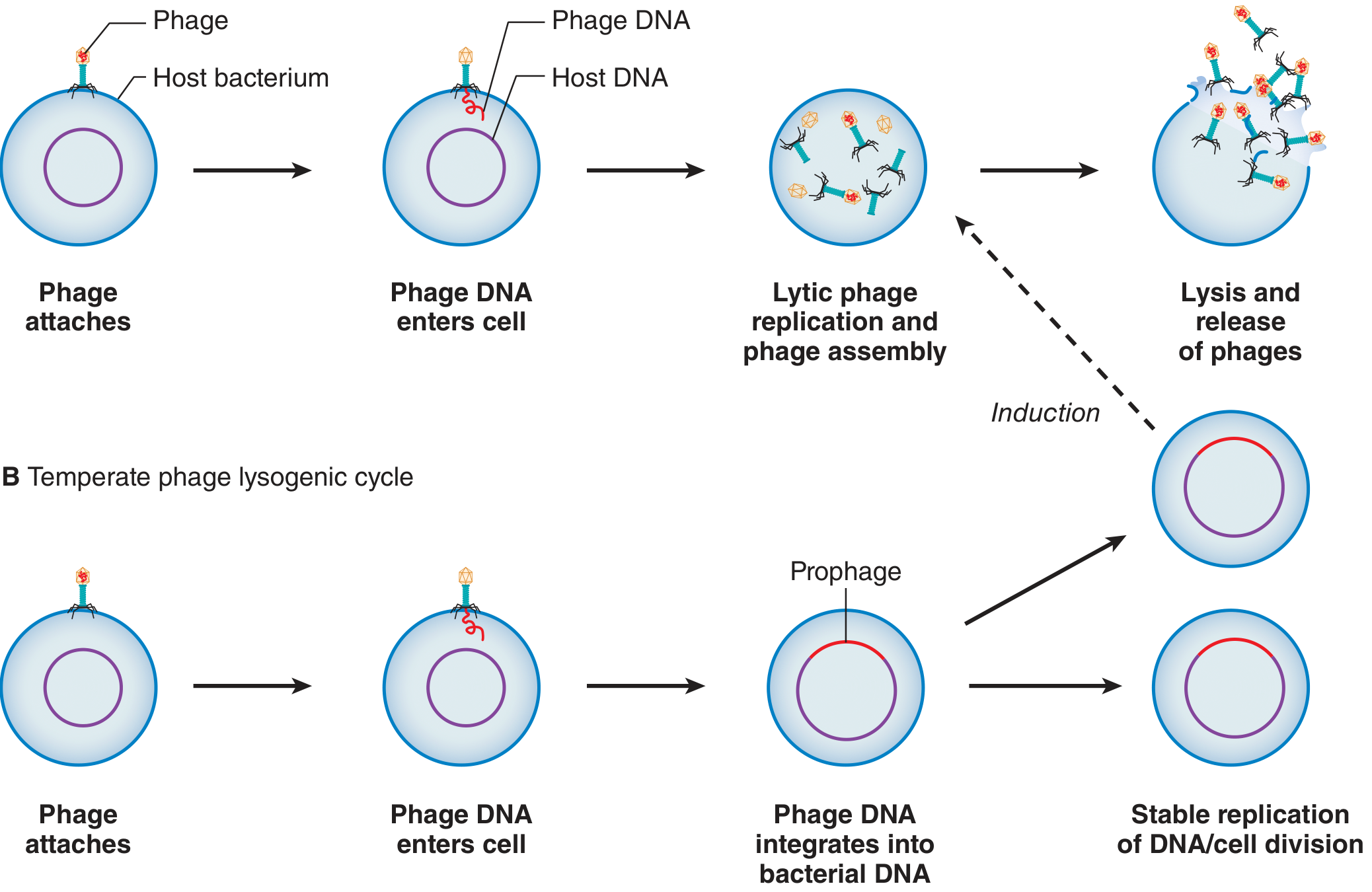
A. Lytic Cycle (Virulent Phage)
-
Adsorption (Attachment) - Tail fibers bind to specific receptors on the bacterial cell wall. This interaction is highly specific and determines host range.
-
Penetration (Injection) - The tail contracts and injects the phage DNA through the hollow tail structure into the bacterial cytoplasm. The capsid remains outside. This is the "syringe" mechanism.
-
Eclipse Phase - Infectious phage particles transiently disappear. The phage genome takes over the host's replicative machinery. Host genome is inactivated ("hijacked"). Phage genes are transcribed and translated.
-
Biosynthesis - New phage genomes are replicated, and structural proteins (capsid, tail components) are synthesized separately.
-
Assembly (Maturation) - New phage genomes are packaged into new capsids and assembled into complete virions.
-
Lysis and Release - Phage enzymes lyse the bacterial cell wall, releasing a burst of ~100 new phage progeny that can infect further bacteria.
- Goodman & Gilman's, p. 1194-1195
- Sherris & Ryan's, p. 207-208
B. Lysogenic Cycle (Temperate Phage)
-
Attachment + DNA Injection - Same initial steps as the lytic cycle.
-
Circularization - The injected linear phage DNA circularizes within the host cell.
-
Integration (Lysogeny) - Instead of immediate replication, the phage DNA integrates into the host chromosome as a prophage. The bacterium is now called a lysogen.
-
Stable Replication - The prophage is replicated passively as part of the bacterial chromosome each time the bacterium divides - passed to all daughter cells silently.
-
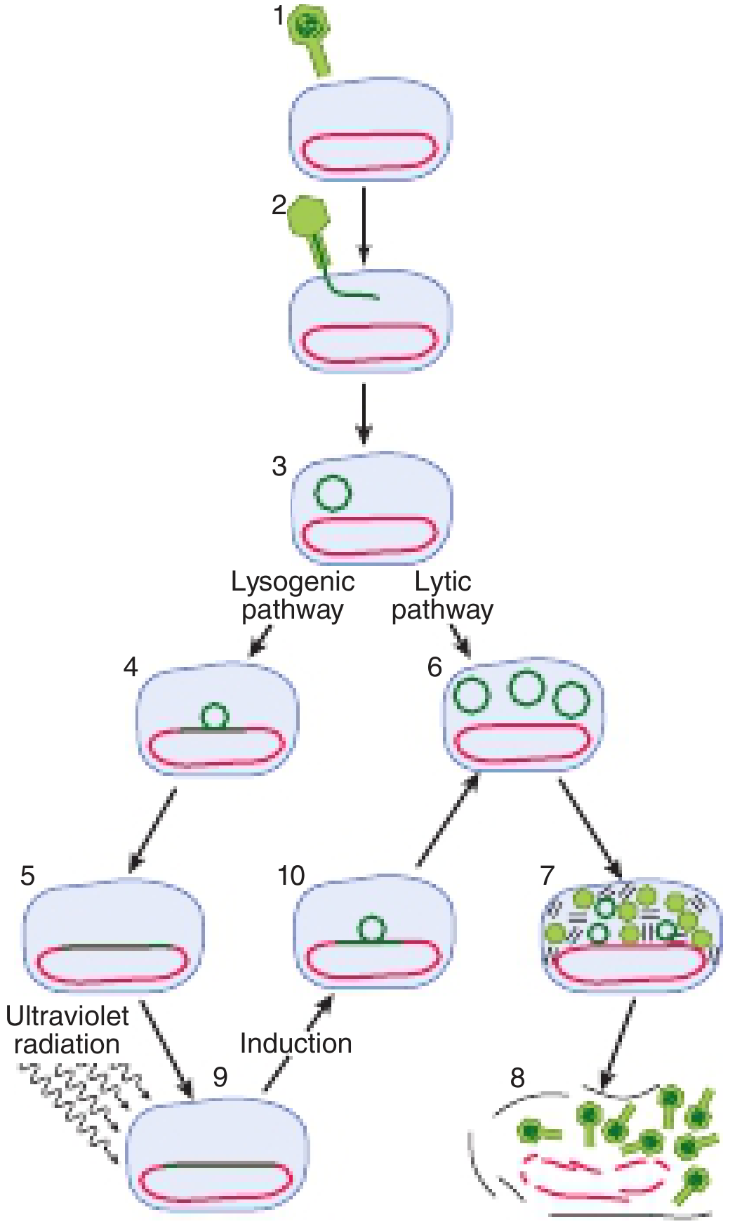
Induction (Excision) - When the lysogen encounters DNA-damaging stimuli (e.g., UV radiation, chemical mutagens), the SOS response is triggered. The dormant prophage becomes induced, excises from the host chromosome, and enters the lytic cycle.
4. The Lambda (λ) Genetic Switch
- Lysogenic state: The cI repressor gene is active. It represses lytic genes (including the cro gene), keeping the phage dormant.
- Lytic state: The cro gene is active. It represses the cI repressor, committing the phage to lysis.
- Poor growth conditions (nutrient stress) favor lysogeny; good growth conditions favor the lytic pathway.
- This switch is analogous to regulatory switches seen in eukaryotic viruses like HIV and herpes simplex virus.
- Harper's Illustrated Biochemistry, 32nd Ed., p. 437
5. Medical Significance of Lysogeny
- Toxin genes carried by prophages: Lysogenic bacteriophages can encode toxins. For example:
- Corynephage beta carries the gene for diphtheria toxin - a bacterium is only toxigenic if infected by this phage
- Other phage-encoded toxins include cholera toxin (V. cholerae) and certain Shiga toxins
- Horizontal gene transfer (Transduction): Phages can accidentally package bacterial DNA and transfer it between bacteria, spreading antibiotic resistance genes and virulence factors
- Medical Microbiology 9e, p. 538
6. Phage Therapy (Clinical Relevance)
- Goodman & Gilman's, p. 1194
Summary Table
| Feature | Lytic Phage | Temperate Phage |
|---|---|---|
| Outcome of infection | Always lyses the host | Can lyse OR integrate |
| Prophage formation | No | Yes |
| Host cell survival | No | Yes (until induction) |
| Induction trigger | N/A | UV, DNA damage (SOS) |
| Clinical example | T4 phage | Lambda phage, corynephage beta |
| Toxin carriage | Rare | Yes (diphtheria, cholera) |
Bacteriophage handmade diagram
A handmade hand-drawn sketch style diagram of a bacteriophage structure, drawn with pencil or pen on white paper look. Label all parts clearly with handwritten-style annotations and arrows: Head (icosahedral capsid containing DNA), Collar, Tail sheath, Tail tube (core), Baseplate, Tail fibers, and DNA inside the head. Show the classic T4 bacteriophage shape with a hexagonal head and long contractile tail. Include a small inset showing the phage injecting DNA into a bacterial cell wall. Use a clean, educational biology textbook sketch aesthetic with neat hand-lettered labels.
404 Not Found. {'message': '{\n "error": {\n "code": 404,\n "message": "Publisher model `projects/engaged-beaker-474810-u2/locations/global/publishers/google/models/gemini-3-pro-image-preview` was not found or your project does not have access to it. Ensure you are using a valid model name and that the model is available in the specified region. For more information, see: https://docs.cloud.google.com/gemini-enterprise-agent-platform/resources/locations.",\n "status": "NOT_FOUND"\n }\n}\n', 'status': 'Not Found'}
We encountered an error while processing your request. Please try again or reach out to us for help.
Handmade diagram of bacteriophage
A handmade pencil sketch diagram of a bacteriophage (T4 phage) on white paper, drawn in a biology student notebook style. The diagram shows the classic bacteriophage shape with a large hexagonal head containing coiled DNA strands inside, a collar below the head, a long contractile tail sheath with crosshatched lines, a central tail tube, a hexagonal baseplate at the bottom, and six long spidery tail fibers extending outward from the baseplate. All parts are labeled with neat handwritten arrows pointing to: Head, DNA, Collar, Tail sheath, Tail tube, Baseplate, Tail fibers. The sketch uses pencil shading, looks like a hand-drawn biology diagram, slightly imperfect lines giving it an authentic hand-drawn feel.
404 Not Found. {'message': '{\n "error": {\n "code": 404,\n "message": "Publisher model `projects/engaged-beaker-474810-u2/locations/global/publishers/google/models/gemini-3-pro-image-preview` was not found or your project does not have access to it. Ensure you are using a valid model name and that the model is available in the specified region. For more information, see: https://docs.cloud.google.com/gemini-enterprise-agent-platform/resources/locations.",\n "status": "NOT_FOUND"\n }\n}\n', 'status': 'Not Found'}
We encountered an error while processing your request. Please try again or reach out to us for help.