
Ostiod osteoma
osteoid osteoma X-ray nidus sclerosis
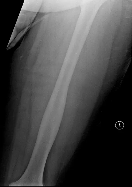
This is a musculoskeletalPlain radiograph (AP view) of the left femur showing cortical thickening and sclerosis within the mid-to-distal femoral cortex. Radiographs alone do not reveal a discrete nidus, which is common for osteoid osteoma on X-ray. The accompanying CT (not shown in this image) demonstrates a small radiolucent nidus within the thickened cortex, with no periosteal reaction or soft-tissue mass, and no aggressive features. The radiographic pattern of focal cortical sclerosis with a nidus on CT is classic for cortical osteoid osteoma, a benign osteogenic tumor that typically presents with localized nighttime pain relieved by NSAIDs in young to middle-aged adults. Differential considerations include osteoblastoma (larger nidus, possible aggression), stress fracture (cortical lucency with sclerosis), and Brodie abscess (infectious nidus-like lesion). Clinically, correlation with pain characteristics and CT nidus confirmation enhances diagnostic accuracy and guides therapy. This image is valuable for radiology education, differential diagnosis practice, and pre-interventional planning (nidus ablation or surgical excision), illustrating imaging-histology correlation and the importance of CT in identifying the nidus when radiographs are inconclusive.
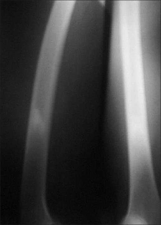
This diagnostic image is a plain radiograph (X-ray) of the right thigh, specifically focusing on the lower shaft of the femur. The radiograph demonstrates a focal area of irregular-shaped, predominantly endosteal sclerosis. The sclerosis is characterized by increased radiopacity (whiteness) within the medullary space of the bone. Notably, there is an absence of an identifiable radiolucent nidus, and there is no visible overlying periosteal reaction or cortical thickening. The surrounding soft tissues appear unremarkable with no signs of inflammation or masses. This visual presentation is typical of certain benign bone lesions, such as an osteoid osteoma where the nidus is obscured by dense reactive sclerosis, though differential diagnoses could include chronic osteomyelitis or a stress fracture. The educational focus is on identifying patterns of bone sclerosis and the importance of further imaging (like SPECT or CT) when a primary lesion is radiographically occult.
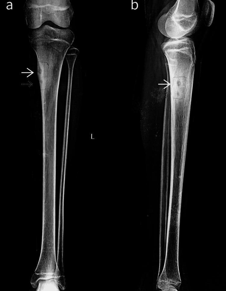
Anteroposterior (a) and lateral (b) plain X-ray radiographs of a left lower leg focusing on the knee and proximal tibial shaft. The diagnostic images demonstrate a multicentric bone lesion located within the proximal meta-diaphyseal region of the medial tibial cortex. Two distinct lytic nidi (indicated by white and dotted arrows) are visible, characterized by central radiolucency. These lesions are surrounded by significant reactive sclerosis, appearing as dense, opaque bone, along with associated cortical thickening (periosteal reaction). The anatomical alignment of the tibia and fibula remains normal, with the knee joint space preserved. These findings are highly characteristic of multicentric osteoid osteoma. This visual material is used in medical education to distinguish benign osteoid lesions and to illustrate the classic presentation of a nidus with surrounding sclerosis on conventional radiography.
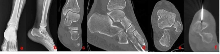
This multi-panel diagnostic figure displays the orthopedic imaging of a 24-year-old male with an osteoid osteoma in the talus. Panels A and B are anteroposterior (AP) and lateral X-ray views of the ankle and foot, showing general bony anatomy including the distal tibia, fibula, talus, and calcaneus. Panels C, D, and E provide multi-planar CT reconstructions in coronal, sagittal, and axial planes, respectively. These views reveal a characteristic small, well-defined radiolucent nidus within the talar bone, surrounded by reactive cortical thickening and sclerosis typical of osteoid osteoma. Panel F is a procedural axial CT scan demonstrating the intraoperative localization and placement of a radiofrequency ablation (RFA) needle directly into the lesion. The series illustrates the diagnostic pathway from conventional radiography to advanced cross-sectional imaging and subsequent minimally invasive therapeutic intervention in a clinical musculoskeletal oncology context.
"osteoid osteoma"[MeSH Terms]