
Lateral venticle
Reading File
Searching sources...
This is a broad anatomical topic - I'll search headings and paragraphs in parallel.
Finding Sources
Finding Sources
Good - the best sources are from Neuroanatomy through Clinical Cases and Gray's Anatomy for Students. Let me read those dedicated sections now.
Finding Sources
Reading File
Let me search specifically for lateral ventricle anatomy in neuroanatomy textbooks and Gray's Anatomy.
Finding Sources
Finding Sources
Excellent! Found dedicated sections. Let me read from Gray's Anatomy, Neuroscience textbook, and Neuroanatomy textbook simultaneously.
Reading File
Reading File
Reading File
Now let me get more detailed content on the walls/relations of the lateral ventricle from the Kaplan Sadock psychiatry textbook, and also retrieve key images.
Reading File
Finding Sources
Loading Image
That image shows the insula, not the lateral ventricle. Let me fetch the correct ventricle diagram and the axial brain scan image.
Loading Image
Finding Sources
Excellent! Now let me read the detailed ventricle anatomy table from Neuroanatomy through Clinical Cases, and also get the ventricle diagram image.
Reading File
Loading Image
I now have comprehensive information. Let me read a bit more from the Neuroanatomy text about the walls of the lateral ventricle in detail.
Reading File
I have enough comprehensive information to write a thorough answer. Let me now compile the complete response.
Lateral Ventricle
The lateral ventricles are the largest components of the ventricular system, one in each cerebral hemisphere. They are C-shaped, fluid-filled cavities derived from the inner lumen of the developing neural tube.
Overview
The ventricular system is the mature derivative of the neural tube cavity. The two lateral ventricles are the most rostral part, lying inferior and lateral to the corpus callosum within the cerebral hemispheres.
Parts of the Lateral Ventricle
The lateral ventricle is subdivided into 5 named parts (formerly called the 1st and 2nd ventricles):
| Part | Location |
|---|---|
| Frontal (Anterior) Horn | Begins anterior to the interventricular foramen of Monro; extends into the frontal lobe |
| Body | Posterior to the foramen of Monro; lies within the frontal and parietal lobes |
| Atrium (Trigone) | Area of convergence of the body, occipital horn, and temporal horn; indented posteromedially by the calcar avis (impression of the calcarine fissure) |
| Occipital (Posterior) Horn | Extends from the atrium posteriorly into the occipital lobe |
| Temporal (Inferior) Horn | Extends from the atrium inferiorly and anteriorly into the temporal lobe |
Neuroanatomy through Clinical Cases, 3rd Edition, p. 161
Shape and Relations
The lateral ventricles follow a distinctive C-shape - mirroring several other C-shaped brain structures: the caudate nucleus, corpus callosum, fornix, and stria terminalis. These structures all curve together because they follow the developmental C-curve of the cerebral hemisphere.
Key structural relationships:
- The head and body of the caudate nucleus bulge into the lateral wall of the lateral ventricle
- The septum pellucidum separates the two lateral ventricles from each other medially
- The corpus callosum forms the roof
- The thalamus forms part of the floor of the body
- The fornix runs along the floor of the body and roof of the temporal horn
- The hippocampus forms the floor of the temporal horn
Gray's Anatomy for Students, p. 1337; Grainger & Allison's Diagnostic Radiology, p. 1348
CSF Pathway
CSF is produced by the choroid plexus - a complex of modified ependymal cells, pia mater, and fenestrated capillaries - that lines the walls of the lateral ventricles. The choroid plexus accounts for approximately 70% of CSF production (about 0.5 L/day in adults).
The flow of CSF proceeds:
Lateral ventricles → Interventricular foramen of Monro → Third ventricle (surrounded by thalamus and hypothalamus) → Cerebral aqueduct of Sylvius (through the midbrain) → Fourth ventricle (between pons/medulla anteriorly and cerebellum posteriorly) → Foramina of Luschka (lateral, ×2) and Foramen of Magendie (midline) → Subarachnoid space → absorbed at arachnoid villi into the superior sagittal sinus.
Kaplan & Sadock's Comprehensive Textbook of Psychiatry, p. 205-206; Gray's Anatomy for Students, p. 1303
The choroid plexus capillaries are fenestrated (unlike brain capillaries), allowing plasma filtrate to pass out - but the overlying choroid epithelial cells have tight junctions forming the blood-CSF barrier. This is distinct from the blood-brain barrier, where the tight junctions are at the endothelial level.
Ventricular System Diagram
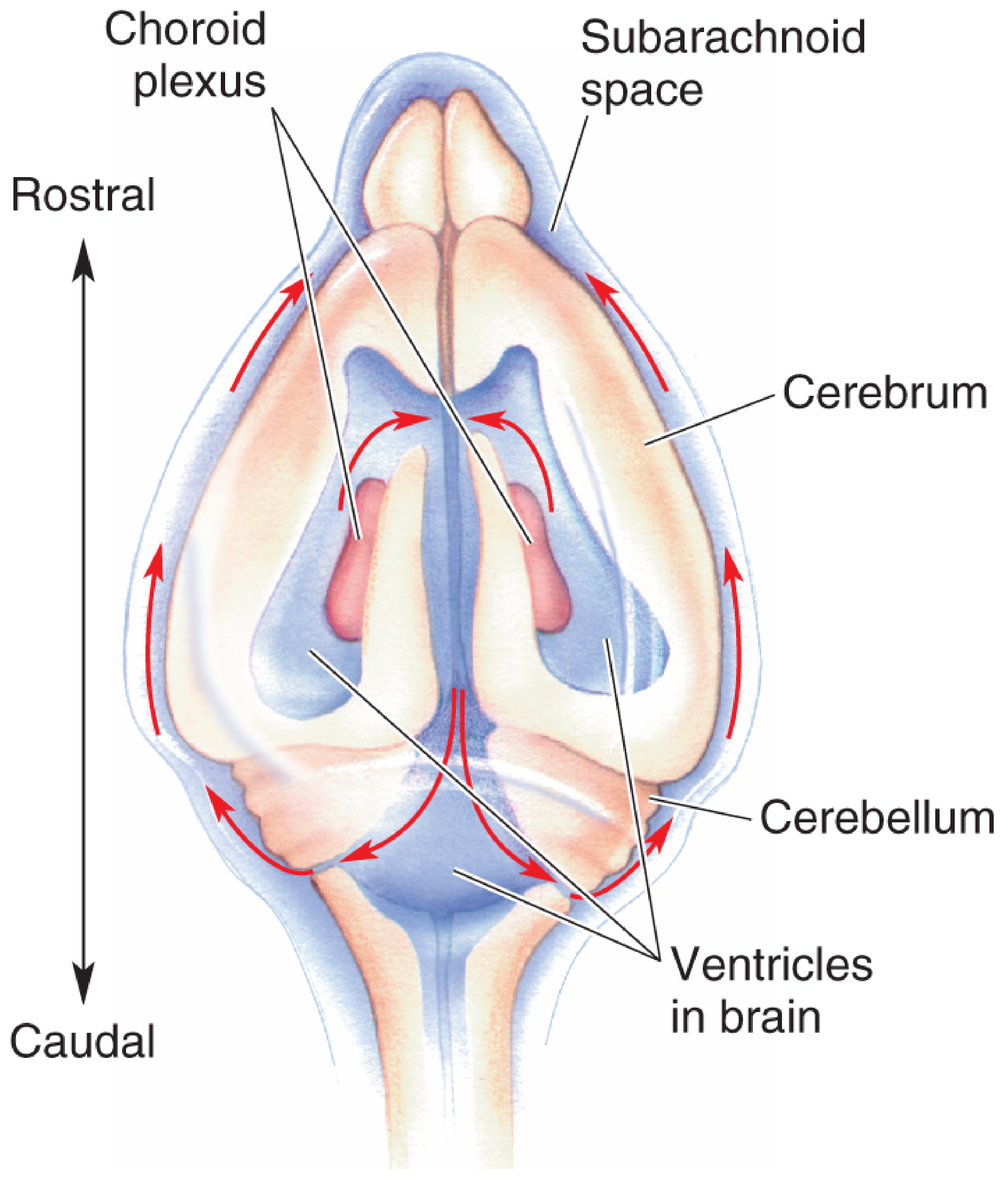
CSF is produced in the lateral ventricles (choroid plexus, shown in pink/red) and flows caudally through the connected ventricular system, exiting into the subarachnoid space. - Neuroscience: Exploring the Brain, 5th Edition
CT Appearance
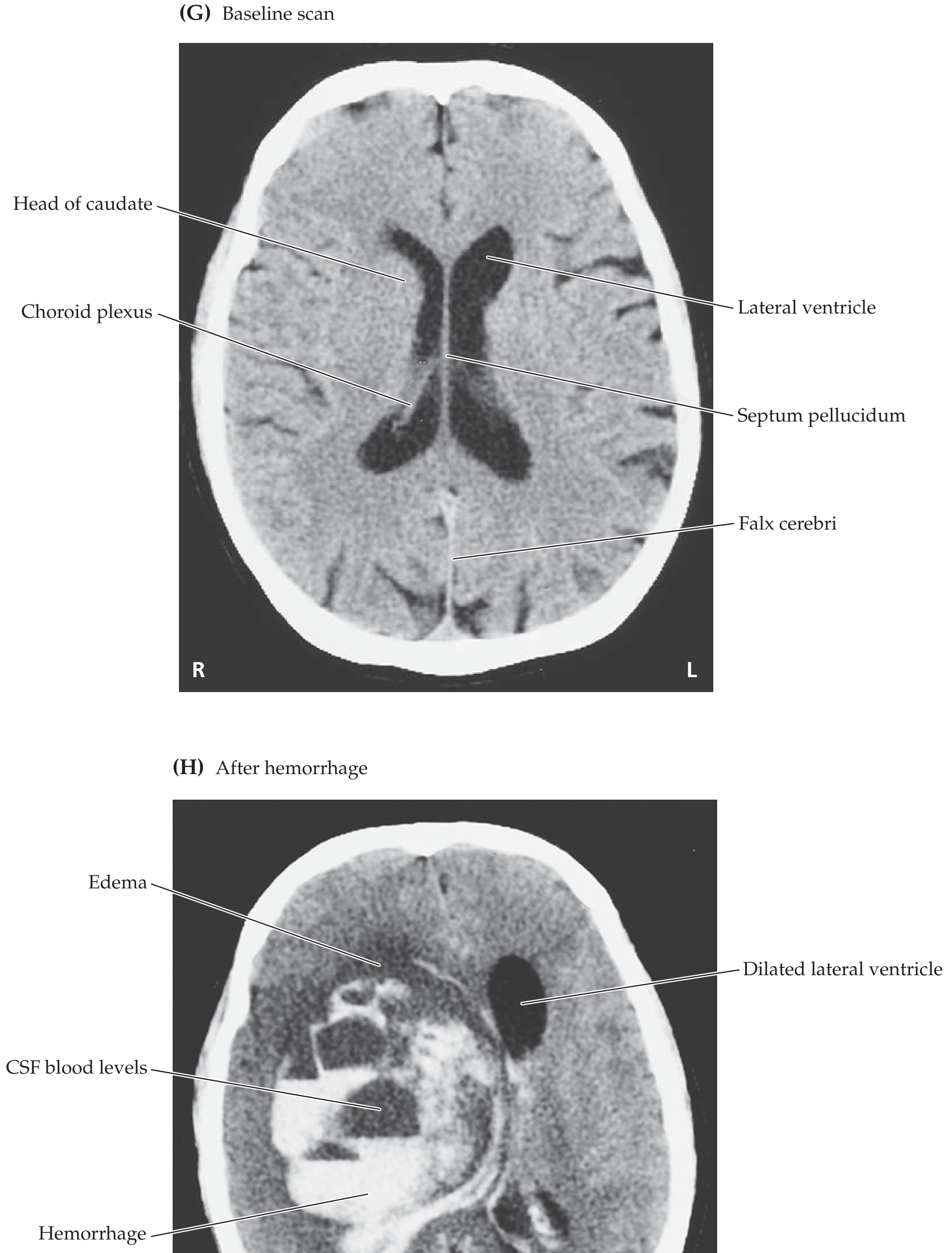
On axial CT, the lateral ventricles appear as bilateral, symmetric, CSF-density (dark) structures flanking the septum pellucidum. The choroid plexus is visible as a hyperdense structure within the trigone and body. If one interventricular foramen is occluded, only the ipsilateral lateral ventricle enlarges - the rest of the ventricular system remains normal size.
Clinical Correlates
| Condition | Relevance to Lateral Ventricle |
|---|---|
| Hydrocephalus | Obstruction of CSF flow causes dilation of the ventricles; obstructive hydrocephalus at the foramen of Monro → unilateral lateral ventricle enlargement |
| Intraventricular hemorrhage | Blood enters the ventricle (e.g., from ruptured AVM, hypertensive hemorrhage in basal ganglia); seen as hyperdensity on CT |
| Choroid plexus papilloma | Intraventricular WHO Grade I tumor; in children, most commonly in the lateral ventricle; in adults, more commonly in the 4th ventricle; causes CSF overproduction and hydrocephalus |
| Ventriculomegaly (prenatal) | Lateral ventricle atrial width ≥10 mm on fetal ultrasound is the threshold for ventriculomegaly (mild = 10-12 mm) |
| Periventricular leukomalacia | Brain US shows frontoparietal hyperechogenic lesion pointing toward the lateral ventricle; liquefaction may cause communication with the ventricle (porencephalic cyst) |
| Thalamic hemorrhage | Can rupture into the anterior horn of the lateral ventricle |
Development
The lateral ventricles develop from the cavities (lumen) of the paired telencephalic vesicles. As the cerebral hemispheres grow and curve, the ventricles follow the same C-shaped trajectory. The caudate nucleus, conforming to this shape, develops an elongated C-shaped body with its head in the floor of the frontal horn and its tail curving into the roof of the temporal horn.
Langman's Medical Embryology; The Developing Human, Clinically Oriented Embryology
Key facts to remember:
- There are two lateral ventricles (one per hemisphere), formerly called the 1st and 2nd ventricles
- They connect to the single midline 3rd ventricle via the interventricular foramina of Monro
- The atrium/trigone is the convergence point of the body, occipital horn, and temporal horn
- The choroid plexus produces ~500 mL of CSF per day and lines the walls (not the frontal and occipital horns)
- Obstruction at the foramen of Monro causes ipsilateral ventricular enlargement only
This is a shared conversation. Sign in to Orris to start your own chat.