
Everything ablout HUS and TTP in pregnancy- reference Williams obstetrics and recent recommended updates
Everything ablout HUS and TTP in pregnancy- reference Williams obstetrics and recent recommended updates In simple languages and flowcharts
thrombotic thrombocytopenic purpura hemolytic uremic syndrome pregnancy
PMID: 38396391
PMID: 37586700
thrombotic microangiopathy TTP HUS pathogenesis ADAMTS13 diagram pregnancy
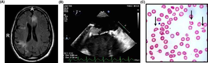
A composite clinical image consisting of three panels illustrating a multi-organ manifestation of Thrombotic Thrombocytopenic Purpura (TTP). Panel A is an axial T2 FLAIR magnetic resonance image of the brain showing hyperintense signals in the left anterior cerebral artery (ACA) distribution, consistent with acute infarction, alongside chronic hypointense areas indicating prior infarcts. Panel B displays a two-dimensional trans-esophageal echocardiogram (TEE) focused on a prosthetic mitral valve; it reveals a 1.5 cm irregular, echogenic mass (vegetation) attached to the atrial surface, indicative of non-bacterial thrombotic endocarditis. Panel C shows a Wright-Giemsa stained peripheral blood smear at high power. The smear demonstrates microangiopathic hemolytic anemia characterized by prominent schistocytes (fragmented red blood cells), indicated by black arrows, against a background of normocytic erythrocytes. Together, these images teach the classic clinical triad of neurological deficits, cardiac involvement, and hematologic evidence of microangiopathy associated with ADAMTS13 deficiency.
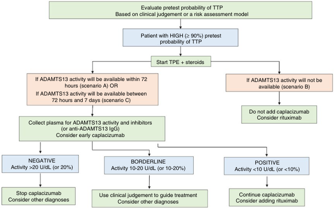
Summary : This flowchart outlines the diagnostic and treatment pathway for patients with a high (≥ 90%) pretest probability of thrombotic thrombocytopenic purpura (TTP), focusing on the role of ADAMTS13 activity testing, therapeutic plasma exchange (TPE), steroids, caplacizumab, and rituximab. flowchart: # Nodes : • Start (rectangle): "Evaluate pretest probability of TTP. Based on clinical judgement or a risk assessment model" • Decision (rectangle): "Patient with HIGH (≥ 90%) pretest probability of TTP" • Action (rectangle): "Start TPE + steroids" • Decision (rectangle): "If ADAMTS13 activity will be available within 72 hours (scenario A) OR If ADAMTS13 activity will be available between 72 hours and 7 days (scenario C)" • Decision (rectangle): "If ADAMTS13 activity will not be available (scenario B)" • Action (rectangle): "Collect plasma for ADAMTS13 activity and inhibitors (or anti-ADAMTS13 IgG). Consider early caplacizumab" • Action (rectangle): "Do not add caplacizumab. Consider rituximab" • Outcome (rectangle): "NEGATIVE Activity >20 U/dL (or 20%)" • Outcome (rectangle): "BORDERLINE Activity 10-20 U/dL (or 10-20%)" • Outcome (rectangle): "POSITIVE Activity <10 U/dL (or <10%)" • Action (rectangle): "Stop caplacizumab. Consider other diagnoses" • Action (rectangle): "Use clinical judgement to guide treatment. Consider other diagnoses" • Action (rectangle): "Continue caplacizumab. Consider adding rituximab" # Connectors : • Top-down arrows connect each step in the process. • After "Start TPE + steroids", the flow splits into two branches: – Left branch: If ADAMTS13 activity will be available (scenarios A or C), proceed to plasma collection and possible early caplacizumab. – Right branch: If ADAMTS13 activity will not be available (scenario B), do not add caplacizumab and consider rituximab. • From plasma collection, three branches based on ADAMTS13 activity result: – NEGATIVE (>20 U/dL): Stop caplacizumab, consider other diagnoses. – BORDERLINE (10-20 U/dL): Use clinical judgement, consider other diagnoses. – POSITIVE (<10 U/dL): Continue caplacizumab, consider adding rituximab. # Layout : • The flowchart is arranged vertically, with initial assessment at the top, followed by treatment initiation, then branching based on ADAMTS13 test availability and results. • The left branch further splits into three outcomes based on test results. • The right branch is a single pathway for when ADAMTS13 activity is unavailable. # Analysis : • The flowchart provides a clear, stepwise approach for managing suspected TTP, emphasizing early treatment and the importance of ADAMTS13 activity testing. • Caplacizumab is considered only if ADAMTS13 testing is available; otherwise, rituximab is considered. • The decision logic is based on test availability and results, guiding whether to continue, stop, or use clinical judgement for caplacizumab, and when to add rituximab. • The structure ensures rapid initiation of TPE and steroids for high-probability cases, with subsequent therapy tailored to diagnostic findings.
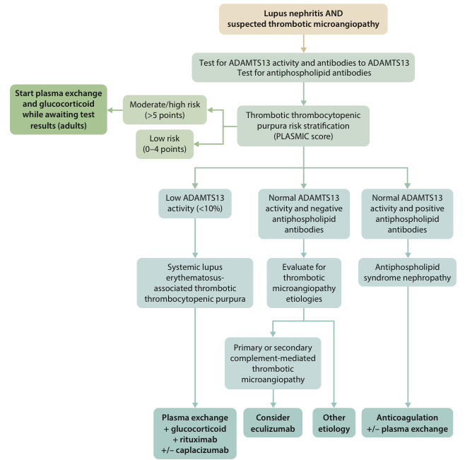
Summary : This flowchart outlines the diagnostic and initial management pathway for adults with lupus nephritis and suspected thrombotic microangiopathy, focusing on risk stratification, laboratory testing, and subsequent treatment options based on ADAMTS13 activity and antiphospholipid antibody results. flowchart: # Nodes : • "Lupus nephritis AND suspected thrombotic microangiopathy" (rounded rectangle, top node) • "Test for ADAMTS13 activity and antibodies to ADAMTS13" (rectangle) • "Test for antiphospholipid antibodies" (rectangle, merged with above) • "Thrombotic thrombocytopenic purpura risk stratification (PLASMIC score)" (rectangle) • "Start plasma exchange and glucocorticoid while awaiting test results (adults)" (rectangle, green highlight, left side) • "Moderate/high risk (>5 points)" (rectangle, left branch) • "Low risk (0–4 points)" (rectangle, left branch) • "Low ADAMTS13 activity (<10%)" (rectangle) • "Systemic lupus erythematosus-associated thrombotic thrombocytopenic purpura" (rectangle) • "Plasma exchange + glucocorticoid + rituximab +/- caplacizumab" (rectangle, green highlight) • "Normal ADAMTS13 activity and negative antiphospholipid antibodies" (rectangle) • "Evaluate for thrombotic microangiopathy etiologies" (rectangle) • "Primary or secondary complement-mediated thrombotic microangiopathy" (rectangle) • "Consider eculizumab" (rectangle, green highlight) • "Other etiology" (rectangle) • "Normal ADAMTS13 activity and positive antiphospholipid antibodies" (rectangle) • "Antiphospholipid syndrome nephropathy" (rectangle) • "Anticoagulation +/- plasma exchange" (rectangle, green highlight) # Connectors : • Downward arrows connect each step, with branches at risk stratification and ADAMTS13/antibody results. • "Start plasma exchange and glucocorticoid..." is a side branch from the initial risk stratification for adults. • Risk stratification (PLASMIC score) splits into "Moderate/high risk" and "Low risk". • Both risk branches converge on ADAMTS13 activity results. • "Low ADAMTS13 activity (<10%)" leads to lupus-associated TTP and then to specific therapy. • "Normal ADAMTS13 activity and negative antiphospholipid antibodies" leads to evaluation for other etiologies, which further splits into complement-mediated TMA (with eculizumab) and other causes. • "Normal ADAMTS13 activity and positive antiphospholipid antibodies" leads to antiphospholipid syndrome nephropathy and anticoagulation therapy. # Layout : • Vertical flow from top to bottom, with horizontal branches for risk stratification and antibody results. • Green-highlighted rectangles indicate urgent or specific therapeutic interventions. • Three main terminal branches: plasma exchange-based therapy, eculizumab consideration, and anticoagulation. # Analysis : • The flowchart provides a clear, stepwise approach for differentiating causes of thrombotic microangiopathy in lupus nephritis, using risk scores and laboratory results to guide therapy. • Immediate plasma exchange and glucocorticoid therapy is recommended for adults at moderate/high risk while awaiting test results. • ADAMTS13 activity and antiphospholipid antibody status are pivotal in determining the diagnosis and subsequent treatment, with distinct pathways for lupus-associated TTP, complement-mediated TMA, and antiphospholipid syndrome nephropathy. • The chart emphasizes rapid initiation of therapy in high-risk cases and tailored treatment based on etiology.
HUS and TTP in Pregnancy - Complete Guide
1. What Are They? (The Big Picture)
| Feature | What it means simply |
|---|---|
| Thrombocytopenia | Low platelets (clotting cells get used up in tiny clots) |
| Microangiopathic Hemolytic Anemia (MAHA) | Red blood cells get shredded as they pass through clogged small vessels - seen as schistocytes on blood smear |
| Organ dysfunction | Brain, kidneys, heart affected by microscopic clots |
2. TTP vs. aHUS - How Are They Different?
| Feature | TTP | aHUS (atypical HUS) |
|---|---|---|
| Root cause | ADAMTS13 deficiency (enzyme missing or blocked) | Complement system goes haywire (overactivated) |
| Which organs hit hardest | Brain/neuro (confusion, seizures) | Kidneys (AKI, need for dialysis) |
| When in pregnancy | Any trimester, mostly 2nd/3rd | Peripartum or POSTPARTUM (>50% cases) |
| ADAMTS13 level | < 10-20% (severely low) | Near normal (or mildly low) |
| Platelet drop | Severe (often <20,000) | Moderate |
| Treatment | Plasma exchange + steroids | Eculizumab (anti-C5 antibody) |
3. Pathophysiology Made Simple
TTP Mechanism
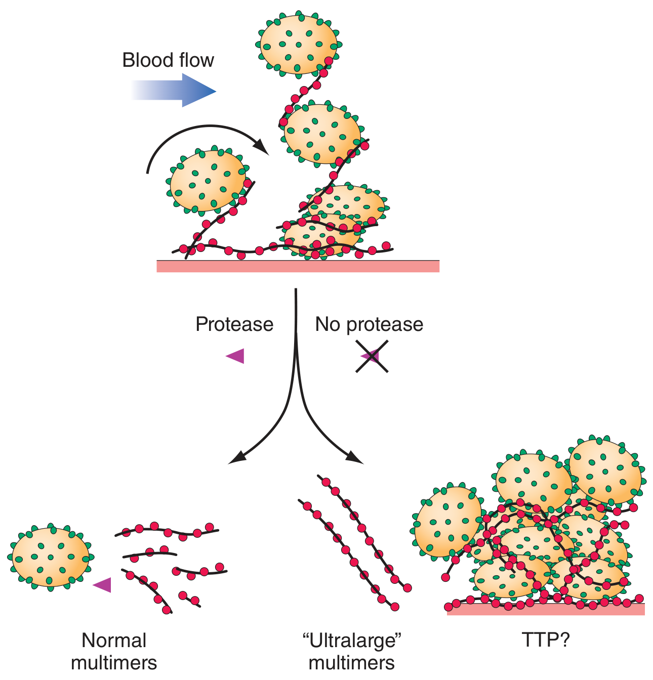
Normal State:
Endothelium releases vWF
        ↓
ADAMTS13 enzyme cleaves ultra-large vWF into small pieces
        ↓
Platelets flow freely ✓

In TTP:
ADAMTS13 is MISSING or BLOCKED by autoantibodies
        ↓
Ultra-large vWF accumulates on vessel walls
        ↓
Platelets stick to these long strands → CLOTS form in microvessels
        ↓
Platelets consumed → Thrombocytopenia
RBCs shredded → Schistocytes + Hemolytic anemia
Organs starved of blood → Brain (neuro sx), Kidney (AKI)
- Pregnancy physiologically REDUCES ADAMTS13 levels
- General hypercoagulable state of pregnancy
- Synergistic features with preeclampsia
aHUS Mechanism
Genetic mutation in complement regulators
(Factor H, Factor I, CD46/MCP, CFH, C3, Factor B)
        ↓
Alternative complement pathway OVERACTIVATED
        ↓
Uncontrolled endothelial cell inflammation and activation
        ↓
Systemic TMA → preferentially DAMAGES KIDNEYS
        ↓
AKI, dialysis-dependent renal failure
4. Epidemiology in Pregnancy
| Condition | Incidence in Pregnancy |
|---|---|
| TTP | 1 in 17,000 to 1 in 200,000 pregnancies |
| aHUS | Even rarer; ~16% of all aHUS cases in women aged 18-45 |
| TTP in pregnancy (% of all TTP) | 10-25% of all TTP cases occur in pregnancy or postpartum |
5. Clinical Features
TTP in Pregnancy - Classic Pentad (rarely all 5 together now)
- Microangiopathic hemolytic anemia (MAHA)
- Thrombocytopenia
- Neurological symptoms (confusion, seizures, stroke, visual changes)
- Renal impairment (mild)
- Fever
aHUS in Pregnancy
- Severe acute kidney injury (dominant feature)
- Moderate thrombocytopenia
- MAHA present
- Hypertension common
- Often peripartum or postpartum onset
- May require dialysis
6. The Most Important Differential - "The Big 4"
| Feature | Preeclampsia/HELLP | TTP | aHUS | DIC |
|---|---|---|---|---|
| Hypertension | +++ | ± | ± | ± |
| Neuro symptoms | + (seizures = eclampsia) | +++ | ± | ± |
| Renal dysfunction | ± | ± | +++ | ± |
| Skin purpura | - | + | - | - |
| Fever | - | ± | ± | ± |
| Platelets | Mildly-mod low | Severely low | Mod low | Low |
| PT/PTT | Normal or slightly ↑ | Normal | Normal | ELEVATED |
| Fibrinogen | Normal or ↑ | Normal/↑ | Normal | DECREASED |
| LDH | ↑ | ↑↑ (very high) | ↑↑ | ↑ |
| AST/ALT | ↑↑ | Normal | Normal | ± |
| ADAMTS13 | Normal | < 10% | Mildly ↓ or normal | Normal |
| Responds to delivery? | YES | NO | NO | YES |
Key Tip (Creasy & Resnik): A high LDH:AST ratio favors TTP over HELLP syndrome. If HELLP/preeclampsia does NOT improve within 48-72 hours after delivery, strongly consider TTP or aHUS.
7. Diagnosis - Step-by-Step
STEP 1: Suspect TMA
━━━━━━━━━━━━━━━━━━━━━━━━━━━━━
Thrombocytopenia + Hemolytic anemia in pregnancy?
→ Get peripheral blood smear → Look for SCHISTOCYTES
→ LDH, Bilirubin, Haptoglobin (↓), Reticulocyte count

STEP 2: Basic Workup
━━━━━━━━━━━━━━━━━━━━━━━━━━━━━
CBC + Smear
LDH, uric acid, AST/ALT, bilirubin
Renal function (creatinine, BUN)
PT, aPTT, fibrinogen (to rule out DIC)
Blood pressure (rule out preeclampsia)
Urinalysis (proteinuria?)

STEP 3: The KEY Test - ADAMTS13 Activity
━━━━━━━━━━━━━━━━━━━━━━━━━━━━━
ADAMTS13 < 10-20%  → TTP confirmed
ADAMTS13 normal    → Think aHUS (after ruling out other causes)
(Also send ADAMTS13 inhibitor/antibody level)

STEP 4: Rule out other causes
━━━━━━━━━━━━━━━━━━━━━━━━━━━━━
aHUS = DIAGNOSIS OF EXCLUSION
(No specific test - rule out TTP, preeclampsia, DIC, infections, drugs)
Consider complement panel (C3, C4, Factor H, genetic panel)
Note: ADAMTS13 results may take days to return. Do NOT wait - start empirical treatment if TTP is suspected.
8. Management Flowchart
OVERALL APPROACH
Pregnant patient with THROMBOCYTOPENIA + HEMOLYSIS (MAHA)
                    ↓
        Is this PREECLAMPSIA/HELLP?
           ↙                ↘
      YES                   NOT SURE / ATYPICAL
        ↓                         ↓
   Deliver (near term)    SEND ADAMTS13 ACTIVITY
   Magnesium sulfate      + FULL TMA WORKUP
   Antihypertensives            ↓
                    ┌───────────┴───────────┐
               ADAMTS13 LOW           ADAMTS13 NORMAL
               (<10-20%)              (or borderline)
                    ↓                         ↓
                  TTP                       aHUS
                    ↓                         ↓
            [TTP Treatment]          [aHUS Treatment]
TTP Treatment (in Pregnancy)
SUSPECT TTP (ADAMTS13 < 10% or pending + clinical features)
                    ↓
    URGENT: PLASMA EXCHANGE (Plasmapheresis) — START SAME DAY
    ━━━━━━━━━━━━━━━━━━━━━━━━━━━━━━━━━━━━━━━━━━━━━━━━━━━━━━
    → Removes autoantibodies against ADAMTS13
    → Replaces missing ADAMTS13
    → Survival improved from <15% to ~80% with this treatment
                    ↓
    ADD: GLUCOCORTICOIDS (Steroids)
    → Reduces anti-ADAMTS13 antibodies
    → For immune-mediated TTP or non-responders
                    ↓
    ⚠️ AVOID PLATELET TRANSFUSIONS
    → Can worsen microvascular thrombosis
    → Use ONLY for life-threatening bleeding or invasive procedure
                    ↓
    Continue until PLATELETS RECOVER (≥150,000) for several days
                    ↓
    REFRACTORY/RELAPSING TTP?
    → Add RITUXIMAB (B-cell depleting antibody)
    → Or other cytotoxic immunosuppressives
    → Consider CAPLACIZUMAB (new anti-vWF agent — see updates below)
                    ↓
    DELIVERY?
    → Delivery alone does NOT cure TTP
    → Consider delivery for refractory cases or viable gestation
    → Up to 33% fetal mortality in severe maternal illness
aHUS Treatment (in Pregnancy)
SUSPECT aHUS (ADAMTS13 normal + severe AKI + TMA)
                    ↓
    URGENT: ECULIZUMAB (Anti-C5 complement inhibitor)
    ━━━━━━━━━━━━━━━━━━━━━━━━━━━━━━━━━━━━━━━━━━━━━━━━━━━━━━
    → Blocks terminal complement activation
    → Current FIRST-LINE treatment (replaced plasmapheresis)
    → Plasma exchange: poor efficacy for aHUS
                    ↓
    BEFORE ECULIZUMAB: Meningococcal vaccination
    (or prophylactic antibiotics if urgent)
                    ↓
    Many patients require DIALYSIS (renal replacement)
                    ↓
    DELIVERY?
    → aHUS is more common POSTPARTUM - delivery may be triggered already
    → Treat maternal disease aggressively
    → Up to 33% fetal mortality in severe cases
9. TTP vs. aHUS Treatment - Quick Compare
| TTP | aHUS | |
|---|---|---|
| First-line | Plasma exchange (TPE) | Eculizumab (anti-C5) |
| Add-on | Steroids + Rituximab | Continue eculizumab |
| Platelet transfusion | AVOID | Avoid (use with caution) |
| Dialysis needed? | Rarely | Often |
| Plasma exchange | YES - mainstay | Poor efficacy, largely replaced |
| New agent | Caplacizumab (anti-VWF) | Ravulizumab (longer-acting anti-C5) |
10. Fetal/Obstetric Implications
- Fetal mortality: up to 33% in severe maternal TTP or aHUS (due to placental insufficiency and premature delivery)
- Neonatal death may result from previable delivery
- Early in pregnancy (< 34 weeks): treat the mother aggressively first; deliver only if refractory
- Near term or refractory: delivery is a valid option
- Preeclampsia/HELLP not improving 48-72h post-delivery = think TTP/aHUS
11. TTP Classification in Pregnancy
TTP
├── CONGENITAL TTP (Upshaw-Schulman Syndrome)
│     → ADAMTS13 gene mutation
│     → Often FIRST EPISODE triggered by PREGNANCY
│     → Very high recurrence risk in every pregnancy
│     → Treat with prophylactic PLASMA INFUSIONS from 1st trimester
│
└── ACQUIRED/IMMUNE TTP
      → Autoantibodies against ADAMTS13
      → Recurrence risk in future pregnancies = uncertain
      → Monitor ADAMTS13 levels every trimester
      → Steroids + Rituximab if relapse

aHUS
├── HEREDITARY (genetic complement mutations)
│     → Factor H, Factor I, MCP, C3, Factor B, Thrombomodulin
│
└── ACQUIRED (anti-Factor H antibodies)
      → DEAP-HUS variant
12. Monitoring in Future Pregnancies
| Condition | Recurrence Risk | Monitoring/Prophylaxis |
|---|---|---|
| Congenital TTP | Extremely high | Prophylactic plasma infusions from 1st trimester, continue postpartum |
| Immune-mediated TTP | Unknown (significant) | Serial ADAMTS13 every trimester; rituximab pre-pregnancy if needed |
| aHUS | ~25% (1 in 4 recur) | Close multidisciplinary monitoring; eculizumab prophylaxis discussed |
13. Recent Updates (2023-2026)
Caplacizumab - New Agent for TTP
- Faster platelet recovery
- Reduces TTP exacerbations
- Limited data in pregnancy - use with clinical judgement

Key 2024 Updates
- Pregnancy-associated TMA classification is now distinct from other TMAs per international consensus - TTP, aHUS, and HELLP are separate entities requiring separate treatments (Giannubilo et al., 2024)
- Eculizumab in pregnancy: Considered safe in pregnancy (Category B); preferred over plasma exchange for aHUS
- Ravulizumab: Longer-acting anti-C5 agent being used increasingly for aHUS
- Terminology shift: "aHUS" now often called complement-mediated TMA (CM-TMA) in newer literature
- ADAMTS13 testing: Still the single most important test to differentiate TTP from aHUS; < 10% activity is diagnostic of TTP (Harrison's 22E, 2025)
14. Peripheral Blood Smear - What You'll See
15. Memory Summary - "TTP vs aHUS at a Glance"
TTP = "The Thinking Person's" disease
  → Mainly BRAIN affected
  → ADAMTS13 very LOW
  → Treat with PLASMA EXCHANGE

aHUS = "Always Hurts the Urine organs" disease
  → Mainly KIDNEYS affected
  → Complement is the problem
  → Treat with ECULIZUMAB

Both:
  → THROMBOCYTOPENIA + HEMOLYSIS (schistocytes)
  → Life-threatening in pregnancy
  → DO NOT cure with delivery alone
  → Up to 33% fetal loss
  → AVOID platelet transfusions
  → Diagnose with ADAMTS13 + clinical picture
  → Multidisciplinary team essential
- Creasy & Resnik's Maternal-Fetal Medicine, 9th ed., Ch. 53 (TTP/aHUS in Pregnancy, pp. 1295-1297)
- Harrison's Principles of Internal Medicine, 22nd ed. (2025), Ch. 120 (Thrombotic Microangiopathies, p. 968)
- BSH Guideline: TTP and TMA Management, 2023 (Scully et al., PMID 37586700)
- HELLP Syndrome vs TMA in Pregnancy, 2024 (Giannubilo et al., PMID 38396391)
Give every thing for quiz on pre eclampsia , eclampsia and HELLP syndrome - reference from Williams Obs, ACOG , figo and other recent updates including incidences , percentage everything
preeclampsia HELLP syndrome management guidelines
ACOG 2024 preeclampsia eclampsia HELLP syndrome guidelines diagnostic criteria
preeclampsia diagnosis criteria aspirin prevention severe features
eclampsia HELLP syndrome incidence maternal mortality outcomes 2022 2023 2024
preeclampsia eclampsia pathophysiology sFlt1 PlGF angiogenic imbalance placenta
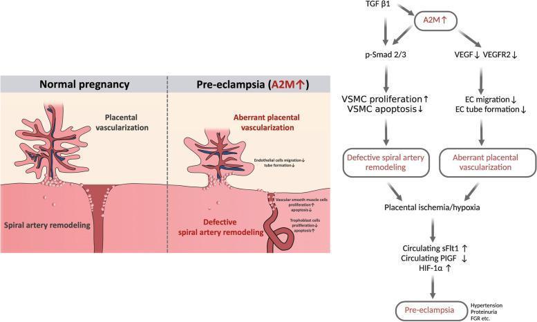
A multi-part medical illustration and signaling pathway diagram detailing the role of Alpha-2-Macroglobulin (A2M) in the pathophysiology of pre-eclampsia (PE). The left panel features a comparative anatomical diagram showing 'Normal pregnancy' versus 'Pre-eclampsia (A2M ↑)'. Normal pregnancy displays healthy placental vascularization with extensive branching and effective spiral artery remodeling (wide, dilated vessels). In contrast, pre-eclampsia exhibits 'aberrant placental vascularization' with sparse branching and 'defective spiral artery remodeling' characterized by narrow, un-remodeled vessels. The right panel outlines a signaling algorithm: TGF ̢1 and increased A2M stimulate p-Smad 2/3, leading to increased vascular smooth muscle cell (VSMC) proliferation and decreased apoptosis, causing defective remodeling. Simultaneously, increased A2M inhibits VEGF/VEGFR2, reducing endothelial cell (EC) migration and tube formation, causing aberrant vascularization. These pathways converge at placental ischemia/hypoxia, which increases circulating sFlt1 and HIF-1̑ and decreases PlGF, culminating in clinical pre-eclampsia symptoms including hypertension, proteinuria, and fetal growth restriction (FGR).
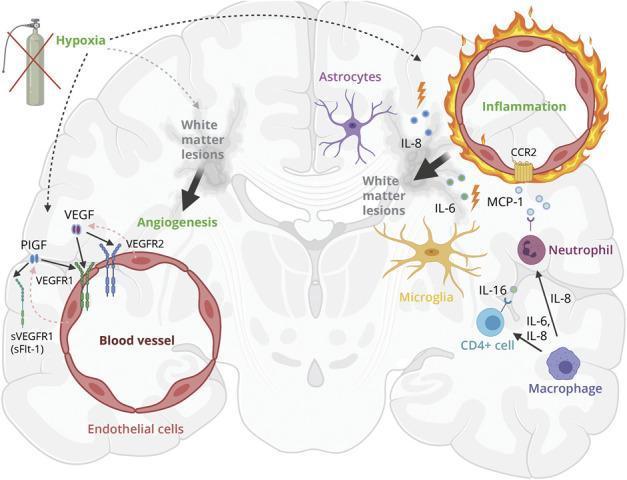
This pathophysiology diagram illustrates the theoretical relationship between hypoxia, neuroinflammation, and pathologic angiogenesis in the development of white matter lesions (WML) in the human brain. The model identifies hypoxia as a primary driver, initiating two parallel cascades. On the left, hypoxia induces an angiogenic response: Placental Growth Factor (PlGF) and Vascular Endothelial Growth Factor (VEGF) are upregulated. PlGF binds to VEGFR1, while VEGF binds to both VEGFR1 and VEGFR2 on endothelial cells. The soluble receptor sVEGFR1 (sFlt-1) acts as an inhibitor by sequestering these growth factors. On the right, hypoxia triggers a neuroinflammatory cascade involving astrocytes and microglia, which release proinflammatory markers IL-8, IL-6, IL-16, and MCP-1. These cytokines facilitate the recruitment of immune cells, including neutrophils, CD4+ T-cells, and macrophages, leading to chronic inflammation and blood-brain barrier (BBB) disruption. The convergence of pathologic angiogenesis and neuroinflammation contributes to the formation and progression of white matter lesions within the brain parenchyma.
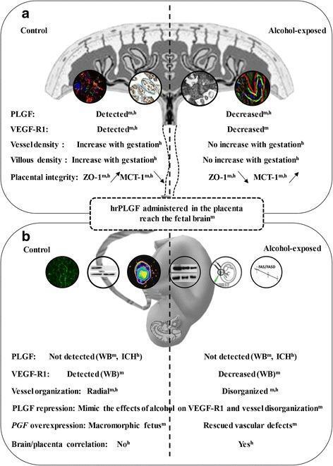
A multi-panel pathophysiology diagram summarizing the comparative effects of in utero alcohol exposure on the placenta (Section A) and fetal brain (Section B) in humans and mouse models. Section A illustrates the placenta, contrasting control and alcohol-exposed states. In the alcohol group, there is a decrease in placental growth factor (PLGF) and VEGF-R1 expression, no gestational increase in vessel or villous density, and impaired placental integrity evidenced by decreased ZO-1 and increased MCT-1 levels. Section B depicts the fetal brain, showing that alcohol exposure leads to decreased VEGF-R1 levels and a shift from radial to disorganized cortical vessel organization. Central to the diagram is the pathway demonstrating that recombinant human PLGF (hrPLGF) administered to the placenta can reach the fetal brain. Experimental results shown include Western blot (WB) and immunohistochemistry (IHC) data indicating that placental PGF repression mimics alcohol's effects on brain vasculature, while PGF overexpression can rescue these defects. The diagram highlights a positive correlation between vascular placental and brain defects in human alcohol-exposed groups, emphasizing the placenta's role in fetal alcohol spectrum disorders (FASD).
PREECLAMPSIA, ECLAMPSIA & HELLP SYNDROME
Complete Quiz Master Guide with Statistics
PART 1: DEFINITIONS & CLASSIFICATION
Hypertensive Disorders of Pregnancy - The Spectrum
GESTATIONAL HYPERTENSION
        ↓ (can progress)
PREECLAMPSIA (without severe features)
        ↓ (can progress)
PREECLAMPSIA WITH SEVERE FEATURES
        ↓                    ↓
   ECLAMPSIA           HELLP SYNDROME
| Condition | Definition |
|---|---|
| Gestational HTN | New-onset SBP ≥140 or DBP ≥90 after 20 weeks, NO proteinuria or other features |
| Preeclampsia | New-onset HTN after 20 weeks + proteinuria OR end-organ involvement |
| Eclampsia | New-onset seizures in woman with preeclampsia, not attributable to other causes |
| HELLP | Hemolysis + Elevated Liver Enzymes + Low Platelets - variant of preeclampsia |
| Superimposed PE | Preeclampsia developing on background of chronic hypertension |
ACOG (2013, reaffirmed 2024): ABANDON the term "mild preeclampsia" - replace with "preeclampsia without severe features." All preeclampsia carries risk.
PART 2: INCIDENCE & EPIDEMIOLOGY
Global and US Statistics
| Statistic | Figure |
|---|---|
| Hypertensive disorders of pregnancy (US) | ~85 per 1,000 deliveries |
| Preeclampsia globally | 2-8% of all pregnancies |
| Global maternal deaths from preeclampsia/eclampsia | >50,000 per year |
| Fetal deaths from preeclampsia/eclampsia globally | >500,000 per year |
| HELLP in all pregnancies | 0.2-0.8% |
| HELLP in severe preeclampsia | Up to 12% |
| Eclampsia incidence (developed countries) | ~1 in 2,000 deliveries |
| Eclampsia incidence (developing countries) | Up to 1 in 100 deliveries |
Risk Factors with % Data
| Risk Factor | Relative Risk / % |
|---|---|
| Nulliparity | Accounts for 32.3% of population attributable fraction for PE |
| Chronic hypertension | 25% of women with chronic HTN develop PE |
| Diabetes mellitus (overall) | ~20% risk |
| - White's class B diabetes | 11-16% |
| - White's class C diabetes | 21-23% |
| - White's class D diabetes | 35-40% |
| - White's class F & R diabetes | Up to 70% |
| Obesity | ~3x increased risk; risk increases with BMI |
| Twin pregnancy | Risk significantly elevated |
| Prior preeclampsia | OR ~7 (previous severe PE carries ~25-65% recurrence risk) |
| Antiphospholipid syndrome | Strong risk factor |
| SLE | High risk, especially with nephropathy |
| Black race | More severe disease; in nulliparas OR = 12.3 (in some studies) |
| Age extremes | Both teenage and advanced maternal age |
| IVF | Independent risk factor |
| Family history | Independent risk factor |
PART 3: DIAGNOSTIC CRITERIA (ACOG/FIGO)
ACOG Diagnostic Criteria for Preeclampsia
- SBP ≥ 140 mmHg OR DBP ≥ 90 mmHg
- On two occasions, at least 4 hours apart
- After 20 weeks' gestation
QUIZ FACT: Incremental rise of 30 mmHg systolic or 15 mmHg diastolic is NO LONGER a diagnostic criterion (ACOG removed this - no increased adverse outcomes in this group alone)
| Feature | Threshold |
|---|---|
| Proteinuria | ≥300 mg in 24h urine specimen, OR protein:creatinine ≥ 0.3, OR dipstick 2+ (only if quantitative unavailable) |
| Thrombocytopenia | Platelet count < 100,000/μL |
| Elevated liver enzymes | AST/ALT > 2× upper limit of normal |
| Renal insufficiency | Serum creatinine > 1.1 mg/dL (in absence of other renal disease) |
| Pulmonary edema | New onset |
| New-onset cerebral/visual disturbances | New headache, visual changes |
QUIZ FACT: Preeclampsia can be diagnosed WITHOUT proteinuria if end-organ involvement is present.
ACOG Criteria for Preeclampsia WITH SEVERE FEATURES
1. SBP ≥ 160 mmHg or DBP ≥ 110 mmHg (on 2 occasions, 4h apart)
2. New-onset cerebral or visual disturbances
3. Pulmonary edema
4. Persistent epigastric/RUQ pain (unresponsive to medications)
5. Impaired liver function: AST/ALT ≥ 2× upper limit of normal
6. Thrombocytopenia: Platelet count < 100,000/μL
7. Progressive renal insufficiency: Creatinine > 1.1 mg/dL
QUIZ FACT (2 removals):
- Proteinuria > 5g is NO LONGER a severe feature criterion
- Fetal growth restriction is NO LONGER a severe feature criterion
- Edema has been abandoned as a diagnostic marker (occurs in 10-15% of normal pregnancies)
PART 4: HELLP SYNDROME - CLASSIFICATION SYSTEMS
HELLP = Hemolysis + Elevated Liver enzymes + Low Platelets
Tennessee Classification (Sibai)
| Criterion | Threshold |
|---|---|
| Hemolysis | Microangiopathic hemolytic anemia - abnormal smear (schistocytes/burr cells), low haptoglobin, elevated LDH |
| LDH | > 600 IU/L (or 2× upper limit of normal) |
| AST | > 70 IU/L (or 2× upper limit of normal) |
| Bilirubin | > 1.2 mg/dL |
| Platelet count | < 100,000/μL |
| Incomplete HELLP | Only 1 or 2 criteria present (may be less severe) |
Mississippi Triple-Class Classification (Martin)
| Class | Platelet Count | Severity |
|---|---|---|
| Class I | ≤ 50,000/mm³ | Most severe |
| Class II | > 50,000 and ≤ 100,000/mm³ | Moderate |
| Class III | > 100,000 and ≤ 150,000/mm³ | Least severe |
ACOG Task Force Criteria for HELLP (Most Current)
- Hemolysis + at least 2 of:
- Schistocytes and burr cells on smear
- Serum bilirubin ≥ 1.2 mg/dL
- Low serum haptoglobin
- Severe anemia unrelated to blood loss
- Elevated liver enzymes: AST or ALT ≥ 2× ULN; LDH ≥ 2× ULN
- Platelets < 100,000/mm³
PART 5: HELLP - CLINICAL FEATURES & STATISTICS
Presenting Symptoms (% of Patients)
| Symptom | % Affected |
|---|---|
| Abdominal pain (RUQ, epigastric) | 65% |
| Nausea or vomiting | 36% |
| Headache | 31% |
| Bleeding | 9% |
| Jaundice | 5% |
Laboratory Values in HELLP (Median with Range)
| Test | Median (Range) |
|---|---|
| Serum AST (normal <40 U/L) | 249 U/L (70-633) |
| Serum bilirubin (normal <1 mg/dL) | 1.5 mg/dL (0.5-25) |
| Platelet count (normal >125×10³) | 57×10³/mm³ (7-99) |
Maternal Complications of HELLP (% Affected)
| Complication | % |
|---|---|
| DIC | 21% |
| Abruptio placentae | 16% |
| Acute kidney injury | 8% |
| Hepatic subcapsular hematoma | 1% |
| Death | 1% |
HELLP - Key Facts for Quiz
- Described in women who are typically older, White, multiparous (differs from typical preeclampsia patient)
- Up to 11% present before 27 weeks gestation
- Up to 30% of HELLP cases present POSTPARTUM (without signs at delivery)
- Hypertension and proteinuria may be absent in HELLP
- HELLP does NOT show the typical renin-angiotensin changes of preeclampsia
- HELLP is treated like preeclampsia with severe features (delivery is definitive treatment)
PART 6: ECLAMPSIA - FEATURES & STATISTICS
Definition
Symptoms Preceding Eclampsia (% of Cases)
| Symptom | % With This Symptom |
|---|---|
| Headache | 83% |
| Hyperreflexia | 80% |
| Proteinuria | 80% |
| Edema | 60% |
| Clonus | 46% |
| Visual signs | 45% |
| Epigastric pain | 20% |
QUIZ TRAPS:
- 17% of eclampsia patients have NO headache
- 80% have NO epigastric pain
- 20% have NORMAL deep tendon reflexes before seizure
- 24% have NO proteinuria before seizure (Chesley)
- Eclampsia can occur ANTEPARTUM, INTRAPARTUM, or POSTPARTUM
- Postpartum eclampsia can occur up to 48 hours after delivery (late postpartum eclampsia up to 4 weeks)
Historical Mortality of Eclampsia
| Era/Treatment | Maternal Mortality |
|---|---|
| Expectant management only | 20-30% |
| Profound maternal sedation | 10-15% |
| Magnesium sulfate (1920s-30s introduction) | ~5% |
| Modern (MgSO₄ + antihypertensives + timely delivery) | Very low (but still leading cause of maternal mortality) |
PART 7: PATHOPHYSIOLOGY (SIMPLIFIED)
Two-Stage Model of Preeclampsia
STAGE 1 (Pre-clinical): Poor placentation
━━━━━━━━━━━━━━━━━━━━━━━━━━━━━━━━━━━━━━━
Defective trophoblast invasion
        ↓
Inadequate spiral artery remodeling
(High-resistance, narrow vessels instead of low-resistance, wide vessels)
        ↓
Relative placental ischemia/hypoxia

STAGE 2 (Clinical): Maternal syndrome
━━━━━━━━━━━━━━━━━━━━━━━━━━━━━━━━━━━━━━━
Ischemic placenta releases factors into maternal circulation:
• ↑ sFLT-1 (soluble VEGF receptor - anti-angiogenic)
• ↑ Endothelin-1
• ↓ Free PlGF (placental growth factor)
• ↓ Prostacyclin, ↑ Thromboxane
        ↓
Widespread maternal ENDOTHELIAL DYSFUNCTION
        ↓
↑ Vasospasm → Hypertension
↑ Vascular permeability → Proteinuria, Edema
Platelet activation → Thrombocytopenia
Hepatic ischemia → HELLP
CNS irritability → Eclampsia
Renal ischemia → Creatinine ↑
QUIZ FACT: Blood pressure normally DECREASES in early pregnancy, reaching a nadir at ~22 weeks, then returns to baseline. This can cause preeclampsia to be missed in women with underlying chronic hypertension.
Angiotensin Sensitivity (Key Pathophysiology)
- Normal pregnant women require ~2.5× more angiotensin II to raise BP (reduced sensitivity = protective)
- Preeclamptic women are far MORE sensitive to angiotensin II (and all pressors) than both normal pregnant AND non-pregnant women
- This increased sensitivity may be detectable as early as 14 weeks before clinical disease
PART 8: MAGNESIUM SULFATE - THE CORNERSTONE DRUG
Serum Magnesium Levels & Effects
| Effect | Serum Level (mEq/L) |
|---|---|
| Therapeutic anticonvulsant range | 4.8 - 8.4 |
| Loss of deep tendon reflexes | 7 - 10 |
| Respiratory depression | > 10 |
| Cardiac arrest | > 15 |
Dosing (Standard Sibai Regimen)
| Phase | Dose |
|---|---|
| Loading dose | 4-6 g IV over 15-20 minutes |
| Maintenance | 1-2 g/hour IV infusion |
| Duration | Continue 24-48 hours after delivery |
| Antidote (toxicity) | Calcium gluconate 1g IV slowly |
Monitoring (the "3 Ms")
- Magnesium level (target 4.8-8.4 mEq/L)
- Minute urinary output (≥ 25-30 mL/hr)
- Muscle reflexes (deep tendon reflexes present = safe)
QUIZ FACTS:
- MgSO₄ is NOT an antihypertensive - it is an ANTICONVULSANT
- It acts by blocking NMDA receptors and reducing CNS irritability
- Excretion is renal - REDUCE dose in renal impairment
- Half-life ~4 hours in normal renal function
MgSO₄ vs. Diazepam (Magpie Trial)
- MgSO₄ reduces risk of eclampsia by 58% compared to placebo
- MgSO₄ is superior to both diazepam and phenytoin for eclampsia prevention and treatment
PART 9: ANTIHYPERTENSIVE TREATMENT
Severe-Range Hypertension (≥160/110)
First-Line Options (ACOG)
| Drug | Dose | Route | Notes |
|---|---|---|---|
| Labetalol | 20 mg IV, then 40-80 mg q10-20 min (max 300 mg) | IV | First-line for acute |
| Hydralazine | 5-10 mg IV q20 min | IV | Can cause reflex tachycardia |
| Nifedipine | 10-20 mg oral, repeat q20 min | Oral | Safe, widely used |
Chronic Antihypertensive in Pregnancy (First-line)
- Labetalol (alpha + beta blocker)
- Nifedipine (calcium channel blocker)
- α-Methyldopa (central alpha agonist)
Contraindicated Antihypertensives in Pregnancy
- ACE inhibitors (fetotoxic - renal tubular dysgenesis)
- ARBs (same mechanism)
- Atenolol (fetal growth restriction risk)
- Nitroprusside (cyanide toxicity risk)
PART 10: PREVENTION - LOW-DOSE ASPIRIN
Evidence Summary
| Study/Analysis | Risk Reduction |
|---|---|
| Cochrane Review (59 RCTs) | 17% reduction in preeclampsia risk |
| Meta-analysis (aspirin before 16 weeks) | 43% reduction when started before 16 weeks |
| Rolnik et al. (150 mg aspirin 11-14 weeks) | 62% reduction in PRETERM preeclampsia |
| Low-dose aspirin (general) | At least 10-24% reduction |
ACOG/FIGO Recommendations for Aspirin Prevention
HIGH RISK - 1 major risk factor = START ASPIRIN
━━━━━━━━━━━━━━━━━━━━━━━━━━━━━━━━━━━━━━━━━━━━━━━
Major risk factors:
• Previous preeclampsia (especially preterm)
• Multifetal gestation
• Chronic hypertension
• Type 1 or Type 2 diabetes
• Kidney disease
• Autoimmune conditions (SLE, APS)

MODERATE RISK - 2+ moderate risk factors = START ASPIRIN
━━━━━━━━━━━━━━━━━━━━━━━━━━━━━━━━━━━━━━━━━━━━━━━
Moderate risk factors:
• Nulliparity
• Obesity (BMI >30)
• Family history (mother or sister)
• Sociodemographic factors
• Age ≥35
• First pregnancy with new partner

DOSE: 81-150 mg/day (ACOG: 81 mg; FIGO/UK: 150 mg preferred)
WHEN TO START: Before 16 weeks (ideally 12-16 weeks)
STOP: At 36 weeks gestation
QUIZ NOTE (FIGO 2019/2023): FIGO recommends first-trimester screening (combined test: maternal factors + uterine artery Doppler + PlGF + PAPP-A) to identify high-risk women for targeted aspirin prophylaxis. This approach identifies >75% of preterm preeclampsia cases.
Calcium Supplementation (WHO Recommendation)
- 1.5-2 g/day calcium supplementation after 20 weeks
- ONLY in women with LOW dietary calcium intake
- Not routinely recommended in high-income countries
PART 11: MANAGEMENT FLOWCHARTS
Flowchart 1: Preeclampsia Without Severe Features
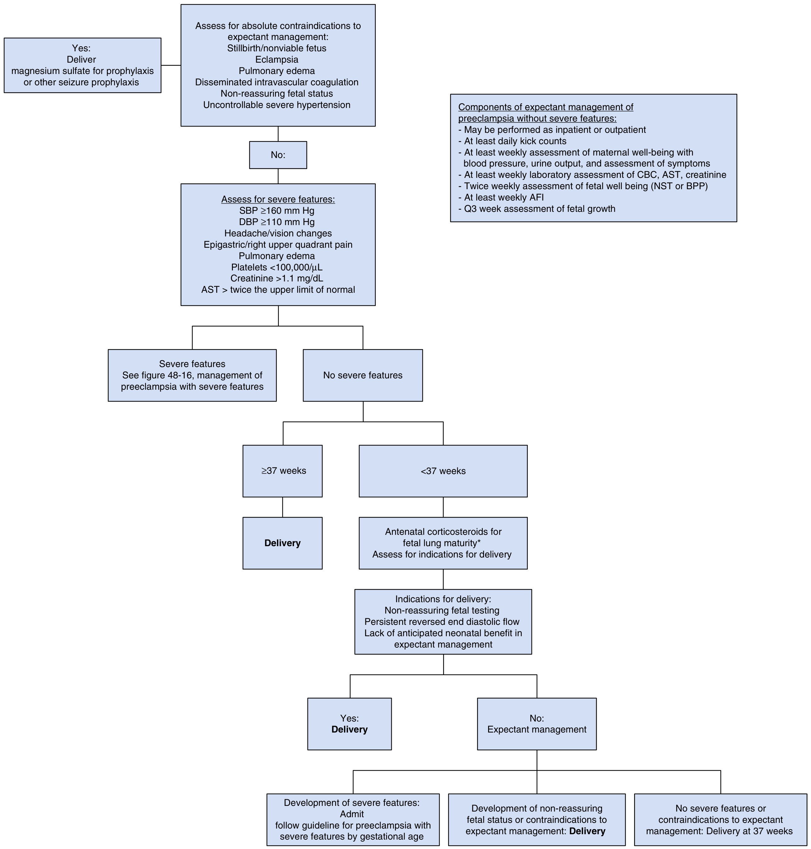
- ≥37 weeks + no severe features → Deliver
- <37 weeks + no severe features → Expectant management with monitoring
- Expectant management components: weekly BP, labs (CBC/AST/creatinine), twice-weekly fetal testing, weekly AFI, every 3-week fetal growth scan
Flowchart 2: Preeclampsia WITH Severe Features
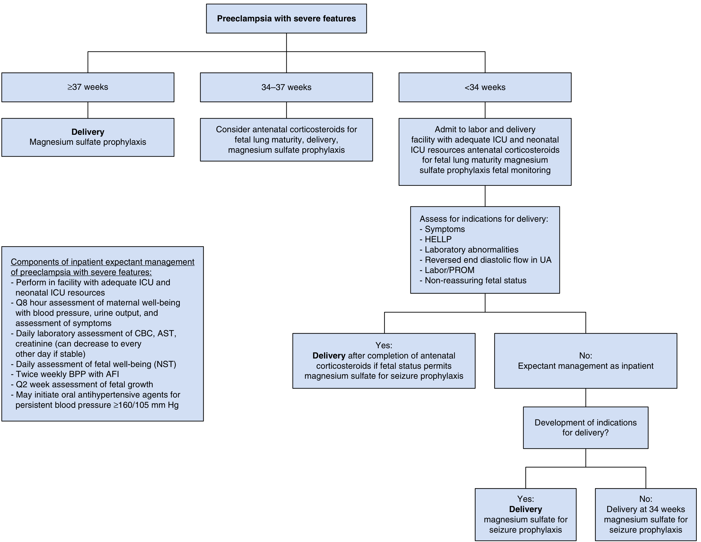
| Gestational Age | Action |
|---|---|
| ≥ 37 weeks | Deliver + MgSO₄ prophylaxis |
| 34-37 weeks | Consider antenatal corticosteroids, then deliver + MgSO₄ |
| < 34 weeks | Admit to facility with ICU/NICU; give corticosteroids; expectant if no emergency indications; deliver at 34 weeks |
PART 12: DELIVERY INDICATIONS (ABSOLUTE)
Contraindications to Expectant Management (= Deliver Now)
✗ Eclampsia
✗ Pulmonary edema
✗ DIC
✗ Non-reassuring fetal status
✗ Uncontrollable severe hypertension
✗ Stillbirth/nonviable fetus
✗ HELLP syndrome (after steroid course if needed)
✗ Placental abruption
✗ Labor/PROM at any gestational age with severe features
PART 13: COMPLICATIONS & FETAL OUTCOMES
Maternal Complications
| Complication | Notes |
|---|---|
| Abruptio placentae | Major cause of maternal and fetal death |
| Hepatic rupture | Rare but life-threatening; from capsular hematoma |
| DIC | Especially in HELLP |
| AKI | Especially aHUS/HELLP |
| Pulmonary edema | CNS cause of maternal death |
| Stroke/cerebral hemorrhage | Severe uncontrolled HTN |
| Blindness (cortical) | PRES (posterior reversible encephalopathy) |
Perinatal Complications
| Outcome | Data |
|---|---|
| Perinatal mortality (preeclampsia vs. normotensive) | OR 5.0 for gestational HTN, 2.7 for chronic HTN vs. normotensive |
| Neonatal death risk | ~2× increased risk |
| Fetal growth restriction | Especially with severe, early-onset disease |
| Stillbirth | Declining over last 35 years due to improved monitoring |
PART 14: LONG-TERM CONSEQUENCES
Cardiovascular Risk After Preeclampsia
| Future Risk | Data |
|---|---|
| Chronic hypertension | Recurrent preeclampsia → more likely to develop |
| Cardiovascular disease | Women with PE have 2-4× increased lifetime CVD risk |
| Women with eclampsia in 2nd+ pregnancy | 2-5× higher mortality over 35 years (Chesley) |
| Recurrence in next pregnancy | ~25% for preeclampsia; up to 65% if previous preterm severe PE |
QUIZ NOTE: Women with eclampsia in their FIRST pregnancy have normal long-term prognosis (no increased CVD risk). It is those with eclampsia in multiparous pregnancies who have increased mortality - suggesting underlying vascular disease that preeclampsia unmasked.
PART 15: BIOMARKERS & RECENT UPDATES (2022-2026)
sFLT-1/PlGF Ratio
- Elevated sFLT-1 / Low PlGF = hallmark of preeclampsia
- sFLT-1:PlGF ratio < 38 has high negative predictive value for ruling out PE in next 1 week (PROGNOSIS trial)
- sFLT-1:PlGF > 85 (or >110 in some studies) = high risk, likely to deliver within 2 weeks
- Now incorporated into FIGO first-trimester screening for preeclampsia prediction
FIGO 2019/2023 First-Trimester Screening Model
Combined First-Trimester Screen (at 11-14 weeks):
━━━━━━━━━━━━━━━━━━━━━━━━━━━━━━━━━━━━━━━━━━━━━━━
• Maternal characteristics and history
• Mean arterial pressure (MAP)
• Uterine artery Doppler (pulsatility index)
• Serum PlGF (↓ = risk)
• Serum PAPP-A (↓ = risk)
        ↓
Detection rate:
• ~75-90% of PRETERM preeclampsia
• ~45-50% of term preeclampsia
(At 10% false-positive rate)

HIGH RISK → Start aspirin 150 mg/day before 16 weeks
Telmisartan/Pravastatin - Investigational
- Statins (particularly pravastatin) being studied as potential preventive agents in very high-risk women
- Not yet standard of care
Esketamine / Hemabate - Not recommended for seizures
- MgSO₄ remains first-line worldwide per WHO, ACOG, FIGO, RCOG, and SOGC
PART 16: QUICK QUIZ SUMMARY TABLE
| Topic | Key Number / Fact |
|---|---|
| PE incidence | 2-8% of pregnancies |
| US hypertensive disorders | 85 per 1,000 deliveries |
| Nulliparity PAF for PE | 32.3% |
| Chronic HTN → PE risk | 25% |
| Diabetes overall PE risk | ~20% |
| DM White class F/R → PE | Up to 70% |
| HELLP incidence | 0.2-0.8% of pregnancies |
| HELLP in severe PE | Up to 12% |
| HELLP postpartum delay | Up to 30% of cases |
| DIC in HELLP | 21% |
| Abruption in HELLP | 16% |
| Headache before eclampsia | 83% |
| No proteinuria before eclampsia | 24% |
| Normal DTR before eclampsia | 20% |
| MgSO₄ therapeutic level | 4.8-8.4 mEq/L |
| DTR loss | 7-10 mEq/L |
| Respiratory depression | >10 mEq/L |
| Cardiac arrest | >15 mEq/L |
| MgSO₄ vs placebo (Magpie) | 58% reduction in eclampsia |
| Aspirin (Cochrane) | 17% reduction in PE |
| Aspirin before 16 weeks | 43% reduction |
| Aspirin 150 mg (Rolnik) | 62% reduction in preterm PE |
| Delivery in non-severe PE | At ≥37 weeks |
| Delivery in severe PE | At ≥34 weeks (if stable) |
| Magnesium antidote | Calcium gluconate 1g IV |
| Term for "mild preeclampsia" | ABANDONED by ACOG |
| Protein >5g = severe feature? | NO - removed by ACOG |
| FGR = severe feature? | NO - removed by ACOG |
| Edema = diagnostic? | NO - abandoned |
| Eclampsia historical mortality | 20-30% without treatment |
| Eclampsia modern mortality | Very low (<1% in developed world) |
- Creasy & Resnik's Maternal-Fetal Medicine, 9th ed., Ch. 45 & 48 (Epidemiology, Pathophysiology, Management)
- Gabbe's Obstetrics, 8th ed., Ch. 40 (HELLP classification, statistics)
- Harrison's Principles 22E (2025)
- ACOG Task Force on Hypertension in Pregnancy (2013, updated criteria reaffirmed 2019-2024)
- FIGO Initiative on Pre-eclampsia (2019/2023)
- Rolnik et al., ASPRE Trial, NEJM 2017 - aspirin 150mg, 62% reduction preterm PE
- BSH/RCOG Hypertension in Pregnancy guidelines (2023)
Make a comparison poster: HELLP vs Preeclampsia vs AFLP vs TTP differential diagnosis
A detailed medical comparison poster titled "Differential Diagnosis in Pregnancy: HELLP vs Preeclampsia vs AFLP vs TTP" designed for medical professionals and students. The poster uses a clean, professional clinical style with a dark navy blue header and white text for the title. Below the title, four vertical color-coded columns compare the four conditions side by side: Column 1 - HELLP Syndrome (red/coral color): - Incidence: 0.2-0.8% all pregnancies, 12% of severe PE - Onset: 3rd trimester, up to 30% postpartum - Key symptoms: RUQ/epigastric pain 65%, nausea/vomiting 36%, headache 31% - BP: Usually elevated (but can be normal) - Proteinuria: May be absent - Platelets: LOW (<100,000) - LDH: Markedly elevated - AST/ALT: Elevated (2x ULN) - Bilirubin: Mildly elevated - PT/PTT: Usually normal - Fibrinogen: Normal - Glucose: Normal - Creatinine: May be elevated - Schistocytes: Present (mild hemolysis) - ADAMTS13: Normal - Liver biopsy: Periportal fibrin deposits - Treatment: Deliver, MgSO4, steroids for platelets - Key distinguishing: Triad - hemolysis + elevated LFTs + low platelets Column 2 - Preeclampsia (blue color): - Incidence: 2-8% pregnancies - Onset: After 20 weeks - Key symptoms: Headache, visual changes, edema - BP: ALWAYS elevated (≥140/90) - Proteinuria: Usually present (≥300mg/24h) - Platelets: Mildly low (if severe: <100,000) - LDH: Mildly elevated - AST/ALT: Mildly elevated (severe: 2x ULN) - Bilirubin: Normal or mildly elevated - PT/PTT: Normal - Fibrinogen: Normal or elevated - Glucose: Normal - Creatinine: May be elevated (>1.1 mg/dL in severe) - Schistocytes: Absent (unless HELLP) - ADAMTS13: Normal - Liver biopsy: Periportal hemorrhage - Treatment: Deliver ≥37wks, MgSO4, antihypertensives - Key distinguishing: Hypertension + proteinuria + end-organ damage Column 3 - AFLP - Acute Fatty Liver of Pregnancy (green/teal color): - Incidence: 1 in 7,000-16,000 pregnancies - Onset: 3rd trimester (28-40 weeks), rarely postpartum - Key symptoms: Nausea, vomiting, abdominal pain, jaundice, encephalopathy - BP: Variable (may have PE features) - Proteinuria: May be present (30-50%) - Platelets: LOW (moderate) - LDH: Elevated - AST/ALT: Moderately elevated (usually <500 U/L) - Bilirubin: MARKEDLY elevated (jaundice prominent) - PT/PTT: PROLONGED (coagulopathy) - Fibrinogen: LOW (DIC common) - Glucose: HYPOGLYCEMIA (key feature!) - Creatinine: Elevated (AKI common) - Schistocytes: Present (mild) - ADAMTS13: Normal - Liver biopsy: Microvesicular fatty infiltration (Zone 3) - Treatment: URGENT DELIVERY, supportive ICU care, glucose replacement - Key distinguishing: Jaundice + hypoglycemia + coagulopathy + encephalopathy Column 4 - TTP - Thrombotic Thrombocytopenic Purpura (purple color): - Incidence: 1 in 17,000-200,000 pregnancies - Onset: Any trimester, 10-25% of all TTP cases in pregnancy - Key symptoms: Neurological (confusion, seizures, stroke), fever, purpura - BP: Usually NORMAL (no hypertension typically) - Proteinuria: Mild or absent - Platelets: SEVERELY LOW (<20,000 typical) - LDH: VERY MARKEDLY elevated - AST/ALT: Normal or mildly elevated - Bilirubin: Elevated (hemolysis) - PT/PTT: NORMAL (key distinguishing from DIC) - Fibrinogen: Normal or elevated - Glucose: Normal - Creatinine: Mildly elevated (severe in aHUS variant) - Schistocytes: PROMINENT (hallmark, ++++) - ADAMTS13: <10% (DIAGNOSTIC!) - Liver biopsy: Not indicated - Treatment: PLASMA EXCHANGE (urgent), steroids, avoid platelets transfusion - Key distinguishing: ADAMTS13 <10%, prominent schistocytes, neurological, NO hypertension At the bottom of the poster, a comparison summary row shows the KEY DIFFERENTIATING FEATURES: - A red warning box: "DO NOT deliver for TTP/aHUS - delivery does NOT cure" - A green box: "ALWAYS deliver for HELLP, severe PE, AFLP - delivery IS curative" - Key pearl: "HELLP not improving 48-72h post-delivery = think TTP or aHUS" - LDH:AST ratio high = favors TTP over HELLP Each column has a color-coded header with the condition name in large bold text. The overall layout is clean, uses medical iconography, and is suitable for printing as an A3/tabloid educational poster. Include subtle background watermarks of molecular/cellular imagery for each condition.
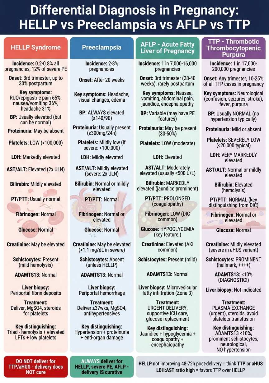
The 4-Column Quick Recall
| Feature | Preeclampsia | HELLP | AFLP | TTP |
|---|---|---|---|---|
| Hypertension | Always (+++) | Usually | Variable | Absent/mild |
| Proteinuria | Always | May be absent | Variable | Absent |
| Platelets | Mildly low | Low (<100k) | Moderately low | SEVERELY low (<20k) |
| LDH | ↑ | ↑↑ | ↑↑ | ↑↑↑↑ |
| AST/ALT | ↑ (if severe) | ↑↑ | ↑↑ (moderate) | Normal |
| Bilirubin | Normal/↑ | Mild ↑ | MARKED ↑ (jaundice) | ↑ (hemolysis) |
| PT/PTT | Normal | Normal | PROLONGED | Normal |
| Glucose | Normal | Normal | HYPOGLYCEMIA | Normal |
| Schistocytes | Absent | Mild (+) | Mild (+) | Prominent (++++) |
| ADAMTS13 | Normal | Normal | Normal | < 10% (DIAGNOSTIC) |
| Encephalopathy | Seizures (eclampsia) | Rare | Yes (liver failure) | Yes (microthrombi) |
| Definitive Rx | Deliver | Deliver | Deliver URGENTLY | Plasma exchange |
3 Critical Quiz Pearls
- Hypoglycemia = think AFLP - not seen in PE, HELLP, or TTP
- ADAMTS13 < 10% = TTP confirmed - test this in any TMA not responding to delivery
- HELLP/PE not improving 48-72h post-delivery = consider TTP or aHUS