
Copd
What would you like to know about COPD?
"COPD" AND "GOLD criteria" AND "diagnosis"
COPD GOLD staging spirometry diagnosis classification
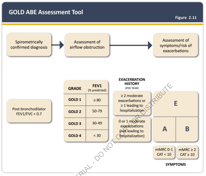
Summary : This figure presents the GOLD ABE Assessment Tool, a flowchart for classifying COPD severity and risk based on spirometric diagnosis, airflow obstruction, and symptoms/exacerbation history. It includes GOLD grades by FEV1, exacerbation history criteria, and a quadrant system (A, B, E) for symptom and risk assessment. flowchart: # Nodes : • "Spirometrically confirmed diagnosis" (rounded rectangle) • "Assessment of airflow obstruction" (rounded rectangle) • "Assessment of symptoms/risk of exacerbations" (rounded rectangle) • "Post-bronchodilator FEV1/FVC < 0.7" (rounded rectangle, side note) • GOLD grade table (rectangle): – "GRADE" column: GOLD 1, GOLD 2, GOLD 3, GOLD 4 – "FEV1 (% predicted)" column: ≥ 80, 50–79, 30–49, < 30 • Exacerbation history table (rectangle): – "≥ 2 moderate exacerbations or ≥ 1 leading to hospitalization" – "0 or 1 moderate exacerbations (not leading to hospitalization)" • Quadrant box (rectangle): – "E" (top right), "A" (bottom left), "B" (bottom right) – mMRC 0–1 / CAT < 10 (left), mMRC ≥ 2 / CAT ≥ 10 (right) – Symptoms axis labeled at bottom # Connectors : • Arrows connect: "Spirometrically confirmed diagnosis" → "Assessment of airflow obstruction" → "Assessment of symptoms/risk of exacerbations" • No explicit arrows between GOLD grade, exacerbation history, and quadrant box, but implied logical flow. # Layout : • Horizontal flow: diagnosis → airflow obstruction → symptoms/risk • GOLD grade and exacerbation history tables are central, feeding into quadrant box for final classification. • Quadrant box divides patients by exacerbation history (vertical) and symptoms (horizontal). # Analysis : • The tool provides a stepwise approach: confirm COPD by spirometry, grade airflow obstruction by FEV1, assess exacerbation risk, and classify symptoms using mMRC/CAT scores. • Patients are classified into A, B, or E groups based on exacerbation history and symptom burden. • GOLD grades (1–4) are determined by FEV1 % predicted, but final risk/symptom group (A/B/E) depends on exacerbation history and symptom scores. • The quadrant system allows for rapid visual categorization of patients for management decisions.
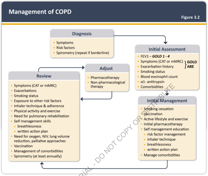
Summary : This flowchart outlines the comprehensive management process for Chronic Obstructive Pulmonary Disease (COPD), detailing the steps from diagnosis through assessment, initial management, ongoing review, and therapy adjustment. flowchart: # Nodes : • Diagnosis (rectangle): Symptoms, Risk factors, Spirometry (repeat if borderline) • Initial Assessment (rectangle): FEV1 – GOLD 1-4, Symptoms (CAT or mMRC), Exacerbation history, Smoking status, Blood eosinophil count, α1-antitrypsin, Comorbidities • Initial Management (rectangle): Smoking cessation, Vaccination, Active lifestyle and exercise, Initial pharmacotherapy, Self management education (risk factor management, inhaler technique, breathlessness, written action plan), Manage comorbidities • Review (rectangle): Symptoms (CAT or mMRC), Exacerbations, Smoking status, Exposure to other risk factors, Inhaler technique & adherence, Physical activity and exercise, Need for pulmonary rehabilitation, Self management skills (breathlessness, written action plan), Need for oxygen, NIV, lung volume reduction, palliative approaches, Vaccination, Management of comorbidities, Spirometry (at least annually) • Adjust (rectangle): Pharmacotherapy, Non-pharmacological therapy # Connectors : • Arrow from Diagnosis to Initial Assessment • Arrow from Initial Assessment to Initial Management • Arrow from Initial Management to Review • Arrow from Review to Adjust • Arrow from Adjust back to Review (forming a cycle) • Arrow from Review back to Initial Assessment (feedback loop) # Layout : • The flowchart is organized in a left-to-right and top-to-bottom sequence, with cyclical feedback between Review and Adjust, and a feedback loop from Review to Initial Assessment. • All nodes are rectangles with section headings and bullet points. • The GOLD classification (GOLD 1-4, GOLD ABE) is referenced in Initial Assessment. # Analysis : • The flowchart emphasizes a cyclical, iterative approach to COPD management, with regular review and adjustment of therapy based on ongoing assessment. • Key management elements include both pharmacological and non-pharmacological interventions, patient education, and comorbidity management. • The process is structured to ensure continuous monitoring and optimization of care, with feedback loops to reassess and refine treatment as needed.
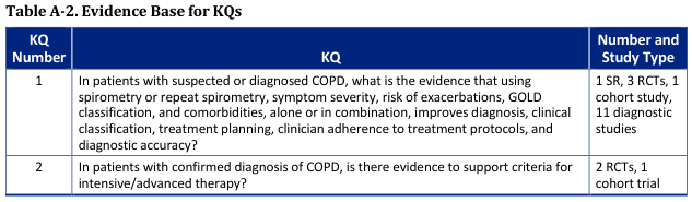
Table A-2. Evidence Base for KQs <table><thead><tr><th>KQ Number</th><th>KQ</th><th>Number and Study Type</th></tr></thead><tbody><tr><td>1</td><td>In patients with suspected or diagnosed COPD, what is the evidence that using spirometry or repeat spirometry, symptom severity, risk of exacerbations, GOLD classification, and comorbidities, alone or in combination, improves diagnosis, clinical classification, treatment planning, clinician adherence to treatment protocols, and diagnostic accuracy?</td><td>1 SR, 3 RCTs, 1 cohort study, 11 diagnostic studies</td></tr><tr><td>2</td><td>In patients with confirmed diagnosis of COPD, is there evidence to support criteria for intensive/advanced therapy?</td><td>2 RCTs, 1 cohort trial</td></tr></tbody></table>
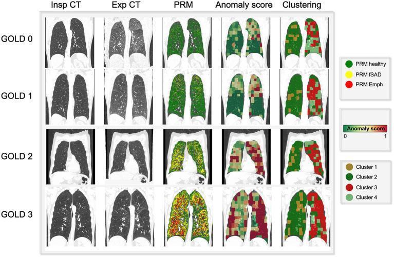
This composite diagnostic image illustrates the progression of Chronic Obstructive Pulmonary Disease (COPD) across GOLD stages 0 to 3 using multiple imaging modalities. The visual is organized into a grid: the first two columns show coronal slices of Inspiratory (Insp) and Expiratory (Exp) CT scans. Subsequent columns show color-coded overlays representing different analytical methods: Parametric Response Mapping (PRM), Anomaly Score, and Clustering. The PRM column tracks disease manifestation: 'PRM healthy' (green) dominates GOLD 0-1, while 'PRM fSAD' (functional small-airway disease, yellow) and 'PRM Emph' (emphysema, red) increase significantly in GOLD 2 and 3. The 'Anomaly Score' column displays a self-supervised learning gradient (green 0 to red 1), showing higher scores (more red) that spatially correlate with PRM-defined diseased areas. The 'Clustering' column identifies four distinct tissue types (Cluster 1-4). This comparison demonstrates how advanced computational mapping correlates with traditional GOLD staging, highlighting the spatial distribution of emphysema and air trapping in the upper and middle lung fields as disease severity advances.
COPD: Diagnosis & GOLD Staging
1. Diagnostic Criteria
Post-bronchodilator FEV1/FVC < 0.7 (fixed ratio criterion)
- Progressive dyspnea
- Chronic cough or sputum production
- Recurrent lower respiratory tract infections
- Multiple risk factors (smoking, occupational/environmental exposures)
2. Spirometry Interpretation
| Finding | Sensitivity | Specificity | LR+ |
|---|---|---|---|
| Absent cardiac dullness at LLSB | 15% | 99% | 15 |
| Early inspiratory crackles | 25-77% | 97-98% | 8-38.5 |
| Any unforced wheeze | 13-56% | 86-99% | 1-56 |
| Subxiphoid cardiac impulse | 4-27% | 97-99% | ~8 |
| Diaphragmatic excursion < 2 cm | 13% | 98% | 6.5 |
3. GOLD Grading of Airflow Limitation
| GOLD Grade | Severity | Post-bronchodilator FEV1 (% predicted) |
|---|---|---|
| GOLD 1 | Mild | ≥ 80% |
| GOLD 2 | Moderate | 50-79% |
| GOLD 3 | Severe | 30-49% |
| GOLD 4 | Very Severe | < 30% |
4. GOLD ABE Assessment Tool (Current System)
- Symptoms - measured by:
- mMRC dyspnea scale: score ≥ 2 = "high symptoms"
- CAT (COPD Assessment Test, 8 items): score ≥ 10 = "high symptoms"
- Exacerbation history in the prior year:
- ≥ 2 moderate exacerbations, OR ≥ 1 leading to hospitalization = high risk (Group E)
- 0 or 1 moderate exacerbation not leading to hospitalization = low exacerbation risk
| Group | Exacerbation history | Symptom burden |
|---|---|---|
| A | Low risk (0-1 moderate, not hospitalized) | Low (mMRC 0-1, CAT < 10) |
| B | Low risk | High (mMRC ≥ 2, CAT ≥ 10) |
| E | High risk (≥2 moderate or ≥1 hospitalization) | Any |
5. Chest Imaging Findings
6. Initial Pharmacologic Treatment Based on GOLD Group
| Patient Group | Recommended Initial Treatment |
|---|---|
| A | Short- or long-acting bronchodilator |
| B | Long-acting bronchodilator (LAMA or LABA) |
| C | LAMA |
| D | LAMA, or LAMA + LABA*, or ICS + LABA** |
Key Points to Remember
- Spirometry is mandatory - clinical diagnosis alone is not sufficient
- Use post-bronchodilator values for both the FEV1/FVC ratio and the FEV1 % predicted
- GOLD grade (1-4) reflects airflow severity; the letter group (A/B/E) reflects symptom burden + exacerbation risk
- Both dimensions together guide management decisions
- Spirometry should be repeated at least annually during follow-up
Create a summary document on COPD diagnosis and management
What format would you like the summary document in?
mkdir -p /tmp/workspace/copd-summary && ls /tmp/workspace/copd-summary/
~/copd-summary/create_copd_doc.py
from docx import Document
from docx.shared import Pt, Inches, RGBColor
from docx.enum.text import WD_ALIGN_PARAGRAPH
from docx.oxml.ns import qn
from docx.oxml import OxmlElement
import copy

doc = Document()

# ── Page margins ──────────────────────────────────────────────────────────────
for section in doc.sections:
    section.top_margin    = Inches(1)
    section.bottom_margin = Inches(1)
    section.left_margin   = Inches(1.2)
    section.right_margin  = Inches(1.2)

# ── Default Normal style ──────────────────────────────────────────────────────
normal = doc.styles["Normal"]
normal.font.name = "Calibri"
normal.font.size = Pt(11)

# ── Helper: shade a table row ─────────────────────────────────────────────────
def shade_row(row, hex_color):
    for cell in row.cells:
        tc   = cell._tc
        tcPr = tc.get_or_add_tcPr()
        shd  = OxmlElement("w:shd")
        shd.set(qn("w:val"),   "clear")
        shd.set(qn("w:color"), "auto")
        shd.set(qn("w:fill"),  hex_color)
        tcPr.append(shd)

def set_cell_font(cell, bold=False, size=11, color=None):
    for para in cell.paragraphs:
        for run in para.runs:
            run.font.bold = bold
            run.font.size = Pt(size)
            run.font.name = "Calibri"
            if color:
                run.font.color.rgb = RGBColor(*color)

def add_cell_text(cell, text, bold=False, size=11, color=None, align=None):
    cell.text = text
    for para in cell.paragraphs:
        if align:
            para.alignment = align
        for run in para.runs:
            run.font.bold  = bold
            run.font.size  = Pt(size)
            run.font.name  = "Calibri"
            if color:
                run.font.color.rgb = RGBColor(*color)

# ══════════════════════════════════════════════════════════════════════════════
# TITLE BLOCK
# ══════════════════════════════════════════════════════════════════════════════
title = doc.add_paragraph()
title.alignment = WD_ALIGN_PARAGRAPH.CENTER
run = title.add_run("CHRONIC OBSTRUCTIVE PULMONARY DISEASE (COPD)")
run.bold      = True
run.font.size = Pt(18)
run.font.name = "Calibri"
run.font.color.rgb = RGBColor(0x1F, 0x49, 0x7D)  # dark blue

sub = doc.add_paragraph()
sub.alignment = WD_ALIGN_PARAGRAPH.CENTER
srun = sub.add_run("Diagnosis & Management Summary  |  Based on GOLD Guidelines")
srun.font.size  = Pt(11)
srun.font.name  = "Calibri"
srun.italic     = True
srun.font.color.rgb = RGBColor(0x70, 0x70, 0x70)

doc.add_paragraph()  # spacer

# ══════════════════════════════════════════════════════════════════════════════
# SECTION HEADING helper
# ══════════════════════════════════════════════════════════════════════════════
def section_heading(text):
    p = doc.add_paragraph()
    run = p.add_run(text.upper())
    run.bold = True
    run.font.size = Pt(12)
    run.font.name = "Calibri"
    run.font.color.rgb = RGBColor(0xFF, 0xFF, 0xFF)
    pPr = p._p.get_or_add_pPr()
    shd = OxmlElement("w:shd")
    shd.set(qn("w:val"),   "clear")
    shd.set(qn("w:color"), "auto")
    shd.set(qn("w:fill"),  "1F497D")
    pPr.append(shd)
    p.paragraph_format.space_before = Pt(10)
    p.paragraph_format.space_after  = Pt(4)
    p.paragraph_format.left_indent  = Inches(0.1)
    return p

def sub_heading(text):
    p = doc.add_paragraph()
    run = p.add_run(text)
    run.bold = True
    run.font.size = Pt(11)
    run.font.name = "Calibri"
    run.font.color.rgb = RGBColor(0x1F, 0x49, 0x7D)
    p.paragraph_format.space_before = Pt(6)
    p.paragraph_format.space_after  = Pt(2)
    return p

def body(text):
    p = doc.add_paragraph(text)
    p.style = doc.styles["Normal"]
    p.paragraph_format.space_after = Pt(4)
    return p

def bullet(text, level=0):
    p = doc.add_paragraph(style="List Bullet")
    p.paragraph_format.left_indent   = Inches(0.3 + level * 0.25)
    p.paragraph_format.space_after   = Pt(2)
    run = p.add_run(text)
    run.font.size = Pt(11)
    run.font.name = "Calibri"
    return p

# ══════════════════════════════════════════════════════════════════════════════
# 1. DEFINITION & OVERVIEW
# ══════════════════════════════════════════════════════════════════════════════
section_heading("1. Definition & Overview")
body(
    "COPD is a common, preventable, and treatable disease characterised by persistent respiratory symptoms "
    "and airflow limitation due to airway and/or alveolar abnormalities, usually caused by significant "
    "exposure to noxious particles or gases (most commonly cigarette smoke)."
)

sub_heading("Key Components")
bullet("Chronic bronchitis: productive cough for ≥3 months in each of 2 consecutive years")
bullet("Emphysema: destruction of alveolar walls with permanent enlargement of air spaces")
bullet("Small-airway disease: inflammation and remodelling of small airways")

sub_heading("Major Risk Factors")
bullet("Cigarette smoking (primary cause - dose-dependent relationship)")
bullet("Occupational dusts and chemicals (coal, silica, grain dust, isocyanates)")
bullet("Indoor air pollution (biomass fuel combustion in poorly ventilated areas)")
bullet("Outdoor air pollution")
bullet("Alpha-1 antitrypsin (AAT) deficiency - genetic risk factor")
bullet("Recurrent childhood respiratory infections")

doc.add_paragraph()

# ══════════════════════════════════════════════════════════════════════════════
# 2. CLINICAL FEATURES
# ══════════════════════════════════════════════════════════════════════════════
section_heading("2. Clinical Features")
body("COPD should be suspected in any patient ≥35-40 years with relevant risk factors presenting with:")
bullet("Progressive breathlessness (initially on exertion, later at rest)")
bullet("Chronic cough (may be intermittent, may not be productive)")
bullet("Chronic sputum production")
bullet("Recurrent lower respiratory tract infections")
bullet("Wheeze")

sub_heading("Physical Examination Findings (with likelihood ratios)")

findings = [
    ["Finding", "Sensitivity", "Specificity", "LR+"],
    ["Absent cardiac dullness at LLSB", "15%", "99%", "15"],
    ["Early inspiratory crackles", "25-77%", "97-98%", "8-38.5"],
    ["Any unforced wheeze", "13-56%", "86-99%", "1-56"],
    ["Subxiphoid cardiac impulse", "4-27%", "97-99%", "~8"],
    ["Diaphragmatic excursion <2 cm", "13%", "98%", "6.5"],
    [">55 pack-year + wheeze + patient-reported wheeze", "-", "-", "156"],
]

tbl = doc.add_table(rows=len(findings), cols=4)
tbl.style = "Table Grid"
for i, row_data in enumerate(findings):
    row = tbl.rows[i]
    for j, text in enumerate(row_data):
        add_cell_text(row.cells[j], text, bold=(i == 0), size=10)
    if i == 0:
        shade_row(row, "BDD7EE")  # light blue header
    elif i % 2 == 0:
        shade_row(row, "F5F9FF")  # alternating row

body("\nNote: Absence of wheezing does NOT rule out COPD. Absence of smoking history is the most effective finding to rule out airflow obstruction (LR- 0.18).")
doc.add_paragraph()

# ══════════════════════════════════════════════════════════════════════════════
# 3. DIAGNOSIS
# ══════════════════════════════════════════════════════════════════════════════
section_heading("3. Diagnosis")

sub_heading("Spirometry (Mandatory)")
body(
    "Spirometry is required to confirm the diagnosis. All patients with suspected COPD and respiratory "
    "symptoms should undergo spirometry."
)

# Diagnostic criteria box
diag_para = doc.add_paragraph()
diag_run = diag_para.add_run(
    "  DIAGNOSTIC CRITERION:  Post-bronchodilator FEV1/FVC < 0.7  "
)
diag_run.bold = True
diag_run.font.size = Pt(12)
diag_run.font.name = "Calibri"
diag_run.font.color.rgb = RGBColor(0xFF, 0xFF, 0xFF)
diag_pPr = diag_para._p.get_or_add_pPr()
diag_shd = OxmlElement("w:shd")
diag_shd.set(qn("w:val"),   "clear")
diag_shd.set(qn("w:color"), "auto")
diag_shd.set(qn("w:fill"),  "C00000")  # red
diag_pPr.append(diag_shd)
diag_para.alignment = WD_ALIGN_PARAGRAPH.CENTER
diag_para.paragraph_format.space_before = Pt(6)
diag_para.paragraph_format.space_after  = Pt(6)

bullet("Values must be measured AFTER bronchodilator administration")
bullet("Patients with normal spirometry but chronic symptoms and emphysema on CT are NOT classified as COPD")
bullet("Airflow limitation may worsen over time, especially with continued noxious stimulus exposure")

sub_heading("Chest X-ray")
bullet("Not required for diagnosis, but supports assessment")
bullet("Classic findings: hyperinflated lungs, flattened diaphragm, increased AP diameter (lateral view)")
bullet("May show bullae, attenuated vascular markings in emphysema")

sub_heading("Additional Investigations")
bullet("ABG: assess hypoxaemia (PaO2 <8 kPa) and hypercapnia in severe disease")
bullet("FBC: polycythaemia (secondary to chronic hypoxaemia)")
bullet("Alpha-1 antitrypsin level: screen all patients with COPD, especially if onset <45 years or minimal smoking history")
bullet("CT thorax: not routine, but useful when bullectomy or lung volume reduction surgery is considered")
bullet("ECG/Echo: if right heart disease (cor pulmonale) suspected")
doc.add_paragraph()

# ══════════════════════════════════════════════════════════════════════════════
# 4. GOLD STAGING
# ══════════════════════════════════════════════════════════════════════════════
section_heading("4. GOLD Staging")

body("COPD severity is classified using the GOLD (Global Initiative for Chronic Obstructive Lung Disease) framework in two steps:")

sub_heading("Step 1: GOLD Grade - Airflow Limitation Severity (based on post-BD FEV1 % predicted)")

gold_grades = [
    ["GOLD Grade", "Severity", "FEV1 % Predicted"],
    ["GOLD 1",     "Mild",       "≥ 80%"],
    ["GOLD 2",     "Moderate",   "50-79%"],
    ["GOLD 3",     "Severe",     "30-49%"],
    ["GOLD 4",     "Very Severe","< 30%"],
]
grade_colors = ["BDD7EE", "E2EFDA", "FFE699", "F4B183", "FF7043"]

tbl2 = doc.add_table(rows=len(gold_grades), cols=3)
tbl2.style = "Table Grid"
tbl2.columns[0].width = Inches(1.2)
tbl2.columns[1].width = Inches(1.5)
tbl2.columns[2].width = Inches(2.5)
for i, row_data in enumerate(gold_grades):
    row = tbl2.rows[i]
    for j, text in enumerate(row_data):
        add_cell_text(row.cells[j], text, bold=(i == 0), size=11,
                      align=WD_ALIGN_PARAGRAPH.CENTER)
    shade_row(row, grade_colors[i])

sub_heading("Step 2: GOLD Group - ABE Assessment Tool (symptoms + exacerbation history)")
body("The letter group is determined by combining the mMRC/CAT symptom score with the prior year exacerbation history:")

abe_data = [
    ["Group", "Exacerbation History", "Symptom Burden (mMRC / CAT)", "Key Feature"],
    ["A",
     "0 or 1 moderate (not hospitalised)",
     "Low: mMRC 0-1 / CAT < 10",
     "Low risk, low symptoms"],
    ["B",
     "0 or 1 moderate (not hospitalised)",
     "High: mMRC ≥2 / CAT ≥10",
     "Low risk, more symptoms"],
    ["E",
     "≥2 moderate OR ≥1 hospitalisation",
     "Any",
     "High exacerbation risk"],
]
abe_colors = ["BDD7EE", "E2EFDA", "FFF2CC", "FCE4D6"]

tbl3 = doc.add_table(rows=len(abe_data), cols=4)
tbl3.style = "Table Grid"
for i, row_data in enumerate(abe_data):
    row = tbl3.rows[i]
    for j, text in enumerate(row_data):
        add_cell_text(row.cells[j], text, bold=(i == 0), size=10)
    shade_row(row, abe_colors[i])

body("\nFinal classification uses BOTH dimensions (e.g., 'GOLD 2, Group B'). This guides treatment intensity and monitoring frequency.")
doc.add_paragraph()

# ══════════════════════════════════════════════════════════════════════════════
# 5. ASSESSMENT TOOLS
# ══════════════════════════════════════════════════════════════════════════════
section_heading("5. Symptom Assessment Tools")

sub_heading("mMRC Dyspnea Scale")
mmrc_data = [
    ["Grade", "Description"],
    ["0", "Breathless only with strenuous exercise"],
    ["1", "Breathless when hurrying on level ground or walking up a slight hill"],
    ["2", "Walks slower than people of same age on level ground, or stops for breath on level ground after 100 m"],
    ["3", "Stops for breath after walking ~100 m or after a few minutes on level ground"],
    ["4", "Too breathless to leave the house, or breathless when dressing/undressing"],
]
tbl4 = doc.add_table(rows=len(mmrc_data), cols=2)
tbl4.style = "Table Grid"
tbl4.columns[0].width = Inches(0.8)
tbl4.columns[1].width = Inches(4.5)
for i, row_data in enumerate(mmrc_data):
    row = tbl4.rows[i]
    for j, text in enumerate(row_data):
        add_cell_text(row.cells[j], text, bold=(i == 0), size=10)
    if i == 0:
        shade_row(row, "BDD7EE")
    elif i % 2 == 0:
        shade_row(row, "F5F9FF")

body("\nCOPD Assessment Test (CAT): 8-item questionnaire scoring 0-40. CAT ≥10 = significant symptom burden.")
doc.add_paragraph()

# ══════════════════════════════════════════════════════════════════════════════
# 6. MANAGEMENT - STABLE COPD
# ══════════════════════════════════════════════════════════════════════════════
section_heading("6. Management of Stable COPD")

sub_heading("Goals of Therapy")
bullet("Reduce symptoms: dyspnoea, cough, improve exercise tolerance and health status")
bullet("Reduce future risk: prevent disease progression, prevent/treat exacerbations, reduce mortality")

sub_heading("Non-Pharmacological Interventions")

non_pharm = [
    ["Intervention", "Evidence / Notes"],
    ["Smoking cessation", "Reduces rate of FEV1 decline; improves survival. First priority for ALL smokers. Use NRT, bupropion, or varenicline + counselling"],
    ["Pulmonary rehabilitation", "Improves dyspnoea, exercise capacity, and quality of life. Recommended for all symptomatic patients (especially post-exacerbation)"],
    ["Oxygen therapy (LTOT)", "Indicated if resting PaO2 ≤7.3 kPa (or ≤8.0 kPa with cor pulmonale). Improves survival. Target SpO2 88-92%"],
    ["Vaccination", "Annual influenza vaccine; pneumococcal vaccine; COVID-19 vaccine. Reduces exacerbation risk"],
    ["Physical activity", "Maintain or increase daily activity. Prevents deconditioning"],
    ["Lung volume reduction surgery (LVRS)", "Selected patients with upper-lobe emphysema and low exercise capacity. Improves survival and function"],
    ["Lung transplantation", "Carefully selected end-stage COPD. Improves quality of life and exercise capacity"],
    ["NIV", "Long-term NIV in severe hypercapnia (PaCO2 >7 kPa). Reduces rehospitalisation risk"],
]
tbl5 = doc.add_table(rows=len(non_pharm), cols=2)
tbl5.style = "Table Grid"
tbl5.columns[0].width = Inches(2.0)
tbl5.columns[1].width = Inches(3.5)
for i, row_data in enumerate(non_pharm):
    row = tbl5.rows[i]
    for j, text in enumerate(row_data):
        add_cell_text(row.cells[j], text, bold=(i == 0), size=10)
    if i == 0:
        shade_row(row, "BDD7EE")
    elif i % 2 == 0:
        shade_row(row, "F5F9FF")

doc.add_paragraph()
sub_heading("Initial Pharmacotherapy by GOLD Group")

init_pharm = [
    ["Group", "First Choice", "Notes"],
    ["A", "Short- or long-acting bronchodilator (as needed or regular)", "Any bronchodilator; reassess effect"],
    ["B", "Long-acting bronchodilator\n(LAMA or LABA)", "LAMA preferred; LAMA+LABA if very symptomatic (CAT ≥20)"],
    ["E", "LAMA + LABA\nor ICS + LABA (if eos ≥300)", "Add ICS if eosinophils ≥300 cells/μL or prior hospitalisations. Avoid ICS monotherapy"],
]
tbl6 = doc.add_table(rows=len(init_pharm), cols=3)
tbl6.style = "Table Grid"
tbl6.columns[0].width = Inches(0.8)
tbl6.columns[1].width = Inches(2.5)
tbl6.columns[2].width = Inches(2.2)
for i, row_data in enumerate(init_pharm):
    row = tbl6.rows[i]
    for j, text in enumerate(row_data):
        add_cell_text(row.cells[j], text, bold=(i == 0), size=10)
    if i == 0:
        shade_row(row, "BDD7EE")
    elif i == 2:
        shade_row(row, "FCE4D6")
    elif i % 2 == 0:
        shade_row(row, "F5F9FF")

doc.add_paragraph()
sub_heading("Drug Classes - Key Points")

drugs = [
    ["Drug Class", "Examples", "Key Points"],
    ["SABA (short-acting β2-agonist)", "Salbutamol, terbutaline", "Relief inhaler. Onset <5 min. For acute symptom relief"],
    ["LABA (long-acting β2-agonist)", "Salmeterol, formoterol, indacaterol, olodaterol, vilanterol", "Twice-daily or once-daily. Reduces symptoms and exacerbations"],
    ["SAMA (short-acting muscarinic antagonist)", "Ipratropium", "Alternative/adjunct to SABA. 4x daily dosing"],
    ["LAMA (long-acting muscarinic antagonist)", "Tiotropium, aclidinium, umeclidinium, glycopyrronium", "Once-daily. Superior to LABA for exacerbation prevention. Main side effect: dry mouth"],
    ["ICS (inhaled corticosteroid)", "Fluticasone, budesonide, beclomethasone", "Add to LABA if eos ≥300 or frequent exacerbations. Risk of pneumonia with high doses"],
    ["Triple therapy (LABA+LAMA+ICS)", "Combination inhalers available", "Reduces mortality in selected patients. Use if persistent exacerbations on dual therapy"],
    ["PDE-4 inhibitor", "Roflumilast (oral)", "Add-on in severe COPD with chronic bronchitis phenotype. Reduces exacerbations. GI side effects"],
    ["Methylxanthines", "Theophylline (oral)", "Modest bronchodilation. Narrow therapeutic index. Use with caution"],
    ["Mucolytics", "Carbocisteine, N-acetylcysteine", "Consider in patients with persistent productive cough. Limited evidence"],
    ["Azithromycin (prophylactic)", "Azithromycin 250-500 mg daily/3x weekly", "Reduces exacerbations in selected patients. Risk: hearing loss, cardiac arrhythmia, antibiotic resistance"],
]
tbl7 = doc.add_table(rows=len(drugs), cols=3)
tbl7.style = "Table Grid"
tbl7.columns[0].width = Inches(1.8)
tbl7.columns[1].width = Inches(2.0)
tbl7.columns[2].width = Inches(2.0)
for i, row_data in enumerate(drugs):
    row = tbl7.rows[i]
    for j, text in enumerate(row_data):
        add_cell_text(row.cells[j], text, bold=(i == 0), size=9)
    if i == 0:
        shade_row(row, "BDD7EE")
    elif i % 2 == 0:
        shade_row(row, "F5F9FF")

doc.add_paragraph()

# ══════════════════════════════════════════════════════════════════════════════
# 7. EXACERBATIONS
# ══════════════════════════════════════════════════════════════════════════════
section_heading("7. COPD Exacerbations (AECOPD)")
body(
    "An acute exacerbation (AECOPD) is an acute worsening of respiratory symptoms from the patient's "
    "baseline day-to-day variation that requires additional therapy."
)

sub_heading("Severity Classification")
sev = [
    ["Severity", "Definition", "Management Setting"],
    ["Mild", "Worsening symptoms only; managed with short-acting bronchodilators alone", "Community/self-management"],
    ["Moderate", "Worsening symptoms requiring antibiotics and/or systemic corticosteroids", "Community/GP/outpatient"],
    ["Severe", "Requires emergency department visit or hospitalisation", "Hospital"],
    ["Very Severe", "Requires ICU admission with or without mechanical ventilation", "ICU"],
]
tbl8 = doc.add_table(rows=len(sev), cols=3)
tbl8.style = "Table Grid"
sev_colors = ["BDD7EE", "E2EFDA", "FFE699", "F4B183", "FF7043"]
for i, row_data in enumerate(sev):
    row = tbl8.rows[i]
    for j, text in enumerate(row_data):
        add_cell_text(row.cells[j], text, bold=(i == 0), size=10)
    shade_row(row, sev_colors[i])

sub_heading("Common Triggers")
bullet("Respiratory tract infection (viral most common - rhinovirus; bacterial in 30-50%)")
bullet("Most common bacteria: Haemophilus influenzae, Streptococcus pneumoniae, Moraxella catarrhalis")
bullet("Environmental pollutants and particulate matter")
bullet("Temperature changes")
bullet("Pulmonary embolism (consider if pleuritic chest pain or signs of right heart failure)")
bullet("Non-adherence to maintenance therapy")

sub_heading("Management of Moderate-to-Severe Exacerbations")

mgmt = [
    ["Intervention", "Details"],
    ["Bronchodilators (1st line)", "Increase frequency/dose of SABA. Add SAMA if not already used. Nebulised if severely breathless"],
    ["Systemic corticosteroids", "Prednisolone 30-40 mg oral (or IV methylprednisolone) for 5 days. Reduces recovery time and treatment failure"],
    ["Antibiotics", "Indicated if: increased sputum purulence AND increased sputum volume OR increased dyspnoea. 5-day course: amoxicillin, doxycycline, or clarithromycin (use co-amoxiclav if risk factors for resistant organisms)"],
    ["Controlled oxygen", "Target SpO2 88-92% (NOT 98-100%). Use Venturi mask. Avoid hypercapnic respiratory failure from excess O2"],
    ["NIV (BiPAP)", "Indicated if pH <7.35 with PaCO2 >6 kPa despite initial medical therapy. First-line for hypercapnic respiratory failure"],
    ["Invasive ventilation", "If NIV fails or contraindicated (pH <7.25, haemodynamic instability, reduced consciousness)"],
    ["DVT prophylaxis", "LMWH in all hospitalised patients"],
    ["Monitor & reassess", "ABG at 30-60 min after starting O2/NIV; daily monitoring of blood gases, electrolytes, fluid balance"],
]
tbl9 = doc.add_table(rows=len(mgmt), cols=2)
tbl9.style = "Table Grid"
tbl9.columns[0].width = Inches(2.2)
tbl9.columns[1].width = Inches(3.3)
for i, row_data in enumerate(mgmt):
    row = tbl9.rows[i]
    for j, text in enumerate(row_data):
        add_cell_text(row.cells[j], text, bold=(i == 0), size=10)
    if i == 0:
        shade_row(row, "BDD7EE")
    elif i % 2 == 0:
        shade_row(row, "F5F9FF")

doc.add_paragraph()

# ══════════════════════════════════════════════════════════════════════════════
# 8. DISCHARGE & FOLLOW-UP
# ══════════════════════════════════════════════════════════════════════════════
section_heading("8. Discharge Planning & Follow-up")

sub_heading("Post-Exacerbation Review (within 4-6 weeks)")
bullet("Review inhaler technique and adherence")
bullet("Review maintenance therapy - step up if needed")
bullet("Complete pulmonary rehabilitation (within 3 months of hospital discharge)")
bullet("Repeat spirometry after recovery (at least 3 months post-exacerbation)")
bullet("Assess for LTOT if still hypoxaemic at rest (PaO2 ≤7.3 kPa)")

sub_heading("Annual Monitoring in Stable COPD")
mon = [
    ["Parameter", "Frequency / Detail"],
    ["Spirometry", "At least annually"],
    ["Symptom scores (CAT/mMRC)", "Every visit"],
    ["Exacerbation history", "Every visit; document frequency and severity"],
    ["Smoking status", "Every visit; reinforce cessation"],
    ["Inhaler technique", "Every visit"],
    ["Blood eosinophil count", "Annually (guides ICS use)"],
    ["SpO2 / ABG", "If FEV1 <30% predicted or clinically hypoxaemic"],
    ["BMI / nutritional status", "Annually; low BMI is poor prognostic factor"],
    ["Comorbidity review", "Including cardiovascular disease, osteoporosis, anxiety/depression, metabolic syndrome"],
]
tbl10 = doc.add_table(rows=len(mon), cols=2)
tbl10.style = "Table Grid"
tbl10.columns[0].width = Inches(2.5)
tbl10.columns[1].width = Inches(3.0)
for i, row_data in enumerate(mon):
    row = tbl10.rows[i]
    for j, text in enumerate(row_data):
        add_cell_text(row.cells[j], text, bold=(i == 0), size=10)
    if i == 0:
        shade_row(row, "BDD7EE")
    elif i % 2 == 0:
        shade_row(row, "F5F9FF")

doc.add_paragraph()

# ══════════════════════════════════════════════════════════════════════════════
# 9. PROGNOSIS & SURVIVAL-MODIFYING INTERVENTIONS
# ══════════════════════════════════════════════════════════════════════════════
section_heading("9. Prognosis & Survival-Modifying Interventions")

body("The following interventions have proven survival benefit in COPD:")
bullet("Smoking cessation (most important - returns FEV1 decline rate towards non-smoker trajectory)")
bullet("Long-term oxygen therapy (LTOT) in chronically hypoxaemic patients")
bullet("Lung volume reduction surgery (LVRS) in selected emphysema patients")
bullet("Triple inhaled therapy (LABA + LAMA + ICS) reduces all-cause mortality in selected patients")
bullet("Pulmonary rehabilitation after hospitalisation reduces readmission and mortality")
bullet("Noninvasive positive-pressure ventilation (NIV/BiPAP) in severe hypercapnia")

sub_heading("BODE Index - Multidimensional Prognostic Score")
body("Predicts mortality better than FEV1 alone. Score 0-10 (higher = worse prognosis):")
bullet("B - BMI (0 if >21; 1 if ≤21)")
bullet("O - Obstruction: FEV1 % predicted (GOLD grade)")
bullet("D - Dyspnoea: mMRC score")
bullet("E - Exercise capacity: 6-minute walk test distance")

doc.add_paragraph()

# ══════════════════════════════════════════════════════════════════════════════
# FOOTER / REFERENCES
# ══════════════════════════════════════════════════════════════════════════════
section_heading("References & Sources")
body("1. Global Initiative for Chronic Obstructive Lung Disease (GOLD). Global Strategy for the Diagnosis, Management, and Prevention of Chronic Obstructive Pulmonary Disease. 2024 Report. www.goldcopd.org")
body("2. Murray & Nadel's Textbook of Respiratory Medicine, 2-Volume Set. Elsevier.")
body("3. Harrison's Principles of Internal Medicine, 22nd Edition. McGraw-Hill Medical, 2025.")
body("4. Rosen's Emergency Medicine: Concepts and Clinical Practice. Elsevier.")
body("5. Symptom to Diagnosis: An Evidence-Based Guide, 4th Edition.")

# ── Save ──────────────────────────────────────────────────────────────────────
output_path = "/tmp/workspace/copd-summary/COPD_Diagnosis_and_Management.docx"
doc.save(output_path)
print(f"Saved: {output_path}")
cd /tmp/workspace/copd-summary && python3 create_copd_doc.py
python3 -c " from docx import Document doc = Document('/tmp/workspace/copd-summary/COPD_Diagnosis_and_Management.docx') headings = [p.text for p in doc.paragraphs if p.text.strip() and p.style.name.startswith('Heading') or (p.runs and p.runs[0].bold and len(p.text) < 80)] print('Sections found:') for h in headings[:30]: print(' -', h) print(f'Total paragraphs: {len(doc.paragraphs)}') print(f'Total tables: {len(doc.tables)}') "
COPD Diagnosis and Management
Word Document · DOCX
| Section | Content |
|---|---|
| 1. Definition & Overview | Disease components, major risk factors |
| 2. Clinical Features | Symptoms, physical exam findings with LR+ values |
| 3. Diagnosis | Spirometry criterion (FEV1/FVC <0.7), CXR, additional investigations |
| 4. GOLD Staging | Grade 1-4 (FEV1 % predicted) + ABE group tool |
| 5. Symptom Assessment Tools | mMRC scale (grades 0-4), CAT score |
| 6. Management - Stable COPD | Non-pharmacological interventions + pharmacotherapy table by GOLD group + drug class reference |
| 7. COPD Exacerbations | Severity classification, triggers, management (O2 targets, NIV, steroids, antibiotics) |
| 8. Discharge & Follow-up | Post-exacerbation review checklist, annual monitoring table |
| 9. Prognosis | Survival-modifying interventions, BODE index |
My patient was never a smoker and has copd from last 9 months
"COPD" AND "never smoker" AND "etiology"
PMID: 35142054
COPD in a Never-Smoker: Key Considerations
Step 1: Verify the Diagnosis
- Was spirometry done post-bronchodilator? (FEV1/FVC < 0.7 is required)
- Is the obstruction fixed (not fully reversible with bronchodilator)?
- Was there adequate technique (3 acceptable manoeuvres, best 2 within 150 mL)?
- A 9-month history is unusually short for COPD - consider whether symptoms were present longer and only recently investigated
Step 2: Consider Differential Diagnoses First
| Condition | Distinguishing Features |
|---|---|
| Asthma | Significant bronchodilator reversibility (>200 mL + >12% FEV1 rise), atopy, episodic symptoms, younger onset, good response to ICS |
| Asthma-COPD Overlap (ACO) | History of asthma + fixed obstruction; high eosinophils; responds well to ICS |
| Bronchiectasis | Daily purulent sputum, clubbing, HRCT shows dilated bronchi; can coexist with COPD |
| Constrictive bronchiolitis (bronchiolitis obliterans) | Post-infection, post-transplant, connective tissue disease; CT shows mosaic attenuation on expiration |
| Hypersensitivity pneumonitis (chronic) | Antigen exposure history (birds, mould, hay); CT shows ground glass, fibrosis; BAL lymphocytosis |
| Diffuse panbronchiolitis | East Asian patients; chronic sinusitis; HRCT centrilobular nodules |
| Lymphangioleiomyomatosis (LAM) | Young women; cysts on CT; chylous pleural effusion |
| Sarcoidosis | Hilar lymphadenopathy, systemic features |
Step 3: Causes of COPD in Never-Smokers
1. Alpha-1 Antitrypsin (AAT) Deficiency - MOST IMPORTANT TO EXCLUDE
- Autosomal codominant; PiZZ genotype is the most severe form
- AAT normally inhibits neutrophil elastase - deficiency leads to progressive alveolar destruction
- Suspect strongly in this patient because: non-smoker, COPD onset, possibly young age
- Classic features: lower lobe predominant emphysema, family history of emphysema/liver disease, onset <45-50 years
- The ATS recommends testing ALL patients with persistent airflow obstruction for AAT deficiency
- Diagnostic threshold: serum AAT < 11 micromol/L (~50 mg/dL); confirm with genotyping
- Treatment implication: AAT augmentation therapy (IV infusion) is available for severe deficiency
2. Biomass/Indoor Air Pollution
- Most common cause of COPD globally in never-smokers
- Burning of wood, dung, crop residues, coal for cooking/heating in poorly ventilated spaces
- Highly relevant in South Asia, Sub-Saharan Africa, Latin America
- Produces a similar inflammatory cascade to cigarette smoke
3. Occupational Exposures
- Coal dust, silica, grain dust, isocyanates, cadmium, welding fumes
- Even without smoking, chronic high-level occupational dust exposure causes airflow obstruction
- Take a detailed occupational history spanning decades
4. Previous Tuberculosis (Post-TB COPD)
- TB causes airway destruction, bronchiectasis, fibrosis, and obstructive physiology
- A major cause of COPD in high-TB-burden countries (India, Southeast Asia, Sub-Saharan Africa)
- Ask about previous TB treatment; look for apical scarring/fibrosis on CXR/CT
5. Childhood Respiratory Disease / Lung Development
- Severe or recurrent childhood respiratory infections (pneumonia, whooping cough)
- Reduced maximal lung function attained at age 20-25 - never reached normal "peak" FEV1
- Premature birth, bronchopulmonary dysplasia
- These patients may develop COPD at lower absolute FEV1 levels earlier in life
6. Passive Smoking (Secondhand Smoke)
- Long-term household or workplace passive smoke exposure increases COPD risk
- Important to quantify years of exposure even if the patient never actively smoked
7. Outdoor Air Pollution
- Chronic exposure to high particulate matter (PM2.5), nitrogen dioxide, ozone
- Relevant in high-pollution urban environments
Step 4: Investigations to Order
| Test | Why |
|---|---|
| Serum AAT level | Screen for AAT deficiency (mandatory per ATS guidelines) |
| AAT phenotype/genotype | If AAT level low (<50 mg/dL or <11 micromol/L) |
| HRCT thorax | Identify distribution of emphysema (lower lobe = AAT), bronchiectasis, hyperinflation pattern, mosaic attenuation |
| Full PFTs including DLCO | DLCO reduced in emphysema; helps distinguish bronchitic vs emphysematous phenotype |
| Bronchodilator reversibility | Assess for asthma or ACO (>200 mL + >12% = significant) |
| Blood eosinophil count | Elevated eosinophils (>300 cells/μL) suggests ACO, guides ICS use |
| Total IgE + RAST/skin prick tests | Assess atopy (supports asthma/ACO) |
| Sputum culture | If chronic productive cough - exclude bronchiectasis with chronic infection |
| Mantoux/IGRA + CXR | Evidence of prior TB, especially if from endemic region |
| Echocardiogram | If significant hypoxaemia - exclude cor pulmonale, pulmonary hypertension |
| Detailed occupational history | Decades-long exposure history |
Step 5: Clinical Clues by Age
| Age group | Most likely cause in a never-smoker |
|---|---|
| <45 years | AAT deficiency, congenital lung anomaly, childhood lung disease, obliterative bronchiolitis |
| 45-60 years | AAT deficiency, biomass exposure, occupational, post-TB, ACO |
| >60 years | Biomass, occupational, passive smoking cumulative effect, post-infectious |
Key Message
Procalcitonin rapid positive in this patient is for
"procalcitonin" AND "COPD exacerbation" AND "antibiotic"
"procalcitonin" AND "COPD" AND "antibiotic"
PMID: 35549921
PMID: 40113161
Procalcitonin (PCT) Rapid Test Positive in Your COPD Patient
What a Positive PCT Means in This Context
- Decide to start antibiotics - and gives you objective justification
- Distinguish bacterial from viral exacerbation - the majority of AECOPD exacerbations are viral (especially rhinovirus), so PCT helps avoid unnecessary antibiotic use
- Gauge severity - a very high PCT suggests systemic bacterial infection or pneumonia, not just airway colonization
How PCT Works as a Biomarker
| Scenario | PCT level | Interpretation |
|---|---|---|
| Healthy / viral infection | < 0.1 ng/mL | No bacterial infection |
| Possible bacterial infection | 0.1 - 0.25 ng/mL | Unlikely bacterial; antibiotics not encouraged |
| Probable bacterial infection | 0.25 - 0.5 ng/mL | Antibiotics encouraged |
| Definite bacterial infection / sepsis | > 0.5 ng/mL | Antibiotics strongly recommended |
| Severe sepsis / septic shock | > 2 ng/mL | High bacterial load, systemic sepsis |
- PCT is produced by virtually all tissues in response to bacterial infection (especially gram-negative endotoxin and gram-positive exotoxins)
- In viral infections, interferon-gamma suppresses PCT release - so PCT stays low even during severe viral illness
- In fungal or parasitic infections, PCT can also rise, but less dramatically
Evidence Specifically in AECOPD
- Moderate quality evidence that serum PCT is associated with bacterial pathogens in sputum during AECOPD
- Bacterial AECOPD had a mean PCT 0.76 ng/mL higher than non-bacterial exacerbations
- PCT and CRP were the only two biomarkers with consistent evidence for distinguishing bacterial from non-bacterial AECOPD
- Sputum IL-8 and TNF-alpha also showed association but evidence was weaker
What to Do With a Positive PCT in Your COPD Patient
Start Antibiotics
| Severity | Setting | Antibiotic choice |
|---|---|---|
| Mild-moderate exacerbation | Outpatient | Amoxicillin, doxycycline, or clarithromycin (5 days) |
| Moderate exacerbation, risk factors* | Outpatient/hospital | Co-amoxiclav or levofloxacin |
| Severe (hospitalised) | Hospital | Co-amoxiclav IV or piperacillin-tazobactam + cover for Pseudomonas if risk factors present |
Also Add
- Prednisolone 30-40 mg for 5 days (reduces recovery time regardless of PCT)
- Increased bronchodilator frequency (SABA ± SAMA nebulised)
- Controlled oxygen - target SpO2 88-92% (not higher)
- Send sputum culture before starting antibiotics if possible
Important Nuances
-
PCT is not the only trigger for antibiotics in AECOPD - classic clinical criteria still apply: increased sputum purulence + increased volume + increased dyspnoea (Anthonisen criteria). If all three are present, antibiotics are indicated regardless of PCT.
-
PCT can be falsely elevated in non-bacterial conditions:
- Severe trauma or surgery
- Cardiogenic shock
- Medullary thyroid carcinoma (PCT is normally produced by thyroid C-cells)
- Some fungal infections
- Major burns
-
PCT can be falsely low despite bacterial infection in:
- Localised infections (empyema, abscess) without systemic spread
- Very early infection (PCT peaks at 24-48 hours)
- Immunosuppressed patients
-
Serial PCT monitoring is useful - a falling PCT (>80% drop from peak) confirms treatment response and can guide antibiotic de-escalation or stopping.
-
In the context of never-smoker COPD (your patient's situation) - if PCT is positive, also consider pneumonia as the presenting illness (rather than "just" a COPD exacerbation), especially if the patient has new consolidation on CXR or significant fever. Get a chest X-ray.
Summary
Which bacterial infection is more comoon
COPD exacerbation bacterial pathogens common organisms
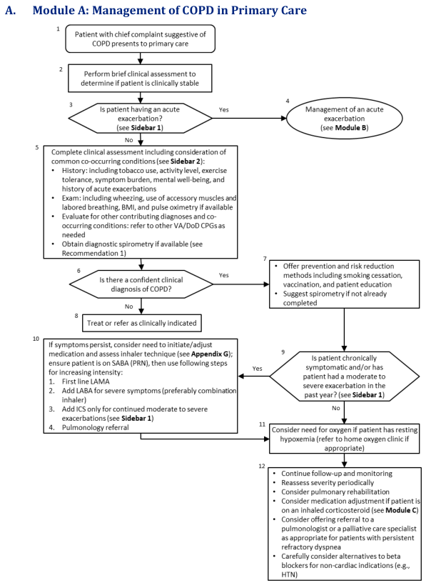
Summary : This flowchart outlines the stepwise management of Chronic Obstructive Pulmonary Disease (COPD) in primary care, including initial assessment, diagnosis, risk reduction, medication adjustment, and referral decisions. flowchart: # Nodes : • Start (rectangle): "Patient with chief complaint suggestive of COPD presents to primary care" • Rectangle: "Perform brief clinical assessment to determine if patient is clinically stable" • Diamond: "Is patient having an acute exacerbation? (see Sidebar 1)" • Ellipse: "Management of an acute exacerbation (see Module B)" • Rectangle: "Complete clinical assessment including consideration of common co-occurring conditions (see Sidebar 2): History, Exam, Evaluate for other contributing diagnoses, Obtain diagnostic spirometry if available" • Diamond: "Is there a confident clinical diagnosis of COPD?" • Rectangle: "Offer prevention and risk reduction methods including smoking cessation, vaccination, and patient education; Suggest spirometry if not already completed" • Rectangle: "Treat or refer as clinically indicated" • Diamond: "Is patient chronically symptomatic and/or has patient had a moderate to severe exacerbation in the past year? (see Sidebar 1)" • Rectangle: "If symptoms persist, consider need to initiate/adjust medication and assess inhaler technique (see Appendix G); ensure patient is on SABA (PRN), then use following steps for increasing intensity: 1. First line LAMA, 2. Add LABA for severe symptoms (preferably combination inhaler), 3. Add ICS only for continued moderate to severe exacerbations (see Sidebar 1), 4. Pulmonology referral" • Rectangle: "Consider need for oxygen if patient has resting hypoxemia (refer to home oxygen clinic if appropriate)" • Rectangle: "Continue follow-up and monitoring; Reassess severity periodically; Consider pulmonary rehabilitation; Consider medication adjustment if patient is on an inhaled corticosteroid (see Module C); Consider offering referral to a pulmonologist or a palliative care specialist as appropriate for patients with persistent refractory dyspnea; Carefully consider alternatives to beta blockers for non-cardiac indications (e.g., HTN)" # Connectors : • Arrows connect nodes in a top-down sequence, with diamonds representing decision points. • From "Is patient having an acute exacerbation?" (diamond): Yes → "Management of an acute exacerbation (ellipse)"; No → "Complete clinical assessment" (rectangle). • From "Is there a confident clinical diagnosis of COPD?" (diamond): Yes → "Offer prevention and risk reduction methods" (rectangle); No → "Treat or refer as clinically indicated" (rectangle). • From "Offer prevention and risk reduction methods" (rectangle) → "Is patient chronically symptomatic and/or has patient had a moderate to severe exacerbation in the past year?" (diamond). • From "Is patient chronically symptomatic..." (diamond): Yes → "If symptoms persist, consider need to initiate/adjust medication..." (rectangle); No → "Consider need for oxygen if patient has resting hypoxemia" (rectangle). • Both "If symptoms persist..." and "Consider need for oxygen..." nodes lead to "Continue follow-up and monitoring..." (rectangle). # Layout : • The flowchart is arranged vertically, with decision diamonds branching to different management paths. • Merges occur after medication adjustment and oxygen consideration, leading to a unified follow-up and monitoring node. • Sidebars and appendices are referenced for further detail at several steps. # Analysis : • The workflow prioritizes acute exacerbation management first, then proceeds to comprehensive assessment and diagnosis. • Preventive strategies and risk reduction are emphasized early for confirmed COPD cases. • Medication adjustment and specialist referral are considered for persistent symptoms or recent exacerbations. • Oxygen therapy is considered for patients with resting hypoxemia. • Ongoing follow-up, severity reassessment, and consideration of rehabilitation or specialist referral are recommended for all patients. • The flowchart supports a systematic, evidence-based approach to COPD management in primary care, with clear decision points and escalation pathways.
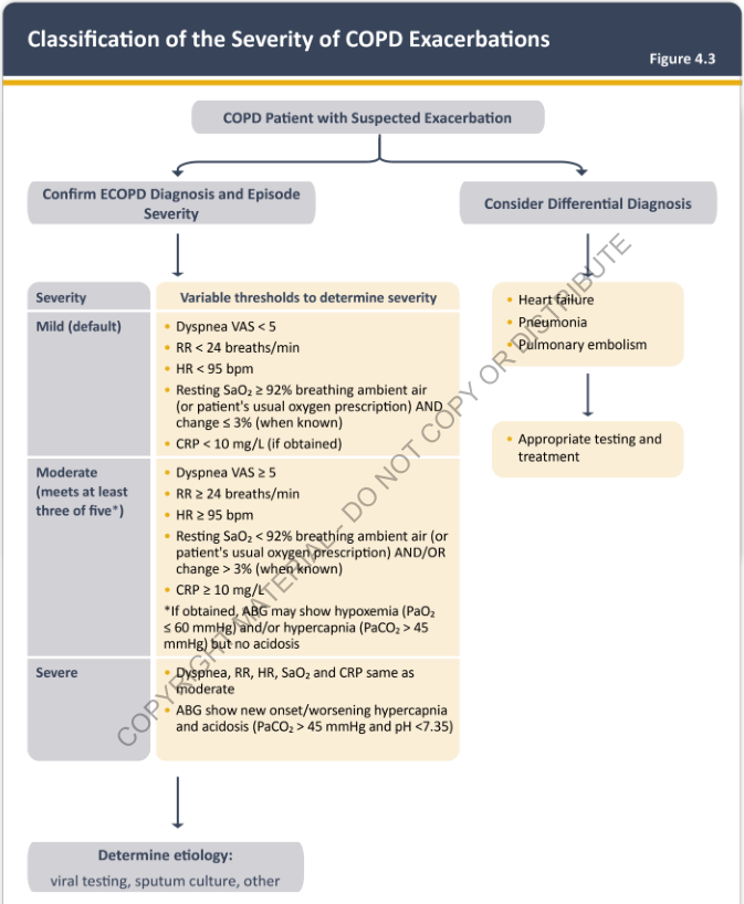
Summary : This flowchart provides a structured approach for classifying the severity of COPD exacerbations, guiding clinicians through diagnosis, severity assessment, differential diagnosis, and etiology determination using specific clinical thresholds. flowchart: # Nodes : • Start (rectangle): "COPD Patient with Suspected Exacerbation" • Decision (split): "Confirm ECOPD Diagnosis and Episode Severity" (left branch) / "Consider Differential Diagnosis" (right branch) • Severity classification (rectangle, left branch): "Severity" with three sub-nodes: – Mild (default) – Moderate (meets at least three of five*) – Severe • Variable thresholds (rectangle, left branch): For each severity level, lists criteria: – Mild: Dyspnea VAS < 5, RR < 24 breaths/min, HR < 95 bpm, Resting SaO₂ ≥ 92%, CRP < 10 mg/L – Moderate: Dyspnea VAS ≥ 5, RR ≥ 24 breaths/min, HR ≥ 95 bpm, Resting SaO₂ < 92%, CRP ≥ 10 mg/L, ABG may show hypoxemia/hypercapnia but no acidosis – Severe: Same as moderate, plus ABG shows new onset/worsening hypercapnia and acidosis (PaCO₂ > 45 mmHg and pH < 7.35) • Etiology determination (rectangle, left branch): "Determine etiology: viral testing, sputum culture, other" • Differential diagnosis (rectangle, right branch): "Heart failure", "Pneumonia", "Pulmonary embolism" • Testing/treatment (rectangle, right branch): "Appropriate testing and treatment" # Connectors : • Main flow starts at "COPD Patient with Suspected Exacerbation" and splits into two branches. • Left branch flows downward through severity classification, variable thresholds, and etiology determination. • Right branch flows downward through differential diagnosis and appropriate testing/treatment. • Severity classification is hierarchical: mild, moderate, severe, each with its own criteria. • Moderate severity requires meeting at least three of five listed criteria. • Severe is defined by ABG findings in addition to moderate criteria. # Layout : • Horizontal split after initial node: left for ECOPD diagnosis/severity, right for differential diagnosis. • Left branch is vertically organized: severity → thresholds → etiology. • Right branch is vertically organized: differential diagnosis → testing/treatment. • Colour coding: yellow boxes for variable thresholds and differential diagnosis. # Analysis : • The flowchart provides a clear, stepwise method for classifying COPD exacerbation severity using objective clinical criteria (dyspnea VAS, RR, HR, SaO₂, CRP, ABG). • Moderate severity is defined by a combination of symptoms and lab findings, requiring at least three criteria. • Severe exacerbation is distinguished by the presence of acidosis on ABG. • The chart emphasizes the importance of considering alternative diagnoses (heart failure, pneumonia, pulmonary embolism) and appropriate testing. • The final step is to determine the etiology of the exacerbation using laboratory tests.
Bacteria Most Commonly Causing AECOPD
The "Big Three" (most common overall)
| Rank | Organism | Key Features |
|---|---|---|
| 1st | Haemophilus influenzae | Most frequent - gram-negative rod; non-typeable strains predominate in COPD; produces beta-lactamase (20-30% resistant to amoxicillin) |
| 2nd | Streptococcus pneumoniae | Gram-positive diplococcus; all disease severities; pneumonia risk also high |
| 3rd | Moraxella catarrhalis | Gram-negative diplococcus; >90% produce beta-lactamase; often in milder disease |
Additional Organisms by Disease Severity
| COPD Severity | Additional Pathogens |
|---|---|
| Mild-moderate (FEV1 >50%) | Big Three above + Chlamydia pneumoniae (atypical) |
| Severe (FEV1 30-50%) | + Pseudomonas aeruginosa, Enterobacteriaceae (Klebsiella) |
| Very severe (FEV1 <30%) + bronchiectasis | Pseudomonas aeruginosa becomes the dominant concern |
| Any severity + prior antibiotic use | MRSA, resistant gram-negatives |
- FEV1 < 30% predicted
- Coexisting bronchiectasis (your patient should be screened for this)
- Previous isolation of Pseudomonas in sputum
- ≥4 antibiotic courses in the past year
- Recent hospitalisation (>2 days in past 3 months)
Special Note for Your Patient (Never-Smoker COPD)
- Non-typeable H. influenzae - persists in damaged airways
- Pseudomonas aeruginosa - if bronchiectasis is present
- Atypical organisms (Chlamydia pneumoniae, Mycoplasma pneumoniae) - particularly if subacute onset, dry cough, no response to standard beta-lactams
Antibiotic Choice Mapped to Organisms
| Scenario | First-line | Why |
|---|---|---|
| Uncomplicated mild-moderate | Amoxicillin or doxycycline | Covers S. pneumoniae, sensitive H. influenzae |
| Beta-lactamase concern | Co-amoxiclav | Covers H. influenzae and M. catarrhalis beta-lactamase producers |
| Atypical cover needed | Clarithromycin or doxycycline | Covers Chlamydia, Mycoplasma |
| Severe / hospitalised | Co-amoxiclav IV or levofloxacin | Broad gram-positive and gram-negative cover |
| Pseudomonas risk | Ciprofloxacin or pip-tazo | Must cover Pseudomonas |