What are the parts included in abdomen teach me with a diagram
abdomen parts regions anatomy diagram labeled
The Abdomen - Parts & Anatomy
- Superiorly - the diaphragm (at vertebral level T12 posteriorly, xiphoid process anteriorly)
- Inferiorly - the pelvic inlet
- Anteriorly - the abdominal wall muscles
- Posteriorly - the lumbar vertebrae (L1-L5)
📍 DIAGRAM 1: The Nine Abdominal Regions
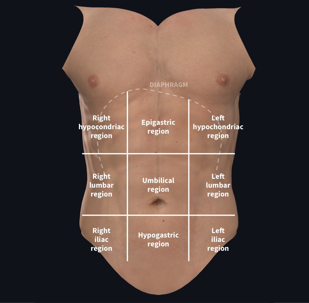
| Region | Location | Key Organs |
|---|---|---|
| Right Hypochondriac | Upper right | Right lobe of liver, gallbladder, right kidney, small intestine |
| Epigastric | Upper middle | Stomach, left lobe of liver, duodenum, head of pancreas |
| Left Hypochondriac | Upper left | Spleen, left kidney, stomach, tail of pancreas, part of colon |
| Right Lumbar | Middle right | Ascending colon, right kidney, part of liver |
| Umbilical | Middle center | Small intestine (jejunum/ileum), transverse colon, aorta |
| Left Lumbar | Middle left | Descending colon, left kidney, parts of small intestine |
| Right Iliac (Inguinal) | Lower right | Cecum, appendix, right ovary (females) |
| Hypogastric (Pubic) | Lower middle | Urinary bladder, uterus (females), sigmoid colon, rectum |
| Left Iliac (Inguinal) | Lower left | Sigmoid colon, left ovary (females) |
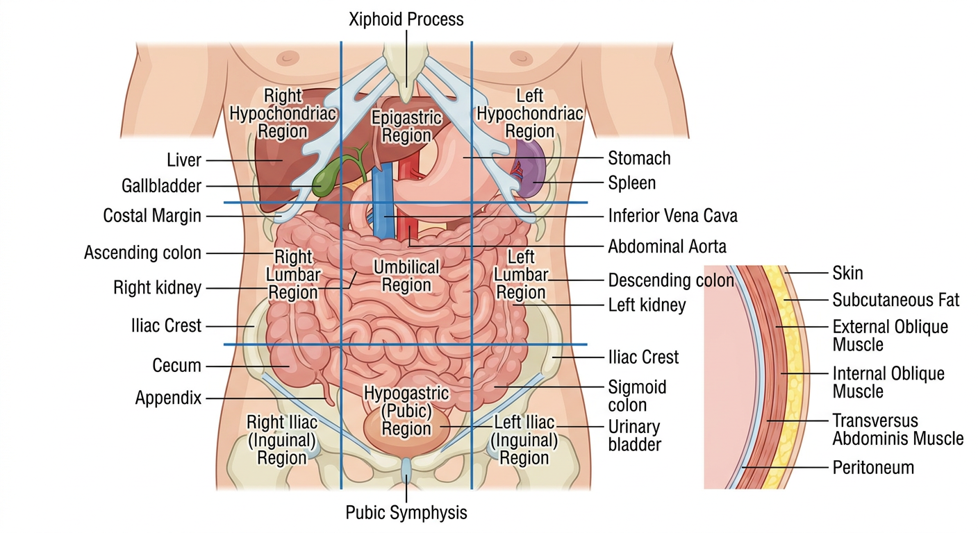
📍 DIAGRAM 2: Nine Regions with Planes

📍 DIAGRAM 3: Custom Labeled Anatomy Diagram
🔲 Alternative: 4-Quadrant Division
| Quadrant | Abbreviation | Contents |
|---|---|---|
| Right Upper Quadrant | RUQ | Liver, gallbladder, right kidney, duodenum, pancreas head, hepatic flexure of colon |
| Left Upper Quadrant | LUQ | Stomach, spleen, left kidney, pancreas body/tail, splenic flexure of colon |
| Right Lower Quadrant | RLQ | Cecum, appendix, right ovary/tube, right ureter |
| Left Lower Quadrant | LLQ | Sigmoid colon, left ovary/tube, left ureter |
🏗️ Layers of the Abdominal Wall
- Skin
- Subcutaneous fat (Camper's fascia)
- Scarpa's fascia (membranous layer of superficial fascia)
- External oblique muscle
- Internal oblique muscle
- Transversus abdominis muscle
- Transversalis fascia
- Preperitoneal fat
- Parietal peritoneum
🫀 Major Vessels in the Abdomen
- Abdominal aorta - runs down the midline, gives off celiac trunk (L1), superior mesenteric artery (L1), renal arteries (L1-L2), inferior mesenteric artery (L3), and bifurcates into common iliac arteries at L4
- Inferior vena cava (IVC) - runs to the right of the aorta, drains into the right atrium through the diaphragm at T8
📌 Key Landmarks
| Landmark | Vertebral Level |
|---|---|
| Xiphoid process | T9-T10 |
| Transpyloric plane | L1 |
| Subcostal plane | L3 |
| Umbilicus | L3-L4 |
| Transtubercular plane | L5 |
| Pubic symphysis | S3-S4 |
What are the parts included in the pelvis what should be learnt
pelvis anatomy parts regions bones muscles organs what to learn medical students diagram
What are the topic included in the pelvis
pelvis anatomy topics syllabus medical students Gray's anatomy curriculum
topics included in pelvis anatomy list
What are the topics included in the pelvis anatomy
pelvis anatomy complete topic list medical curriculum bony pelvis muscles organs nerves vessels perineum
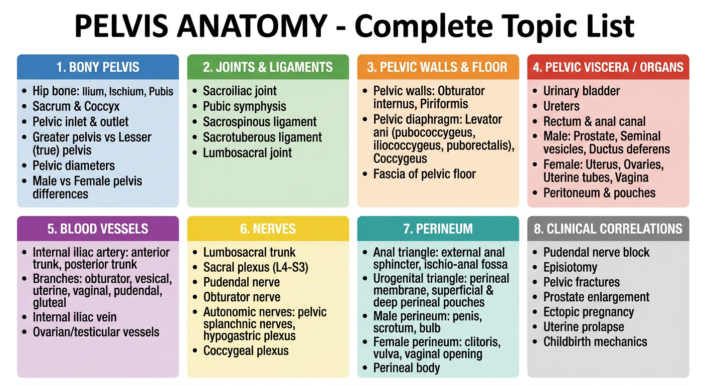
📚 Complete Topics in Pelvis Anatomy
1. 🦴 BONY PELVIS
- Hip bone (Os coxae) - made of 3 fused bones:
- Ilium - iliac crest, ASIS, AIIS, PSIS, iliac fossa, arcuate line
- Ischium - ischial spine, ischial tuberosity, lesser & greater sciatic notch
- Pubis - pubic symphysis, superior & inferior pubic ramus, obturator foramen
- Sacrum - sacral promontory, sacral foramina, sacral canal
- Coccyx
- Greater pelvis (false) vs Lesser pelvis (true)
- Pelvic inlet (brim) and pelvic outlet
- Pelvic diameters (obstetric conjugate ~11 cm, transverse ~13 cm)
- Male vs Female pelvis differences
2. 🔗 JOINTS & LIGAMENTS
- Sacroiliac joint - fibrocartilaginous, very strong
- Pubic symphysis - secondary cartilaginous joint
- Sacrospinous ligament - divides greater & lesser sciatic foramen
- Sacrotuberous ligament
- Lumbosacral joint
3. 💪 PELVIC WALLS & FLOOR
- Pelvic walls:
- Obturator internus
- Piriformis
- Pelvic floor (Pelvic diaphragm):
- Levator ani (3 parts):
- Pubococcygeus
- Iliococcygeus
- Puborectalis
- Coccygeus
- Levator ani (3 parts):
- Obturator canal and obturator membrane
- Fascia of pelvic floor and walls
4. 🫀 PELVIC VISCERA (Organs)
- Urinary bladder - trigone, detrusor muscle, neck
- Ureters - pelvic course, relation to uterine artery ("water under the bridge")
- Rectum & Anal canal
- Male organs: Prostate, Seminal vesicles, Ductus deferens, Ejaculatory ducts
- Female organs: Uterus, Ovaries, Uterine (Fallopian) tubes, Vagina
- Peritoneum - rectouterine pouch (of Douglas), vesicouterine pouch, rectovesical pouch (male)
5. 🩸 BLOOD VESSELS
- Internal iliac artery - main artery of the pelvis
- Posterior trunk: Iliolumbar, lateral sacral, superior gluteal arteries
- Anterior trunk: Umbilical, obturator, vesical, uterine/vaginal, middle rectal, internal pudendal, inferior gluteal arteries
- Internal iliac vein and tributaries
- Median sacral artery
- Ovarian/testicular vessels (from aorta)
- Lymphatics - internal, external, common iliac nodes
6. 🧠 NERVES
- Lumbosacral trunk (L4-L5)
- Sacral plexus (L4-S3):
- Superior & inferior gluteal nerves
- Sciatic nerve (largest nerve in the body)
- Pudendal nerve (S2-S4) - main nerve of perineum
- Nerve to obturator internus, piriformis, quadratus femoris
- Posterior cutaneous nerve of thigh
- Coccygeal plexus
- Autonomic nerves:
- Pelvic splanchnic nerves (parasympathetic S2-S4) - control erection & bladder
- Hypogastric plexus (sympathetic) - controls ejaculation
- Inferior hypogastric (pelvic) plexus
7. 🔺 PERINEUM
- Ischio-anal fossae
- External anal sphincter (deep, superficial, subcutaneous parts)
- Anococcygeal body
- Perineal membrane
- Deep perineal pouch: external urethral sphincter, deep transverse perineal muscle
- Superficial perineal pouch: erectile tissues, ischiocavernosus, bulbospongiosus muscles
- In men: crura of penis, bulb of penis, corpus cavernosum, corpus spongiosum
- In women: crura of clitoris, bulbs of vestibule, Bartholin's (greater vestibular) glands
- Perineal body - central fibromuscular node where everything converges
8. 🏥 CLINICAL CORRELATIONS
| Topic | Relevance |
|---|---|
| Pudendal nerve block | Analgesia in childbirth at ischial spine |
| Episiotomy | Cut at perineal body during delivery |
| Pelvic fractures | Risk to bladder, urethra, pudendal vessels |
| Prostate enlargement (BPH) | Urethral compression, urinary retention |
| Ectopic pregnancy | Rupture into rectouterine pouch |
| Uterine prolapse | Levator ani weakness |
| Ureteric injury | Risk in hysterectomy - ureter crosses under uterine artery |
| Varicocele | Dilated pampiniform plexus |
| Digital rectal exam | Palpates prostate posteriorly through rectum |
| Childbirth mechanics | Pelvic diameters, fetal head engagement |
📋 Study Order Recommendation (from Gray's)
1. Bony pelvis & landmarks
2. Joints & ligaments
3. Pelvic walls & floor muscles
4. Pelvic viscera (start with bladder → rectum → reproductive organs)
5. Peritoneum & fascial spaces
6. Nerves (somatic first, then autonomic)
7. Blood vessels & lymphatics
8. Perineum (anal triangle → urogenital triangle)
9. Clinical cases
Create a summary PDF of pelvis anatomy for quick revision
Pelvis Anatomy Quick Revision
PDF Document · PDF
What's inside the PDF (4 pages, color-coded):
| Section | Color | Contents |
|---|---|---|
| 1. Bony Pelvis | Navy | Hip bone parts, landmarks, pelvic diameters table, Greater vs Lesser pelvis |
| 2. Joints & Ligaments | Green | Sacroiliac, pubic symphysis, sacrospinous, sacrotuberous |
| 3. Pelvic Walls & Floor | Orange | Obturator internus, piriformis, levator ani (3 parts), coccygeus |
| 4. Pelvic Viscera | Red | Bladder, ureters, rectum, female organs, male organs with key relations |
| 5. Peritoneum & Fascia | Teal | All peritoneal pouches, fascial spaces (including pouch of Douglas) |
| 6. Blood Vessels | Purple | Full internal iliac artery branch table (anterior + posterior trunk), lymphatics |
| 7. Nerves | Blue | Sacral plexus table, autonomic NS (parasympathetic vs sympathetic) |
| 8. Perineum | Teal | Anal triangle, urogenital triangle, perineal pouches (male & female) |
| 9. Male vs Female Pelvis | Orange | 12-point comparison table |
| 10. Clinical High-Yields | Red | 12 clinical scenarios with anatomy basis |
| Mnemonics | Purple | 7 memory aids including "water under the bridge", "point and shoot" |
Make a pdf just like you did in pelvis with abdomen also
Abdomen Anatomy Quick Revision
PDF Document · PDF
What's inside (5 pages, color-coded):
| Section | Color | Contents |
|---|---|---|
| 1. Regions & Boundaries | Navy | 9 abdominal regions table with organs, 4-quadrant system with clinical uses |
| 2. Abdominal Wall | Green | 9 layers (skin to peritoneum), rectus sheath above/below arcuate line, all 5 muscles (origin/insertion/nerve/action), inguinal canal in full detail, hernia types |
| 3. Peritoneum | Teal | Intraperitoneal vs retroperitoneal organs, greater/lesser omentum, epiploic foramen, peritoneal pouches, all key ligaments |
| 4. GI Tract | Orange/Red | Stomach parts & blood supply, duodenum 4 parts with relations, jejunum vs ileum features, large intestine from caecum to rectum, Meckel's diverticulum |
| 5. Liver, Biliary & Pancreas | Brown | Liver lobes & Couinaud segments, biliary system, Calot's triangle, pancreas parts/ducts/zones |
| 6. Spleen & Kidneys | Purple | Spleen "1×3×5×7×9×11" rule, kidney relations, 3 ureteric narrowings, adrenal gland zones |
| 7. Blood Vessels | Red | Full aortic branch table (T12→L4), portal venous system + 4 porto-systemic anastomoses, IVC tributaries |
| 8. Nerves | Blue | T7–T12 dermatomes, lumbar plexus table, autonomic supply (vagus vs pelvic splanchnics, splenic flexure watershed) |
| 9. Posterior Wall | Green | Psoas, iliacus, quadratus lumborum; vertebral level landmarks (T8 to L5) |
| 10. Clinical High-Yields | Red | 12 clinical scenarios (appendicitis, pancreatitis, AAA, hernias, portal HTN, etc.) |
| Mnemonics | Purple | 10 memory aids including SAD PUCKER, Rule of 2's, T8/T10/T12 diaphragm openings |