


From cutaneous skin infections mentioned in the first picture till the last image of the three images, give me a notes, short notes, a made-up notes, which I can present tomorrow to my ma'am. Only cutaneous skin infections. Make it concise.
Cutaneous Skin Infections: Short Notes
1. Impetigo, Ecthyma and Furunculosis
- It is a common superficial bacterial infection.
- Causes: S. aureus (most often) and group A β-hemolytic streptococci.
- The primary lesion is a superficial pustule that ruptures and leaves a honey-colored (yellow-brown) crust.
- It can occur on normal skin (primary) or on already diseased skin (secondary).
- Bullous impetigo is the less common form, with tense, clear bullae.
- The bullae are caused by exfoliative toxin of S. aureus phage type II.
- This is the same toxin responsible for staphylococcal scalded-skin syndrome (SSSS).
- SSSS is more common in children. In adults, consider it along with TEN and severe drug eruptions when there is widespread blistering.
- It is a deep, non-bullous variant of impetigo.
- It causes punched-out ulcerative lesions.
- It is more often caused by S. pyogenes.
- It is deeper than impetigo and heals with scarring.
- Gentle debridement of adherent crusts, using soaks.
- Topical antibiotics plus appropriate oral antibiotics.
- Cause: S. aureus. It has become more prominent because of CA-MRSA.
- A furuncle (boil) is a painful, erythematous nodule that can occur on any cutaneous surface.
- Lesions may be solitary but are most often multiple.
- Patients often think they were bitten by a spider or insect.
- Family or close contacts may also be affected.
- Treatment:
- Incision and drainage may be enough for a small solitary furuncle without cellulitis or systemic symptoms.
- Send lesional material for culture whenever possible.
- Use antibiotics according to MSSA or CA-MRSA coverage.
- Warm compresses and nasal mupirocin help.
- Severe infection may need IV antibiotics.
2. Erysipelas and Cellulitis
- The page only refers to Chap. 134 for these.
3. Dermatophytosis (Ringworm)
- Organisms: Trichophyton, Microsporum, Epidermophyton. They infect skin, hair and nails.
- Typical lesion: erythematous, scaly, annular plaque. This gives the name "ringworm".
- Deep inflammatory nodules or granulomas can occur, usually after inappropriate treatment with mid- to high-potency topical steroids.
| Type | Key points |
|---|---|
| Tinea corporis | Glabrous (hairless) skin. Variable appearance. |
| Tinea cruris | Groin. More common in males. Scaling, erythematous, spares the scrotum. |
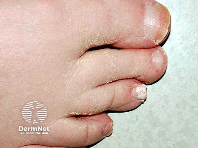
| Tinea pedis | Most common dermatophyte infection. Often chronic. Erythema, scaling, itching, occasional vesicles. Usually involves the web space between the 4th and 5th toes. |
| Tinea unguium (onychomycosis) | Thickened, opacified nails with subungual debris. The distal-lateral variant is most common. Proximal subungual type may be a marker of HIV or other immunocompromise. |
| Tinea capitis | Mainly inner-city children, also immunocompromised adults. Main organism: T. tonsurans, which gives mild scaling and hair loss. T. tonsurans and Microsporum canis can cause a boggy inflammatory mass called a kerion. |
- Skin, nail or hair scrapings.
- Culture or direct microscopy with KOH.
- Nail clippings with PAS stain.
- Topical therapy is enough for uncomplicated tinea corporis, cruris and limited pedis.
- Apply twice daily and continue for 1 week beyond clinical cure.
- Tinea pedis needs longer courses and often relapses.
- Oral antifungals are needed for hair and nail infection and for disease that fails topical therapy.
- Topicals are not effective alone for tinea capitis or onychomycosis.
- Confirm the fungus by microscopy or culture before giving oral agents.
- All oral agents may cause hepatotoxicity. Avoid them in pregnancy and breast-feeding.
- Nystatin is not active against dermatophytes.
- Inflammatory tinea capitis may scar and cause hair loss. Add a systemic antifungal, plus steroids if needed.
| Drug | Points |
|---|---|
| Griseofulvin | Approved for skin, hair and nail dermatophytosis. Side effects: GI upset, headache, urticaria. |
| Itraconazole | Approved for onychomycosis. Many CYP450 drug interactions. Avoid in ventricular dysfunction or CHF. |
| Terbinafine | Approved for onychomycosis, and the granule form for tinea capitis. Fewer interactions than itraconazole. |
| Ketoconazole (oral) | Not first line for any fungal infection (FDA warning on hepatotoxicity). Topical ketoconazole is not affected. |
4. Tinea (Pityriasis) Versicolor
- Organism: Malassezia furfur. It is a dimorphic, non-dermatophytic fungus and a normal skin inhabitant.
- It is promoted by heat and humidity.
- Lesions: oval scaly macules, papules and patches on the chest, shoulders and back. The face and distal extremities are rarely involved.
- On dark skin they look hypopigmented.
- On light skin they look slightly erythematous or hyperpigmented.
- KOH prep: short hyphae plus round spores, described as "spaghetti and meatballs".
- Treatment:
- Topical lotions or shampoos containing sulfur, salicylic acid, selenium sulfide, or azoles.
- Use daily for 1 to 2 weeks, then weekly.
- Wash off completely, because selenium is irritating if left on for more than 10 minutes.
- Oral agents work but give no lasting result and are not FDA approved for this.
5. Candidiasis
- Organism: usually Candida albicans, a normal GI saprophyte.
- Predisposing factors:
- Broad-spectrum antibiotics.
- Diabetes mellitus.
- Immunosuppression.
- Chronic moisture and maceration.
- Poorly fitting dentures.
- It is a very common infection in HIV patients.
- Sites and features:
- Oral cavity: thrush (white plaques on the tongue or buccal mucosa).
- Corners of the mouth: perleche (fissured, macerated lesions).
- Nail folds and nails: paronychia and onycholysis.
- Intertriginous areas: edematous, erythematous, scaly lesions with satellite pustules.
- Males: penis and scrotum may be involved. Dermatophyte infection spares the scrotum.
- Candidal infections are painful with marked inflammation. Dermatophyte infections are less so.
- Diagnosis: clinical pattern, plus yeast (pseudohyphae) on KOH or culture.
- Treatment:
- Remove predisposing factors.
- Topical nystatin or azoles (miconazole, clotrimazole, econazole, ketoconazole).
- A mild steroid (2.5% hydrocortisone) can be added for inflammation on glabrous skin.
- Systemic therapy is for immunosuppressed patients or those who fail topical therapy.
- Oral fluconazole is most commonly used for cutaneous candidiasis.
- Oral nystatin works only for GI tract candidiasis.
6. Warts (Papillomavirus)
- Warts are cutaneous neoplasms caused by HPV. More than 100 types are known.
- Types:
- Verruca vulgaris: sessile, dome-shaped, about 1 cm, hyperkeratotic surface.
- Plantar warts: endophytic with thick keratin. Paring shows keratin debris and punctate bleeding points.
- Filiform warts: face, neck and skinfolds, on a narrow base.
- Flat warts (verruca plana): velvety, slightly elevated. Face, arms, legs. Often spread by shaving.
- Genital warts: small papillomas that can grow into large fungating lesions.
- HPV and cancer:
- HPV 16 and 18 are major risk factors for intraepithelial neoplasia and squamous cell carcinoma (cervix, anus, vulva, penis).
- Risk is higher in transplant recipients and HIV patients.
- Patients on long-term immunosuppression should be monitored for SCC and other cutaneous malignancies.
- Treatment:
- Most warts in normal individuals resolve spontaneously in 1 to 2 years.
- No single therapy works for all warts, and recurrence is common.
- Cryotherapy with liquid nitrogen is the most useful and convenient method.
- Keratolytics such as salicylic acid plasters need more compliance.
- Genital warts:
- Podophyllin solution (in-office).
- Topical imiquimod (induces local cytokine release).
- Sinecatechins (green tea extract).
- Recalcitrant warts may need conventional or laser surgery.
- The HPV vaccine reduces anogenital warts and cervical carcinoma.
7. Herpes Simplex and Herpes Zoster
- The page only refers to Chap. 197 (herpes simplex) and Chap. 198 (herpes zoster).
Quick Revision Table (Table 60-5)
| Infection | Clinical features | Agent | Treatment |
|---|---|---|---|
| Impetigo | Honey-colored crusted papules, plaques or bullae | Group A Streptococcus, S. aureus | Systemic or topical antistaphylococcal and antistreptococcal antibiotics |
| Dermatophytosis | Annular scaly plaques, hair loss, groin spares scrotum, hyphae on KOH | Trichophyton, Epidermophyton, Microsporum | Topical azoles, systemic griseofulvin, terbinafine or azoles |
| Candidiasis | Papules and plaques with satellite pustules, intertriginous, may involve scrotum, pseudohyphae on KOH | C. albicans and other Candida spp. | Topical nystatin or azoles, systemic azoles for resistant disease |
| Tinea versicolor | Hyper- or hypopigmented scaly patches on trunk, "spaghetti and meatballs" on KOH | Malassezia furfur | Topical selenium sulfide lotion or azoles |
Make a PPT from these notes for tomorrow's presentation
ls -la /tmp/workspace/; python -c "import pptx;print(pptx.__version__)"
Quick answer
Slide 1: Title
Slide 2: Overview
- Bacterial: impetigo, ecthyma, furunculosis
- Fungal: dermatophytosis, tinea versicolor, candidiasis
- Viral: warts
- Erysipelas, cellulitis, herpes simplex and zoster are covered in other chapters (134, 197, 198)
Slide 3: Impetigo
- Superficial bacterial infection
- Causes: S. aureus (most often) and group A β-hemolytic streptococci
- The pustule ruptures and leaves a honey-colored crust
- Occurs on normal skin (primary) or already diseased skin (secondary)
- Bullous impetigo: tense, clear bullae caused by exfoliative toxin of S. aureus phage type II. This is the same toxin as in SSSS.
Slide 4: Ecthyma
- Deep, non-bullous variant of impetigo
- Punched-out ulcerative lesions
- More often caused by S. pyogenes
- Deeper than impetigo, so it heals with scarring
- Treatment for both:
- Gentle debridement of crusts, using soaks
- Topical antibiotics plus appropriate oral antibiotics
Slide 5: Furunculosis
- Cause: S. aureus, more prominent now because of CA-MRSA
- A painful, erythematous nodule (boil); lesions are most often multiple
- Patients often think it was a spider bite; close contacts may be affected
- Treatment:
- Incision and drainage for small solitary lesions
- Send material for culture
- Antibiotics according to MSSA or CA-MRSA
- Warm compresses and nasal mupirocin
- IV antibiotics if severe
Slide 6: Dermatophytosis (Ringworm)
- Fungi: Trichophyton, Microsporum, Epidermophyton
- Lesion: erythematous, scaly, annular plaque
- Deep granulomas can follow inappropriate steroid use
- Diagnosis:
- KOH microscopy or culture
- PAS stain for nail clippings
Slide 7: Types of Tinea
| Type | Key point |
|---|---|
| Corporis | Glabrous skin |
| Cruris | Groin, males; spares the scrotum |
| Pedis | Most common; web space between the 4th and 5th toes |
| Unguium | Thick, opacified nails; proximal type may signal HIV |
| Capitis | Children; T. tonsurans; kerion (boggy inflammatory mass) |
Slide 8: Treatment of Dermatophytosis
- Topical for corporis, cruris and limited pedis
- Twice daily, continued 1 week after clinical cure
- Oral for hair, nails and failures of topical treatment
- Confirm by microscopy or culture first
- Hepatotoxicity risk; avoid in pregnancy and breast-feeding
- Nystatin is not active against dermatophytes
Slide 9: Oral Antifungals
| Drug | Key point |
|---|---|
| Griseofulvin | Skin, hair, nails; GI upset, headache, urticaria |
| Itraconazole | Onychomycosis; CYP450 interactions; avoid in CHF |
| Terbinafine | Onychomycosis, tinea capitis (granules); fewer interactions |
| Ketoconazole (oral) | Not first line (FDA hepatotoxicity warning) |
Slide 10: Tinea Versicolor
- Organism: Malassezia furfur, a normal skin inhabitant
- Promoted by heat and humidity
- Site: chest, shoulders and back
- Dark skin: hypopigmented; light skin: erythematous or hyperpigmented
- KOH: "spaghetti and meatballs"
- Treatment:
- Selenium sulfide, sulfur, salicylic acid or azoles
- Daily for 1 to 2 weeks, then weekly
- Wash off within 10 minutes (irritant)
Slide 11: Candidiasis
- Organism: Candida albicans (normal GI saprophyte)
- Predisposing factors: broad-spectrum antibiotics, diabetes, immunosuppression, moisture, dentures; common in HIV
- Sites:
- Thrush (oral)
- Perleche (corners of the mouth)
- Paronychia
- Intertriginous areas with satellite pustules
- Painful with marked inflammation; may involve the scrotum
- KOH: pseudohyphae
Slide 12: Treatment of Candidiasis
- Remove predisposing factors
- Topical nystatin or azoles (miconazole, clotrimazole, econazole, ketoconazole)
- Mild steroid (2.5% hydrocortisone) for inflammation on glabrous skin
- Systemic therapy is for immunosuppressed patients or failures of topical treatment
- Oral fluconazole for cutaneous disease
- Oral nystatin works only for the GI tract
Slide 13: Warts (HPV)
- Cutaneous neoplasms caused by papillomavirus (more than 100 types)
- Types: verruca vulgaris, plantar, filiform, flat (often spread by shaving), genital
- HPV 16 and 18 are linked to cervical, anal, vulvar and penile carcinoma
- Higher risk in transplant and HIV patients; monitor for SCC
Slide 14: Treatment of Warts
- Most resolve spontaneously in 1 to 2 years
- Cryotherapy (liquid nitrogen) is the most convenient method
- Salicylic acid plasters
- Genital warts: podophyllin, imiquimod, sinecatechins
- Surgical or laser removal for recalcitrant warts
- Recurrence is common
- HPV vaccine reduces anogenital warts and cervical carcinoma
Slide 15: Quick Revision (Table 60-5)
| Infection | Agent | Treatment |
|---|---|---|
| Impetigo | Group A Strep, S. aureus | Antistaphylococcal and antistreptococcal antibiotics |
| Dermatophytosis | Trichophyton, Epidermophyton, Microsporum | Topical azoles; griseofulvin, terbinafine |
| Candidiasis | Candida spp. | Nystatin or azoles |
| Tinea versicolor | Malassezia furfur | Selenium sulfide or azoles |
Slide 16: Summary and Thank You
- Impetigo has honey-colored crusts; ecthyma scars
- Tinea cruris spares the scrotum; candidiasis may involve it
- Take a KOH prep before treating fungal infections
- Most warts clear on their own
- Questions?
So the response that you have sent written above, I want you to make it in an easy language that can make me understand in a easy way, and also explain it in an easy way. Also try to include images of the respective infections.
impetigo honey-colored crust
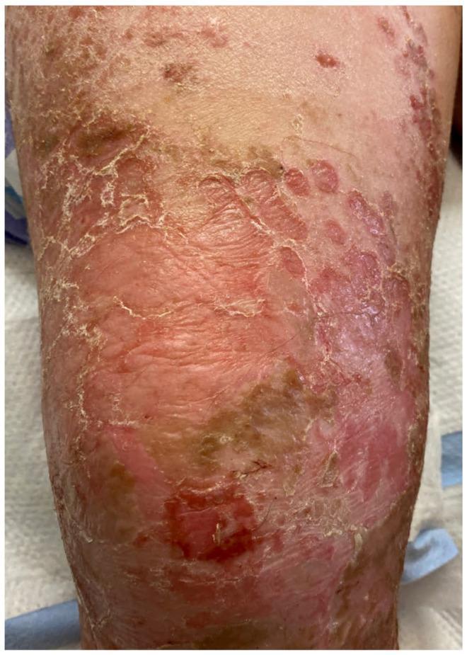
A clinical photograph displaying bullous impetigo on an extremity. The image shows the characteristic progression following the rupture of flaccid bullae. Key features include multiple superficial erosions with raw, erythematous bases, surrounded by prominent peripheral collarettes of scale representing the remnants of the blister roof. A thin, light brown (varnish-like) crust is visible in several areas, distinct from the thicker, honey-colored crust associated with non-bullous impetigo. The lesions are confluent in some regions, demonstrating the highly contagious spread of the infection within the superficial epidermis. This visual representation is an educational example of bacterial skin infection caused by Staphylococcus aureus, highlighting the diagnostic features of post-bullous erosion and scaling suitable for medical training in dermatology and pediatrics.
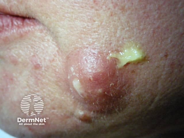
This is a high-resolution clinical photograph of a localized facial lesion on the left cheek/perioral region. Imaging modality: standard dermatology photography; viewpoint: anterior-lateral view. The lesion shows a red, erythematous plaque with a centered yellow crust, suggestive of a crusted exudate over an eroded surface. The surrounding skin is mildly inflamed with intermittent scaling. The crust appears honey-colored and adherent, characteristic of impetigo, a contagious superficial bacterial infection commonly caused by Staphylococcus aureus or Streptococcus pyogenes. Differential diagnoses include nonbullous impetigo vs bullous impetigo, bacterial folliculitis, contact dermatitis with secondary crusting, or eczema with secondary infection. No evident vesicles beyond the crust; no facial vesicular eruption is visible. If infection is present, treatment typically involves topical antibiotics (mupirocin, fusidic acid) or systemically if extensive, with emphasis on hygiene to prevent spread. The image emphasizes early recognition of impetiginous lesions on the face in adults and children. Clinically correlated signs such as fever, regional lymphadenopathy, or spreading redness would guide management. For radiographic correlation, consider bacterial culture in persistent cases or when confirming etiology. This photograph educates dermatology students on morphological features of crusted erythematous plaques. Note lesion color, crust density, and surrounding edema for assessment.
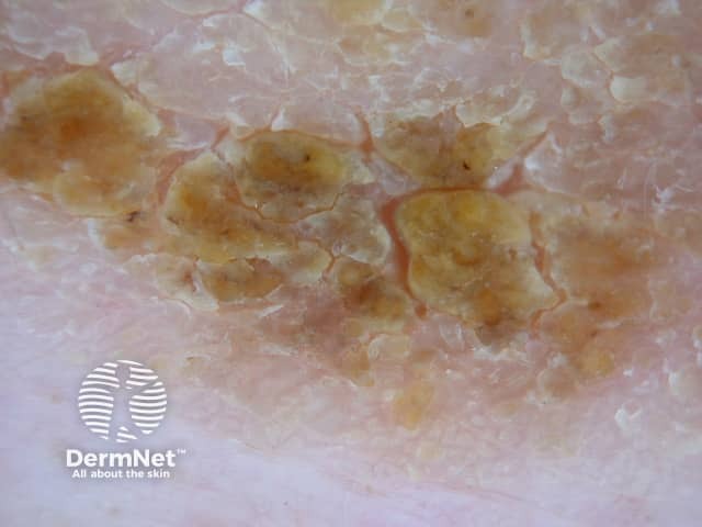
Clinical dermatology photograph illustrating multiple circular or oval plaques on pink, near-normal skin, each with a prominent golden-yellow crust consistent with impetigo. The image is a macroscopic skin photograph captured under diffuse ambient lighting with non-magnified view of the trunk or proximal limb region. Visual features include centrally eroded areas surrounded by a thick crust that appears amber to brown, with mild surrounding erythema and slight scaling. Lesions are clustered in a roughly linear or patchy distribution, lacking deep ulceration or purulent discharge. This presentation is typical of nonbullous impetigo, a superficial bacterial infection most often caused by Staphylococcus aureus or Streptococcus pyogenes; however, crusts may also form in secondary bacterial colonization of dermatitis. The image emphasizes a crusted, honey-colored exudate over nearly intact epidermis, consistent with surface infection, minimal necrosis, and a superficial dermal inflammatory response. Clinically, impetigo is highly contagious, commonly affects children, and may accompany mild pruritus. Differential diagnoses include crusted eczema, tinea corporis with secondary bacterial infection, and dermatitis herpetiformis in other contexts. Recognizing the yellow crust and peripheral erythema supports prompt initiation of topical antiseptics or antibiotics, and assessment for regional lymphadenopathy or systemic signs if dissemination occurs. This image is useful for educational comparison, dermatology atlases, and clinical training in infectious skin diseases.
ecthyma punched-out ulcer
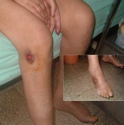
This clinical photograph demonstrates a localized lower extremity ulceration, specifically located on the anterior surface of the right leg distal to the knee. The primary lesion is a circular, punched-out ulcer with a deep red, granulated base and raised, hyperpigmented, indurated borders suggestive of localized inflammation or necrosis. The surrounding perilesional skin displays brownish-yellow hyperpigmentation and xerosis. An inset image provides a close-up of the dorsal aspect of the foot and ankle, highlighting diffuse edema, generalized skin darkening, and shiny, tense skin texture. These findings are clinically significant for chronic venous insufficiency, diabetic foot complications, or infectious processes like cutaneous leishmaniasis or ecthyma. The visual material is useful for dermatology and primary care education regarding the assessment of chronic lower limb ulcers, peripheral edema, and associated stasis-related skin changes.
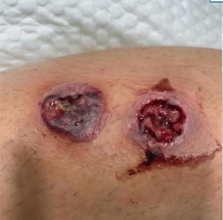
Clinical photograph of two distinct, rapidly progressive skin ulcers on a patient's lower extremity. The lesion on the left is a well-circumscribed, roughly circular ulcer featuring a central area of dark necrotic eschar and peripheral purpuric discoloration. Yellowish fibrinous exudate is visible within the wound bed. The lesion on the right demonstrates a deeper, punched-out ulcerative morphology with irregular, ragged borders and an erythematous, edematous rim. Its base contains raw red granulation tissue, areas of necrosis, and visible serosanguinous fluid streaking inferiorly. Both lesions are approximately 3 to 4 cm in diameter and exhibit signs of severe tissue breakdown and localized inflammation. This visual presentation is characteristic of necrotizing soft tissue infections or aggressive ulcerative conditions such as ecthyma gangrenosum, requiring urgent diagnostic evaluation for underlying pathogens or systemic involvement.
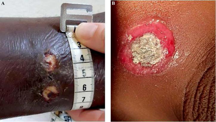
This clinical photograph set displays dermatological manifestations of cutaneous diphtheria (Corynebacterium diphtheriae). Image A shows a close-up of a patient's lower extremity featuring two small, circular ulcerated lesions. The lesions appear well-demarcated with an erythematous, inflammatory border and a central fibrinous exudate. A medical measuring tape is placed alongside the lesions to provide clinical scale, indicating the ulcers are approximately 1 to 1.5 cm in diameter. Image B provides a high-detail view of a characteristic diphtheritic ulcer. This lesion demonstrates a classic 'punched-out' morphology, consisting of a thick, greyish-white necrotic pseudomembrane centrally, which has a slightly textured or leathery appearance. The central necrotic core is surrounded by a prominent, bright red ring of inflamed tissue. These images illustrate the non-specific but clinically significant presentation of cutaneous diphtheria, which often involves multiple inflammatory ulcers that may mimic impetigo or tropical ecthyma, particularly in endemic regions such as French Guiana.
furuncle boil
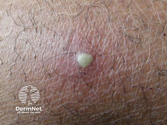
This dermatology clinical photograph documents a solitary boil (furuncle) on the cutaneous surface. Modality: Clinical photography; technique: macro close-up imaging of the skin with standard, noninvasive illumination. The lesion is a dome-shaped, erythematous nodule with a central yellowish-white pustular core representing purulent material within a hair follicle. Surrounding skin shows mild edema and perifollicular erythema. The lesion appears tender on palpation; no drainage is evident although the pus core is visible. The image captures typical signs of acute bacterial folliculitis progressing to furunculosis, most commonly caused by Staphylococcus aureus, with neutrophil-rich inflammatory infiltrate and abscess formation around the follicular unit. Clinically, this finding supports a diagnosis of furuncle rather than a simple acne lesion or epidermal cyst; differential diagnoses include carbuncle, abscess, or inflamed epidermoid cyst. Pathophysiology involves follicular occlusion, microbial colonization, and localized pus accumulation. Management implications include warm compresses, incision and drainage if fluctuance is present, and targeted antibiotics for extensive infection or cellulitis or systemic signs. This image is educational for medical trainees in recognizing cutaneous infections, dermatology case discussions, and demonstrations of localized skin abscesses and follicular infection. Useful for exam prep, clinical decision making, and patient education about boil treatment and outcomes globally.
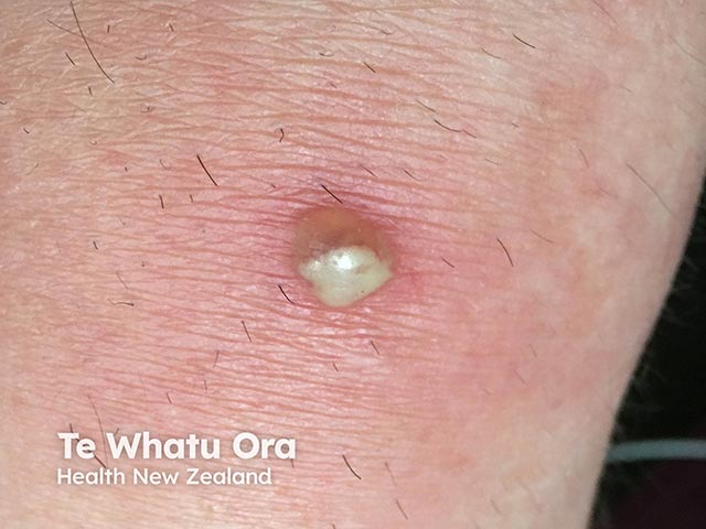
Imaging modality: Clinical photography of a cutaneous lesion. Anatomical location: left forearm skin, superficial dermis and hair follicle involvement. Perspective: close-up, frontal view of a solitary boil. Visual features: round, dome-shaped erythematous plaque with a central pustule and a creamy-white fluctuant core; surrounding perilesional induration; mild surrounding erythema; skin folds visible; no surrounding necrosis. Pathology inference: consistent with furuncle (boil) caused by Staphylococcus aureus infection of a hair follicle with inflammatory abscess formation within the dermis and subcutaneous tissue. In the clinical context, this lesion may arise in patients receiving Bruton tyrosine kinase inhibitors, which can disrupt B-cell function and predispose to bacterial skin infections; however, localization remains cutaneous and localized drainage is often therapeutic. Diagnostic significance: highlights a localized cutaneous infection; differential includes folliculitis, carbuncle, impetigo, abscess; MRSA risk; culture may guide antibiotics. Clinical correlation: correlate with systemic signs (fever, leukocytosis); assess need for wound drainage; versus incision and drainage; consider antibiotic therapy targeting Staphylococcus aureus; review BTK inhibitor therapy with treating oncologist for infection risk mitigation. Potential applications: educational dermatology cases, infection management training, BTK inhibitor adverse effect surveillance, antimicrobial stewardship. Image-guided documentation supports clinical decision-making, antibiotic stewardship, and comparative learning for dermatology and infectious disease education and training.
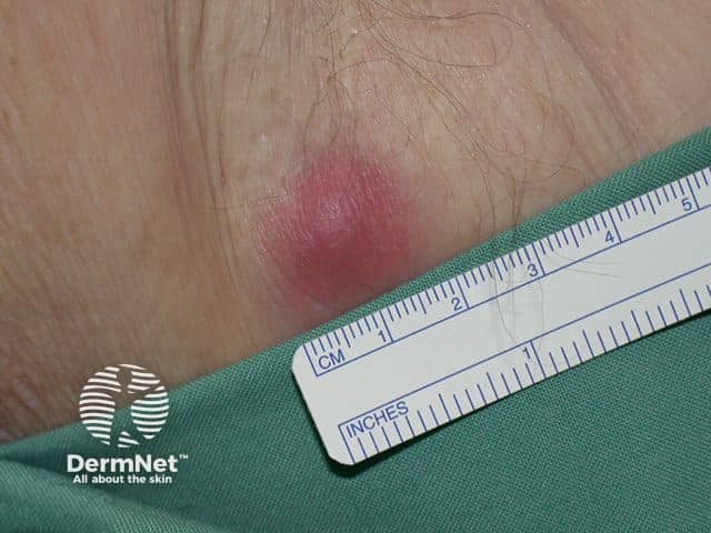
This is a clinical close-up photograph of a single left axillary skin lesion consistent with a boil (furuncle) or localized abscess. Modality: clinical photography; technique: macro close-up with standard color-calibrated lighting; scale is provided by a ruler for size estimation (~1.5–2 cm in diameter). The lesion is erythematous and tender with a raised, dome-shaped nodular configuration in the subcutaneous tissue of the axillary hair-bearing region. Surrounding perilesional erythema and mild edema reflect an acute inflammatory process. A central pus collection may be present; in some furuncles the area can be fluctuant, though drainage is not visible in this still image. The differential includes furuncle/abscess, folliculitis, hidradenitis suppurativa, and infected sebaceous cyst; HS typically shows recurrent painful nodules and sinus tracts in the axilla. Clinically, such lesions arise from bacterial infection of hair follicles, most often Staphylococcus aureus, with perifollicular inflammation. The image provides educational documentation for clinical scenarios, tele-dermatology triage, and surgical planning regarding drainage and antibiotic therapy. It supports visual recognition, teaching for medical students and residents, and correlation with histopathology or dermoscopy when available. Documentation of size, location, and morphology supports monitoring, patient education, and outcome assessment after drainage, incision, or antibiotic therapy, and aids research in cutaneous infectious disease.
tinea corporis ringworm annular scaly plaque
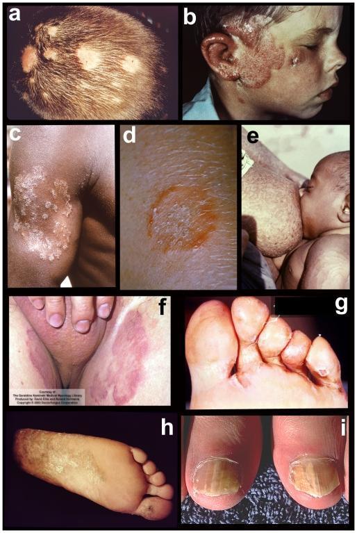
A composite of nine clinical photographs (labeled a-i) illustrating various clinical manifestations of dermatophyte infections (tinea) across different anatomical sites. (a) Tinea capitis on the scalp showing inflammatory patches and alopecia. (b) Tinea faciei presenting as an erythematous, scaly plaque on the cheek and ear. (c) Tinea corporis on the arm displaying an annular morphology with scaly borders. (d) Close-up of tinea corporis (ringworm) emphasizing the characteristic raised, erythematous leading edge and central clearing. (e) Tinea imbricata/corporis on the torso featuring concentric rings. (f) Tinea cruris (jock itch) showing confluent erythematous patches in the inguinal region. (g) Interdigital tinea pedis exhibiting maceration and scaling in the toe webbing. (h) Chronic 'moccasin-type' tinea pedis characterized by diffuse hyperkeratotic scaling on the plantar surface. (i) Onychomycosis presenting as distal subungual thickening, yellowish discoloration, and nail plate dystrophy. The collection serves as an educational reference for dermatological diagnosis based on lesion morphology and distribution.
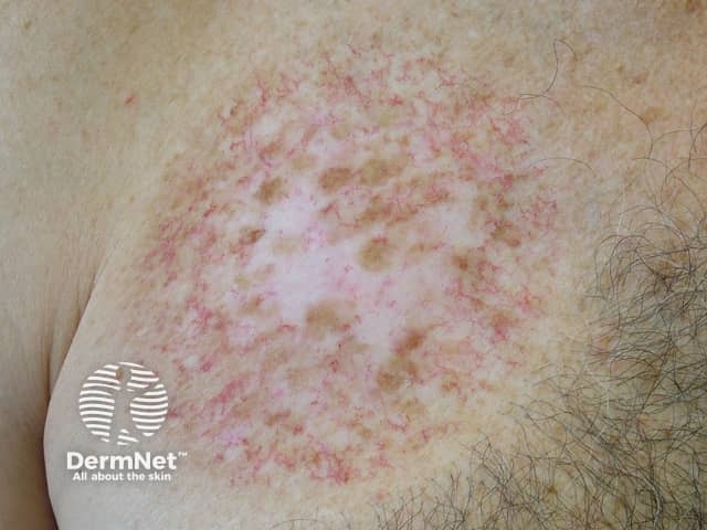
Clinical photograph of a skin lesion on the left shoulder/upper chest region displaying an annular, erythematous plaque with central clearing and fine desquamation. The lesion shows a pink-red, slightly raised border and irregular, patchy hypopigmentation in the center. Surrounding skin is mildly erythematous, with peripheral scale and no obvious vesiculation. The pattern resembles a ring-shaped dermatitis consistent with tinea corporis (dermatophyte infection) or an annular plaque with an active border. Differential diagnoses include tinea corporis, granuloma annulare, discoid eczema, plaque psoriasis with annular configuration, and contact dermatitis. The lesion appears well-demarcated with expansion margins and central clearing; the border may be scaly and demonstrates variable pigmentation. Notable features: peripheral pruritic erythema, annular geometry, scaly edge, central clearing, and mild crusting. Clinical significance: ring-like lesions on trunk or proximal limbs warrant superficial fungal evaluation (KOH preparation) and fungal culture, particularly in individuals without immunosuppression. Management implications include topical antifungals (terbinafine, azoles) and escalation to systemic therapy if widespread or unresponsive. This image is suitable for dermatology education, teledermatology triage, and differential diagnostic training focusing on annular erythematous plaques. Keywords for indexing: annular lesion, dermatophyte infection, ringworm, border scaling, central clearing, papulosquamous, inflammatory dermatology, infection vs. inflammation, imaging modality: clinical photo reference.
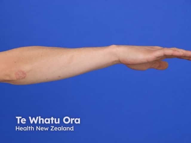
Clinical photography of a single well-circumscribed, erythematous, scaly plaque on the ventral/lateral forearm. The lesion exhibits classic annular configuration with peripheral scaling and a relatively clear central area, consistent with a ring-shaped dermatologic eruption. The observed lesion diameter is approximately 1–2 cm; the erythema is mild-to-moderate, and the scale appears fine, desquamative at the active border. This presentation is most suggestive of a superficial dermatophyte infection, i.e., tinea corporis (ringworm), though annular variants of eczema or psoriasis can mimic this pattern. The image demonstrates a well-demarcated border, thin rim of scale, and central clearing, with no overt vesiculation or ulceration. In a clinical context, one would consider mycological confirmation via potassium hydroxide (KOH) preparation and fungal culture, Wood’s lamp examination, and response to topical antifungal therapy as diagnostic adjuncts. Differential diagnoses include tinea corporis, annular psoriasis, nummular eczema, granuloma annulare, and pityriasis versicolor; however, the morphology here—scaly, active edge with central clearing—favors dermatophyte infection. This photograph is useful for dermatology education, teledermatology triage, pattern-recognition training, and patient counseling about fungal skin infections, contagiousness, and treatment expectations. Clinicians may correlate with pruritus, lesion distribution, exposure history, and lesion evolution over time.
tinea pedis athlete's foot
onychomycosis nail fungal infection
Imaging modality: clinical photography of a single toe. Location: dorsal aspect of the middle toe nails (onychomycosis). Visual features: chalky, opaque white patches on the dorsal nail plate with slight surface roughness; nails appear thickened at the distal edge; surrounding nail folds are unremarkable; no subungual hyperkeratosis is clearly visible in this image, though that feature can accompany fungal nail disease. Pathology: features compatible with superficial white onychomycosis caused by dermatophyte invasion of the dorsal surface of the nail plate. Diagnostic significance: the appearance supports a fungal nail infection; confirmation with potassium hydroxide (KOH) preparation or culture may be pursued if needed. Differential considerations include Candida nail infection, distal lateral subungual onychomycosis, trauma-induced leukonychia, or psoriasis of the nail, which may mimic the white patches but have distinct patterns. Clinical use cases: dermatology and primary care screening for nail fungus, monitoring antifungal therapy response, educational examples of nail plate disorders, and telemedicine assessment of distal digits. Terminology: onychomycosis, tinea unguium, superficial white onychomycosis, toe nail infection. The image is useful for teaching nail anatomy, differential diagnosis of white nail plate changes, and patient counseling on treatment expectations.
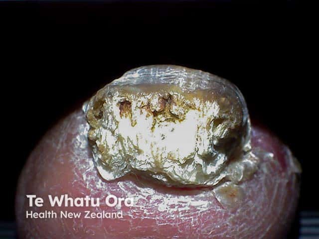
Clinical photography of the right hallux nail demonstrating total nail involvement (total onychomycosis) with marked subungual hyperkeratosis. The nail plate is thickened and opaque, with yellowish-brown crumbly debris filling the subungual space and lifting of the proximal edge. The proximal nail fold remains intact but overlies a markedly dystrophic nail bed. The surface of the nail shows irregular flaking and lamellar cracking consistent with chronic dermatophytic infection; the subungual keratotic mass is prominent. Imaging modality: Clinical photography; close-up, macro view; noninvasive. The anatomical location is the toenail unit, hallux, digit 1 of the foot. The observed features correspond to mycotic nail disease (onychomycosis) with hyperkeratosis, hypertrophy (onychosis), and subungual debris. Differential diagnosis includes psoriasis nail dystrophy, lichen planus, trauma-related nail dystrophy, and non-dermatophyte mold onychomycosis; however, the total involvement and characteristic subungual keratosis favor onychomycosis. Clinically, this finding is associated with prolonged fungal infection, potential discomfort, infection risk, and functional impairment; it may respond to systemic or topical antifungal therapy and debridement. This image is relevant for dermatology education, clinical diagnosis practice, and image-based medical training; useful for AI datasets focusing on nail pathology, fungal infections, and subungual hyperkeratosis. This image aids lesion recognition, grading severity, and treatment planning decisions.
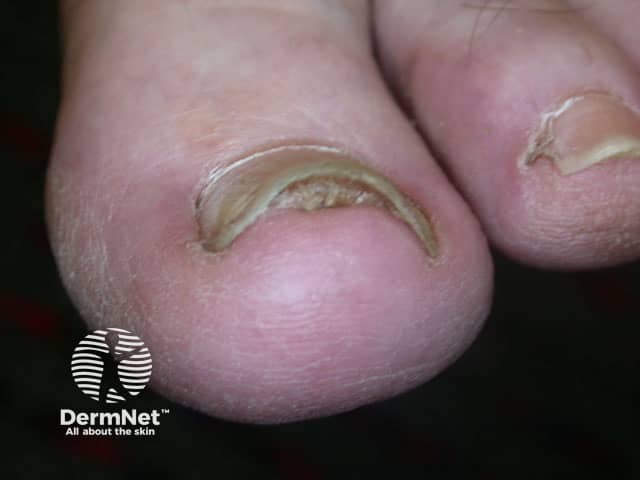
Clinical photography of a dystrophic toenail. Modality: high-resolution close-up digital photograph of the great toe nails in dorsal orientation. Location: toenail unit, plantar/dorsal aspect, with visible nail plate and distal edge. The image demonstrates thickened, yellow-brown discolored nail plate with a rough, crumbly surface and subungual keratinous debris. A crescent-shaped band of discoloration along the inner edge of the distal nail bed is evident, consistent with onychomycosis. Mild onycholysis at the distal margin may be present. Periungual skin appears uninflamed without surrounding erythema. Observed pattern suggests Ungual candidiasis? No; treat as dermatophyte infection. Overall features are typical of tinea unguium (onychomycosis) with nail plate dystrophy and subungual hyperkeratosis. Clinically significant as fungal nail infection that can cause nail thickening, discomfort, and cosmetic concern. Diagnostic significance: supports clinical suspicion of onychomycosis; differential includes nail psoriasis, lichen planus, trauma. Diagnostic workflow would include KOH prep, fungal culture, or PCR to confirm dermatophyte infection. Potential clinical uses include education, tele-dermatology triage, and guiding antifungal therapy decisions (systemic vs topical) with follow-up assessment. This image is suitable for teaching nail anatomy, pathology, and infection patterns in dermatology. Its simplicity aids rapid recognition in primary care and remote consultations.
tinea capitis kerion scalp
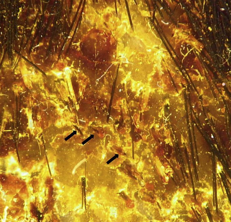
This diagnostic image is a high-magnification dermoscopy of the scalp, illustrating classic features of inflammatory tinea capitis (kerion). The clinical findings include prominent, ill-defined patches of erythema and thick, yellowish-white diffuse scaling (scales) across the scalp surface. Notable trichoscopic markers of fungal infection are present, specifically 'comma hairs'—short, fractured hair shafts that exhibit a distinctive curved, comma-like morphology (indicated by black arrows). Additionally, the field shows several dystrophic broken hairs and the presence of white hair casts (perifollicular keratin cylinders) encircling proximal hair shafts. These features are highly suggestive of ectothrix fungal invasion and are utilized in clinical dermatology to differentiate tinea capitis from other forms of alopecia or inflammatory scalp conditions. The image demonstrates key diagnostic indicators for medical students and clinicians specializing in dermatology and infectious diseases.
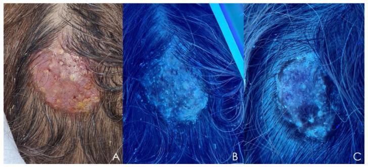
Clinical and diagnostic images illustrating tinea capitis progressing to kerion, caused by Microsporum canis. Panel A displays a clinical photograph of the scalp featuring a well-circumscribed, solitary inflammatory mass. The lesion is characterized by significant alopecia, erythema, an eroded surface, and yellowish crusting, consistent with a kerion. Panels B and C demonstrate Wood’s lamp examinations of the same area. Panel B shows a characteristic bright, intense blue-greenish fluorescence across the entire lesion, a pathognomonic finding for certain ectothrix fungal infections like M. canis. Panel C displays the same lesion after two weeks of fluconazole treatment, showing a significant reduction in fluorescence intensity and area, indicating therapeutic response and a decrease in fungal load. This sequence is used for medical education to demonstrate clinical diagnosis, diagnostic imaging using ultraviolet light (Wood’s lamp), and the objective monitoring of treatment efficacy in inflammatory fungal infections of the scalp.
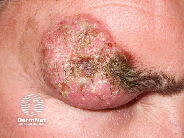
Clinical photography of a scalp lesion consistent with kerion, an inflammatory tinea capitis. Modality: dermatologic photography, close-up, frontal view of the left eyebrow/scalp region. The lesion is a raised, erythematous plaque with conspicuous yellow-brown crusts and surrounding scaling. Central areas show pustules and crusting with focal necrosis potential, and there is hair breakage and non-scarring alopecia at the periphery. The lesion is bilaterally inflamed but focal, with edema of the surrounding dermis producing a boggy, indurated impression. The pattern is unilateral and localized to the frontal-temporal scalp and periorbital area, compatible with fungal infection rather than primarily bacterial. The appearance corresponds to kerion: thick, crusted plaque, painful nodularity, regional lymphadenopathy not visible in image. Dermatoscopic details not applicable; histology would show perifollicular granulomatous inflammation with fungal elements. Clinically, kerion signals active dermatophytosis requiring systemic antifungal therapy and possibly adjunctive topical antifungals, with careful differential from bacterial cellulitis and folliculitis. Early recognition is critical to prevent scarring alopecia. This image, used for medical education, illustrates characteristic features: inflammatory plaque, yellow crusts, scaling perifollicular involvement, and localized hair loss, aiding differential diagnosis, treatment planning, and patient counseling. Note: fungal etiology confirmed by culture or molecular methods guides therapy selection and prognosis monitoring.
tinea versicolor pityriasis versicolor
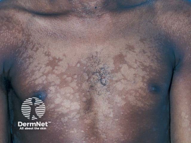
Comprehensive description: This is a high-resolution clinical photograph of the trunk depicting pityriasis versicolor–related hypopigmented patches. Imaging modality: Clinical photography of the anterior torso using standard lighting, color calibration, and digital capture to document surface pigmentary changes. Anatomical localization: Trunk (anterior chest and upper abdomen) within the integumentary system, illustrating cutaneous involvement of the thoracic skin. Visual features: irregular, coalescent hypopigmented macules and patches with fine superficial scale; color contrast against surrounding darker skin; scalloped margins with some central clearing and patchy confluent areas; distribution is predominantly on the chest, extending laterally and toward the upper abdomen. Pathology/disease: Classic presentation of pityriasis versicolor from Malassezia species causing superficial pigmentary disturbance without deep tissue invasion. Diagnostic significance: Supports clinical diagnosis of tinea versicolor; findings are often complemented by Wood’s lamp fluorescence (copper-orange) and potassium hydroxide examination revealing yeast and hyphae; antifungal therapy yields pigment restoration. Clinical use cases: Dermatology education, documentation of pigmentary disorders, monitoring response to topical or systemic therapy, and research into Malassezia-related hypopigmentation. This description emphasizes terminology such as pityriasis versicolor, tinea versicolor, hypopigmented macules, trunk, anterior view, Malassezia pigmentary change, and antifungal management. The image serves as a teaching and archival reference for pigmentary dermatoses in clinics.
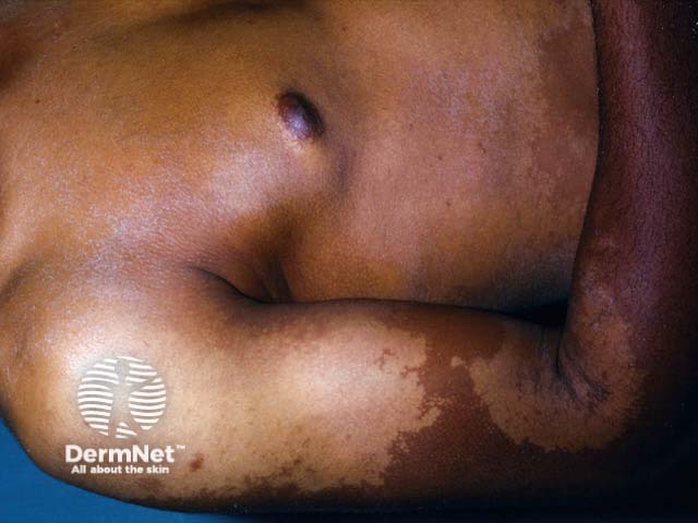
Clinical photograph of in vivo human forearm skin showing hypopigmented, confluent patches compatible with pityriasis versicolor (tinea versicolor). The image demonstrates localized involvement of the volar forearm with pale, irregularly shaped macules that coalesce into larger hypopigmented patches. The lesions display subtle surface scaling and well-defined but variable borders; surrounding skin is darker, giving a contrast typical for this condition. The distribution is on sun-exposed or humid skin regions and may appear more prominent after tanning. Pathophysiology involves superficial Malassezia yeast colonization of the stratum corneum with production of acids that disrupt melanin synthesis, leading to pigmentary heterogeneity. Clinically, pityriasis versicolor presents as nonpruritic hypo- or hyperpigmented patches that fluoresce under Wood's lamp. Differential diagnoses include vitiligo, postinflammatory hypopigmentation, and pityriasis alba; however, tinea versicolor generally preserves skin texture and shows minimal scale on closer inspection. Diagnostic confirmation is often via KOH preparation revealing the spaghetti-and-meatballs pattern of yeast and hyphae. Management is antifungal (topical azoles, selenium sulfide, or oral therapy for extensive disease). This image is valuable for dermatology education, pigmentary disorder assessment, and search-indexing for clinical skin disease repositories, aiding learners in recognition and differential diagnosis of superficial fungal hypopigmentation.
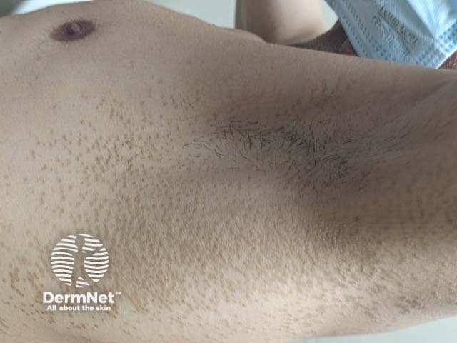
The image depicts a clinical photograph of axillary and proximal chest skin showing pigmented follicular lesions consistent with pityriasis versicolor. Imaging modality is clinical photography using a standard digital color camera to document cutaneous involvement. Visual features include multiple small brown, slightly scaly papules and patches centered on hair follicles, with fine surface scaling and patchy distribution. Lesions appear hypo- or hyperpigmented relative to adjacent skin, with preserved contour and no overt inflammation. The follicular localization suggests involvement of the follicular ostia and sebaceous-rich zones of the upper trunk. Histopathology (if sampled) would typically reveal Malassezia yeast and hyphae within the stratum corneum; KOH preparation may demonstrate the classic spaghetti-and-meatballs pattern. Clinically, pityriasis versicolor is a superficial fungal infection that can mimic other pigmentary disorders such as seborrheic dermatitis or vitiligo; differential diagnoses include post-inflammatory hyperpigmentation and tinea corporis. This image is valuable for dermatology education, telemedicine, and patient counseling, and supports planning of antifungal therapy (azoles, selenium sulfide) with attention to recurrence in hot, humid environments. For educational indexing, include commonly used synonyms such as tinea versicolor, Malassezia infection, pityriasis versicolor; this enhances recognition in dermatology databases, textbooks, and image banks used by clinicians, students, and researchers.
candidiasis oral thrush or intertrigo satellite pustules
verruca vulgaris common wart
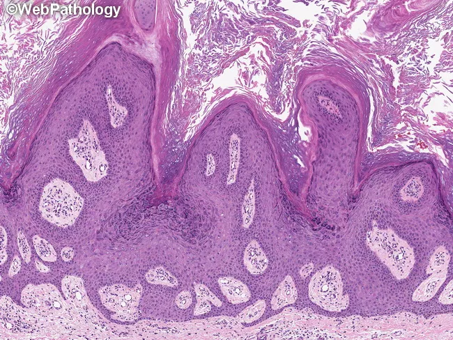
Histopathology of a common cutaneous wart (verruca vulgaris) on a routine hematoxylin and eosin stained skin biopsy. Imaging modality: Light microscopy of a skin biopsy with standard H&E staining. The epidermis shows marked hyperplasia with broad acanthosis and pronounced papillomatosis, producing a corrugated, exophytic surface. Hyperkeratosis with focal parakeratosis is evident at the tips of the projections. Rete ridges extend downward in elongated, serpiginous fashion. Filiform or finger-like papillary extensions project into the dermis with narrow dermal cores. Infected keratinocytes exhibit koilocytosis: enlarged, pale nuclei with perinuclear halos, a hallmark of HPV-related cytopathic effect. The underlying dermis shows mild chronic inflammation without deep invasion. Together, these features define verruca vulgaris and help distinguish it from verruca plana and other papillary lesions. Diagnostic significance: HPV-induced epidermal hyperplasia with papillomatosis and koilocytosis is characteristic; supports clinical diagnosis of a common wart. Differential considerations include verruca plana, seborrheic keratosis, keratoacanthoma, and cutaneous horn. Clinical correlation: rough, hyperkeratotic papules on exposed or frequently traumatized skin, commonly on hands and fingers; treatment options include cryotherapy, salicylic acid preparations, immunotherapy, and recurrence risk is moderate. Correlation with clinical morphology strengthens diagnostic confidence and guides targeted destructive therapies.
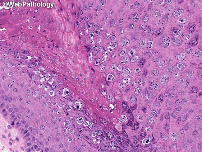
Histology of a common cutaneous wart (Verruca vulgaris) presenting in a hematoxylin and eosin stained slide viewed under light microscopy at high magnification. The specimen originates from skin biopsy with epidermal and superficial dermal involvement. The epidermis shows marked acanthosis with pronounced papillomatosis and irregular thickening of rete ridges. The upper epidermal layers contain numerous koilocytes—keratinocytes with enlarged, raisinoid nuclei surrounded by clear halos—and there is conspicuous cytoplasmic clearing and perinuclear halos. Basophilic keratohyalin granules are abundant in the granular layer, giving the granular layer a dark basophilic appearance. Hyperkeratosis and focal parakeratosis are evident in the stratum corneum, with mild epidermal spongiosis and dermal inflammatory infiltrate composed of lymphocytes and rare neutrophils. The dermal-epidermal junction is not disrupted, consistent with benign HPV-induced hyperproliferation. These features collectively support verruca vulgaris: HPV infection of keratinocytes leading to hyperplasia, koilocytosis, and a papillary surface. Clinically, verruca vulgaris is contagious, often skin-to-skin transmitted, frequently arising in children and young adults, and commonly treated with cryotherapy, salicylic acid, or immunomodulators. Differential diagnoses include seborrheic keratosis and squamous cell carcinoma; however, absence of atypia and invasion supports benign wart. The histopathology correlates with clinical management decisions and prognosis.
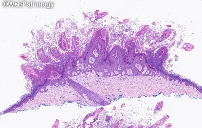
Histopathology of a cutaneous verruca vulgaris (common wart) demonstrated on a hematoxylin and eosin stained skin biopsy. The specimen shows an exophytic, cauliflower-like lesion arising from the epidermis with marked epidermal hyperplasia. Papillomatosis is evident as elongation and upgrowth of the rete ridges with surface projections. Acanthosis is present with diffuse thickening of the squamous cell layer; hyperkeratosis with parakeratosis is observed at the lesion surface, reflecting aberrant keratinization. Filiform papillae extend from the surface and end in finger-like projections. The dermal interface is relatively thin with a sparse inflammatory infiltrate and preserved dermal connective tissue. There is no obvious dysplasia or invasion. The histologic pattern is characteristic for HPV-induced verruca vulgaris, though koilocytosis (perinuclear halos in squamous cells) may be variably present and not always conspicuous in all sections. This morphology supports diagnosis of a benign, HPV-associated cutaneous wart rather than a corn, seborrheic keratosis, or other hyperkeratotic lesion. Clinically, verruca vulgaris presents as discrete, rough, hyperkeratotic papules or plaques on exposed or non-exposed skin, commonly in children and young adults. The diagnostic significance lies in recognizing papillomatosis with acanthosis and parakeratosis as classic features, guiding management such as topical therapies, cryotherapy, or surgical removal and reducing misdiagnosis with keratotic lesions.
mkdir -p /tmp/workspace/skin-infections-ppt/assets && cd /tmp/workspace/skin-infections-ppt && cat > fetch.py <<'EOF' import json, subprocess, base64 B="https://cdn.orris.care/cdss_images/" names={ "impetigo":"DermNetNZ_1760033337240_4cf5579e-5a2d-495a-9832-5c293cb4c510.jpg", "bullous":"pmc_clinical_VQA_2ecd82fac8e5dd6e79d5dc56f883e7c37456943086336dcfa801a9596cf0f9cb.jpg", "furuncle":"DermNetNZ_1760029731882_f2c902eb-ecb2-420a-ab90-0c9651207d1b.jpg", "corporis":"DermNetNZ_1760030298420_d8589b6e-8d5a-4838-b6c7-08660997a2ab.jpg", "pedis":"DermNetNZ_1760031359498_06d65124-aecd-4122-a0e5-16f6b3588729.jpg", "nail":"DermNetNZ_1760033830613_a6f4cac1-f552-4e65-9afd-e17397095c67.jpg", "kerion":"DermNetNZ_1760031092566_9309023a-90f1-4e8a-bcae-969386117f8f.jpg", "versicolor":"DermNetNZ_1760034273761_6b933656-dcbc-4e42-b9f3-675230b52466.jpg", "thrush":"DermNetNZ_1760034949085_834a7b74-6f80-4b1f-897e-8d47484a5160.jpg", "perleche":"DermNetNZ_1760031953434_23c98f3e-7104-40f6-a9b6-7c697b63b288.jpg", "wart_hand":"Pathology_1760048440657_51e94e59-2636-493f-8989-c4ff3ec3d7f1.jpg", "wart_plantar":"pmc_clinical_VQA_0e01837dc310c599f76e2743497819f45ad0243dc1054d46fb58d866133f318c.jpg", } keys=list(names) out=json.loads(subprocess.check_output(["python","/tmp/skills/shared/scripts/fetch_images.py"]+[B+names[k] for k in keys])) for k,o in zip(keys,out): if o["base64"]: raw=base64.b64decode(o["base64"].split("base64,")[1]) open(f"assets/{k}.jpg","wb").write(raw); print(k,len(raw)) else: print(k,"ERR",o["error"]) EOF python fetch.py; python -c " from PIL import Image;import glob for f in sorted(glob.glob('assets/*.jpg')):print(f,Image.open(f).size)"
cd /tmp/workspace/skin-infections-ppt && cat > build.py <<'EOF' from pptx import Presentation from pptx.util import Inches, Pt from pptx.dml.color import RGBColor from pptx.enum.text import PP_ALIGN, MSO_ANCHOR from pptx.enum.shapes import MSO_SHAPE from PIL import Image NAVY=RGBColor(0x1F,0x3A,0x5F); TEAL=RGBColor(0x14,0x8F,0x87); LIGHT=RGBColor(0xF2,0xF6,0xFA) CORAL=RGBColor(0xD9,0x48,0x2B); DARK=RGBColor(0x22,0x2B,0x36); WHITE=RGBColor(255,255,255) YEL=RGBColor(0xFF,0xF4,0xD6) prs=Presentation(); prs.slide_width=Inches(13.333); prs.slide_height=Inches(7.5) BL=prs.slide_layouts[6] def rect(s,x,y,w,h,fill,line=None): sh=s.shapes.add_shape(MSO_SHAPE.RECTANGLE,Inches(x),Inches(y),Inches(w),Inches(h)) sh.fill.solid(); sh.fill.fore_color.rgb=fill if line: sh.line.color.rgb=line else: sh.line.fill.background() sh.shadow.inherit=False return sh def runs(p,text,size,color=DARK,bold=False): parts=text.split("**") for i,t in enumerate(parts): if not t: continue r=p.add_run(); r.text=t; r.font.size=Pt(size); r.font.name="Calibri" r.font.bold = bold or (i%2==1); r.font.color.rgb = CORAL if (i%2==1 and not bold) else color def text(s,x,y,w,h,lines,size=18,color=DARK,bullet=True,anchor=MSO_ANCHOR.TOP,align=PP_ALIGN.LEFT,space=6,bold=False): tb=s.shapes.add_textbox(Inches(x),Inches(y),Inches(w),Inches(h)); tf=tb.text_frame tf.word_wrap=True; tf.vertical_anchor=anchor tf.margin_left=tf.margin_right=Inches(0.08); tf.margin_top=tf.margin_bottom=Inches(0.04) for i,l in enumerate(lines): p=tf.paragraphs[0] if i==0 else tf.add_paragraph() p.alignment=align; p.space_after=Pt(space) runs(p,("• "+l) if bullet else l,size,color,bold) return tb def header(s,title,sub=None): rect(s,0,0,13.333,1.05,NAVY); rect(s,0,1.05,13.333,0.07,TEAL) text(s,0.5,0.12,12.3,0.85,[title],size=32,color=WHITE,bullet=False,anchor=MSO_ANCHOR.MIDDLE,bold=True) def bg(s): rect(s,0,0,13.333,7.5,WHITE) def simple(s,msg,y=6.3,h=0.95): rect(s,0.5,y,12.33,h,YEL,RGBColor(0xE8,0xC5,0x6A)) rect(s,0.5,y,0.12,h,CORAL) text(s,0.75,y,11.9,h,["**In simple words:** "+msg],size=17,bullet=False,anchor=MSO_ANCHOR.MIDDLE) def img(s,name,x,y,w,h,cap=None): im=Image.open(f"assets/{name}.jpg"); iw,ih=im.size ch=0.35 if cap else 0 bh=h-ch; r=min(w/iw,bh/ih); pw,ph=iw*r,ih*r px=x+(w-pw)/2 s.shapes.add_picture(f"assets/{name}.jpg",Inches(px),Inches(y),Inches(pw),Inches(ph)) if cap: text(s,x,y+ph+0.02,w,0.33,[cap],size=12,color=TEAL,bullet=False,align=PP_ALIGN.CENTER,bold=True) def table(s,x,y,w,rows,colw,size=15,rowh=0.5): nr,nc=len(rows),len(rows[0]) t=s.shapes.add_table(nr,nc,Inches(x),Inches(y),Inches(w),Inches(rowh*nr)).table for j,cw in enumerate(colw): t.columns[j].width=Inches(cw) for i,row in enumerate(rows): for j,val in enumerate(row): c=t.cell(i,j); c.fill.solid() c.fill.fore_color.rgb = TEAL if i==0 else (LIGHT if i%2 else WHITE) c.margin_left=c.margin_right=Inches(0.08); c.vertical_anchor=MSO_ANCHOR.MIDDLE tf=c.text_frame; tf.word_wrap=True; p=tf.paragraphs[0] runs(p,val,size,WHITE if i==0 else DARK, bold=(i==0 or j==0)) return t def notes(s,t): s.notes_slide.notes_text_frame.text=t # 1 Title s=prs.slides.add_slide(BL); rect(s,0,0,13.333,7.5,NAVY); rect(s,0,4.9,13.333,0.1,TEAL) text(s,0.8,1.9,11.7,1.4,["Cutaneous Skin Infections"],size=54,color=WHITE,bullet=False,bold=True) text(s,0.8,3.3,11.7,1.2,["Bacterial, fungal and viral infections of the skin explained simply"],size=26,color=RGBColor(0xBF,0xE3,0xE0),bullet=False) text(s,0.8,5.3,11.7,1.2,["Presented by: [Your name]","Source: Chapter 60, Table 60-5 (textbook pages)"],size=20,color=WHITE,bullet=False) notes(s,"Introduce the topic: these are infections that live in or on the skin. We will go through them by germ type: bacteria, fungi, then virus.") # 2 Big picture s=prs.slides.add_slide(BL); bg(s); header(s,"The Big Picture: 3 Types of Germs") cards=[("BACTERIA","Impetigo\nEcthyma\nFuruncle (boil)","Mainly Staph aureus and Strep",NAVY), ("FUNGI","Ringworm (tinea)\nTinea versicolor\nCandidiasis","Dermatophytes, Malassezia, Candida",TEAL), ("VIRUS","Warts","Human papillomavirus (HPV)",CORAL)] for i,(h,l,g,c) in enumerate(cards): x=0.5+i*4.18 rect(s,x,1.5,3.95,0.7,c); text(s,x,1.5,3.95,0.7,[h],size=24,color=WHITE,bullet=False,anchor=MSO_ANCHOR.MIDDLE,align=PP_ALIGN.CENTER,bold=True) rect(s,x,2.2,3.95,3.6,LIGHT) text(s,x+0.2,2.4,3.6,2.2,l.split("\n"),size=22,bullet=True,space=10) text(s,x+0.2,4.7,3.6,1.0,["**Germ:** "+g],size=17,bullet=False) simple(s,"We sort skin infections by the germ that causes them. The germ decides the treatment: antibiotics for bacteria, antifungals for fungi.") notes(s,"Erysipelas, cellulitis, herpes simplex and herpes zoster are only cross-referenced to other chapters (134, 197, 198) on these pages, so they are not covered here.") # 3 Impetigo & ecthyma s=prs.slides.add_slide(BL); bg(s); header(s,"Impetigo and Ecthyma (Bacterial)") text(s,0.5,1.3,6.6,5.0,[ "**Impetigo** = a surface skin infection", "Causes: **Staph aureus** (most often) and **Group A Strep**", "A small pustule bursts and dries into a **honey-colored crust**", "**Bullous impetigo:** clear, tense blisters. A Staph toxin splits the top skin layer", "**Ecthyma** = deeper impetigo. **Punched-out ulcers**, heals with a **scar**", "Treatment: remove crusts gently with soaks + topical and oral antibiotics"],size=19,space=9) img(s,"impetigo",7.4,1.3,5.4,2.4,"Impetigo: golden-yellow crusts") img(s,"bullous",7.4,3.8,5.4,2.45,None) s.shapes[-1].left=Inches(9.1); simple(s,"Impetigo is a crusty, honey-colored skin infection. Ecthyma is the same thing but deeper, so it leaves scars.") notes(s,"Primary infection means it starts on healthy skin. Secondary means it starts on skin already damaged, for example by eczema. The same toxin causes staphylococcal scalded skin syndrome, which is more common in children. In adults with widespread blistering, also think of toxic epidermal necrolysis and severe drug reactions. No clear picture of ecthyma was available in the image library, so only impetigo images are shown.") # fix bullous placement: re-do cleanly (remove last picture and re-add with caption) sp=s.shapes[-3] if False else None # 4 Furuncle s=prs.slides.add_slide(BL); bg(s); header(s,"Furunculosis (Boils)") text(s,0.5,1.3,6.9,5.0,[ "A **furuncle (boil)** = a painful red lump around a hair follicle", "Cause: **Staph aureus**. Now more common because of **CA-MRSA** (resistant Staph)", "Can be single, but usually **several**", "Patients often think it is a **spider bite**", "Family members may get it too", "Small boil: **incision and drainage** may be enough", "Send pus for **culture**; antibiotics depend on MSSA or MRSA", "Helpers: **warm compresses** and **nasal mupirocin**. Severe: IV antibiotics"],size=18,space=7) img(s,"furuncle",7.7,1.4,5.1,4.5,"Furuncle: red dome-shaped nodule with a pus core") simple(s,"A boil is a Staph infection of a hair root. Drain it, culture it, and treat with antibiotics if it is big or spreading.") notes(s,"CA-MRSA means community-acquired methicillin-resistant Staph aureus. Because MRSA is resistant to many common antibiotics, culture helps pick the right drug.") # 5 Dermatophytosis s=prs.slides.add_slide(BL); bg(s); header(s,"Dermatophytosis (Ringworm / Tinea)") text(s,0.5,1.3,6.9,5.0,[ "**Dermatophytes** = fungi that eat keratin in **skin, hair and nails**", "Three genera: **Trichophyton, Microsporum, Epidermophyton**", "Typical lesion: red, scaly, **ring-shaped (annular)** plaque with a clearer center. That is why it is called **ringworm**. There is no worm!", "Wrong use of strong steroid creams can cause deep **inflammatory nodules (granulomas)**", "**Diagnosis:** scrape and look under the microscope with **KOH**, or culture. Nail clippings: **PAS stain**"],size=19,space=9) img(s,"corporis",7.7,1.5,5.1,4.5,"Tinea corporis: ring with scaly edge") simple(s,"Ringworm is a fungal infection that makes a ring. Always confirm with a KOH test before starting strong tablets.") notes(s,"KOH dissolves skin cells so the fungal threads (hyphae) become visible under the microscope.") # 6 Types of tinea s=prs.slides.add_slide(BL); bg(s); header(s,"Types of Tinea by Body Site") table(s,0.5,1.35,7.6,[ ["Type","Site","Key point"], ["Corporis","Body (hairless skin)","Classic ring"], ["Cruris","Groin","Mostly males. **Spares the scrotum**"], ["Pedis","Foot","**Most common**. Between 4th and 5th toes. Often chronic"], ["Unguium","Nails","Thick, yellow, crumbly. Proximal type may signal **HIV**"], ["Capitis","Scalp","Children. *T. tonsurans*. Hair loss; **kerion** if severe"]], [1.4,2.0,4.2],size=15,rowh=0.75) img(s,"pedis",8.4,1.3,4.4,2.35,"Tinea pedis: scaling between toes") img(s,"nail",8.4,3.75,4.4,2.35,"Onychomycosis: thick, yellow nail") simple(s,"Name = site. Pedis is foot, cruris is groin, unguium is nail, capitis is scalp. Remember: groin tinea spares the scrotum.") notes(s,"Tinea cruris sparing the scrotum is a useful way to tell it from candidiasis, which often involves the scrotum. Tinea pedis is the most common dermatophyte infection. A proximal subungual onychomycosis in a young adult can be a clue to HIV or other immune problems.") # 7 Capitis / kerion s=prs.slides.add_slide(BL); bg(s); header(s,"Tinea Capitis and Kerion (Scalp)") text(s,0.5,1.3,6.9,5.0,[ "Common in **inner-city children**, also in immunocompromised adults", "Main germ: ***Trichophyton tonsurans***", "Usually mild: **scaling and patchy hair loss**", "*T. tonsurans* and *Microsporum canis* can cause a **kerion**: a boggy, swollen, inflamed mass with pustules", "Kerion can cause **scarring and permanent hair loss**", "Needs an **oral antifungal**. Sometimes steroids are added to reduce inflammation"],size=19,space=9) img(s,"kerion",7.7,1.5,5.1,4.5,"Kerion: inflamed, crusted scalp mass with hair loss") simple(s,"Scalp ringworm needs tablets, not cream. A kerion is an angry, swollen scalp patch that can leave a bald scar if not treated.") notes(s,"The slide text shows asterisks around italics are not rendered; species names are in italics in the textbook. Topical drugs cannot reach the hair root, which is why oral therapy is needed.") # 8 Treatment tinea s=prs.slides.add_slide(BL); bg(s); header(s,"Treating Tinea: Cream or Tablet?") rect(s,0.5,1.4,6.0,0.65,TEAL); text(s,0.5,1.4,6.0,0.65,["TOPICAL (creams)"],size=22,color=WHITE,bullet=False,anchor=MSO_ANCHOR.MIDDLE,align=PP_ALIGN.CENTER,bold=True) rect(s,0.5,2.05,6.0,3.9,LIGHT) text(s,0.65,2.2,5.7,3.7,["For **corporis, cruris** and **limited pedis**","Apply **twice daily**","Continue **1 week after** the skin looks normal","Pedis needs longer treatment and often **relapses**"],size=19,space=10) rect(s,6.83,1.4,6.0,0.65,NAVY); text(s,6.83,1.4,6.0,0.65,["ORAL (tablets)"],size=22,color=WHITE,bullet=False,anchor=MSO_ANCHOR.MIDDLE,align=PP_ALIGN.CENTER,bold=True) rect(s,6.83,2.05,6.0,3.9,LIGHT) text(s,6.98,2.2,5.7,3.7,["For **scalp (capitis)** and **nails**, or if cream fails","**Confirm the fungus first** (microscopy or culture)","All can harm the liver (**hepatotoxicity**)","**Avoid in pregnancy and breast-feeding**","**Nystatin does not work** on dermatophytes"],size=19,space=10) simple(s,"Cream for small, simple skin patches. Tablets for hair, nails and stubborn cases. Never use nystatin for ringworm.") notes(s,"Topical drugs alone do not work on tinea capitis or onychomycosis. If an asymptomatic toenail infection is present, weigh risk against benefit before using systemic drugs.") # 9 Drugs s=prs.slides.add_slide(BL); bg(s); header(s,"Oral Antifungal Drugs") table(s,0.5,1.4,12.33,[ ["Drug","Used for","Remember"], ["**Griseofulvin**","Skin, hair and nail dermatophytes","Side effects: stomach upset, headache, hives"], ["**Itraconazole**","Onychomycosis","Many **drug interactions** (CYP450). **Avoid in heart failure / ventricular dysfunction**"], ["**Terbinafine**","Onychomycosis; granules for tinea capitis","**Fewer interactions** than itraconazole. Still be careful with many medicines"], ["**Ketoconazole (oral)**","Not first line for any fungal infection","FDA warning: **liver toxicity**. Topical ketoconazole is fine"]], [2.6,4.0,5.73],size=17,rowh=0.95) simple(s,"Griseofulvin: older drug, tummy upset. Itraconazole: heart failure warning. Terbinafine: safest on interactions. Oral ketoconazole: avoid.") notes(s,"Hepatotoxicity is a class concern for oral antifungals, so ask about liver disease and other medicines before prescribing.") # 10 Versicolor s=prs.slides.add_slide(BL); bg(s); header(s,"Tinea (Pityriasis) Versicolor") text(s,0.5,1.3,6.9,5.0,[ "Cause: ***Malassezia furfur***, a yeast that **normally lives on skin**", "It causes disease in **heat and humidity**", "Where: **chest, shoulders, back**. Rarely face or limbs", "Color: **pale (hypopigmented)** on dark skin; **pink or brown** on light skin", "**KOH test:** short threads + round spores = **\"spaghetti and meatballs\"**", "Treatment: **selenium sulfide**, sulfur, salicylic acid or azole lotion/shampoo, daily 1 to 2 weeks, then weekly", "**Wash off within 10 minutes**: selenium irritates"],size=18,space=7) img(s,"versicolor",7.7,1.4,5.1,4.5,"Pale scaly patches on the chest") simple(s,"A normal skin yeast overgrows in hot, humid weather and leaves pale or dark patches on the upper body. Lotion clears it.") notes(s,"Pigment returns slowly after the fungus is treated, so patches may look pale for weeks. Oral drugs work but results do not last and are not FDA approved for this.") # 11 Candidiasis s=prs.slides.add_slide(BL); bg(s); header(s,"Candidiasis (Yeast Infection)") text(s,0.5,1.3,6.6,5.0,[ "Germ: **Candida albicans**, a normal gut yeast", "Grows when there are **antibiotics, diabetes, low immunity** (very common in **HIV**), or **moisture**", "**Thrush:** white plaques in the mouth", "**Perleche:** cracked corners of the mouth (common with ill-fitting dentures)", "Skin folds: red, scaly, **satellite pustules**. May involve **scrotum** and nails", "**Painful and very inflamed**", "**KOH:** yeast and **pseudohyphae**"],size=18,space=7) img(s,"thrush",7.3,1.3,5.5,2.45,"Oral thrush: white curd-like plaques") img(s,"perleche",7.3,3.85,5.5,2.4,"Perleche: cracked mouth corners") simple(s,"Candida likes warm, wet, moist places. Look for satellite spots around a red patch in skin folds.") notes(s,"Candidiasis is usually more painful and inflamed than dermatophyte infection. Chronically wet and macerated sites, such as around the nails and between skin folds, are favorite spots. An image of skin-fold (intertriginous) candidiasis was not found in the image library.") # 12 Candida tx + compare s=prs.slides.add_slide(BL); bg(s); header(s,"Treating Candidiasis, and Tinea vs Candida") text(s,0.5,1.3,6.0,4.9,[ "**Step 1:** fix the cause (stop unnecessary antibiotics, keep area dry)", "**Topical:** nystatin or azoles (miconazole, clotrimazole, econazole, ketoconazole)", "Add mild steroid (**2.5% hydrocortisone**) for inflammation on smooth skin", "**Systemic** only if immunosuppressed or topical fails: **oral fluconazole**", "**Oral nystatin** works only in the **gut**"],size=18,space=8) table(s,6.8,1.4,6.03,[ ["Feature","Tinea","Candida"], ["Shape","Ring","Red patch + satellite pustules"], ["Scrotum","Spared","Often involved"], ["Pain / inflammation","Less","More"], ["KOH","Hyphae","Pseudohyphae"], ["Nystatin","No","Yes"]],[1.9,1.5,2.63],size=15,rowh=0.7) simple(s,"Dry the area, use cream, and keep tablets for hard cases. Tinea spares the scrotum; Candida does not. Nystatin works on Candida only.") notes(s,"Use the comparison table to answer 'how do you tell tinea cruris from candidal intertrigo?'") # 13 Warts s=prs.slides.add_slide(BL); bg(s); header(s,"Warts (HPV)") text(s,0.5,1.3,6.6,5.0,[ "Caused by **human papillomavirus (HPV)**; more than 100 types", "**Verruca vulgaris:** rough, dome-shaped, about 1 cm", "**Plantar:** on the sole, grows inward; paring shows black dots (bleeding points)", "**Flat:** smooth and slightly raised; spread by **shaving**", "**Filiform:** finger-like, on face and neck", "**Genital:** small papillomas that can become large", "**HPV 16 and 18** raise cancer risk (cervix, anus, vulva, penis)", "Higher risk in **transplant and HIV** patients"],size=17,space=6) img(s,"wart_hand",7.3,1.3,5.5,2.6,"Common warts on the hand") img(s,"wart_plantar",7.3,4.0,5.5,2.25,"Plantar and periungual warts") simple(s,"Warts are a viral skin growth. Most are harmless, but some HPV types can lead to cancer.") notes(s,"The plantar wart image is a tall picture, so it appears small on this slide. Squamous cell carcinoma with HPV has also been seen outside the genital area, mostly in immunosuppressed organ transplant patients, so they should be checked regularly.") # 14 Wart tx s=prs.slides.add_slide(BL); bg(s); header(s,"Treating Warts") text(s,0.5,1.3,12.3,4.9,[ "Most warts in healthy people **go away on their own in 1 to 2 years**", "No single treatment is perfect, and **recurrence is common**", "**Cryotherapy (liquid nitrogen):** most useful and convenient method", "**Salicylic acid plasters or solutions:** as good for non-genital warts, but the patient must apply them regularly", "**Genital warts:** podophyllin (clinic or home form), **imiquimod** (boosts local immune response), sinecatechins (green tea extract)", "**Stubborn warts:** conventional or laser surgery", "**HPV vaccine** lowers anogenital warts and cervical cancer"],size=20,space=11) simple(s,"Wait, freeze, or use acid. Genital warts have special creams. The HPV vaccine is the best prevention.") notes(s,"Choice of treatment depends on location, extent, age and immune status, and what the patient prefers.") # 15 Quick revision s=prs.slides.add_slide(BL); bg(s); header(s,"Quick Revision (Table 60-5)") table(s,0.5,1.35,12.33,[ ["Infection","What you see","Germ","Treatment"], ["Impetigo","Honey-colored crusted papules, plaques or bullae","Group A Strep, Staph aureus","Antibiotics (topical or oral)"], ["Dermatophytosis","Ring-shaped scaly plaques, hair loss, groin spares scrotum, hyphae on KOH","Trichophyton, Epidermophyton, Microsporum","Topical azoles; oral griseofulvin, terbinafine or azoles"], ["Candidiasis","Papules and plaques with satellite pustules, folds, may involve scrotum, pseudohyphae","Candida albicans and other Candida","Nystatin or azoles; oral azoles for resistant cases"], ["Tinea versicolor","Pale or dark scaly patches on trunk, spaghetti and meatballs","Malassezia furfur","Selenium sulfide lotion or azoles"]], [2.1,4.6,2.9,2.73],size=15,rowh=1.0) simple(s,"Four infections to know cold: crust (impetigo), ring (dermatophyte), satellite spots (Candida), pale patches (versicolor).") notes(s,"This is the textbook summary table from page 1 of the images.") # 16 Summary s=prs.slides.add_slide(BL); rect(s,0,0,13.333,7.5,NAVY); rect(s,0,1.2,13.333,0.08,TEAL) text(s,0.6,0.25,12,0.9,["Key Takeaways"],size=36,color=WHITE,bullet=False,bold=True) text(s,0.7,1.6,12,4.6,[ "**Impetigo** = honey crust. **Ecthyma** = deeper ulcer with scar", "**Boils** = Staph (watch for MRSA). Drain and culture", "**Tinea** = ring; confirm with KOH; groin type **spares the scrotum**", "**Candida** = satellite pustules, painful; likes moisture", "**Versicolor** = \"spaghetti and meatballs\"; selenium sulfide", "**Warts** = HPV; most resolve alone; cryotherapy is the go-to treatment"],size=24,color=WHITE,space=14) text(s,0.7,6.3,12,0.8,["Thank you. Any questions?"],size=30,color=RGBColor(0xBF,0xE3,0xE0),bullet=False,bold=True) notes(s,"Close with the key take-aways and invite questions.") prs.save("Cutaneous_Skin_Infections.pptx") print("ok",len(prs.slides)) EOF python build.py
cd /tmp/workspace/skin-infections-ppt && python - <<'EOF' import glob from PIL import Image for f in glob.glob("assets/*.jpg"): im=Image.open(f); print(f,im.format) im.convert("RGB").save(f,"JPEG",quality=90) src=open("build.py").read() src=src.replace('img(s,"bullous",7.4,3.8,5.4,2.45,None)\ns.shapes[-1].left=Inches(9.1); \n','img(s,"bullous",7.4,3.85,5.4,2.4,"Bullous impetigo: eroded blisters")\n') src=src.replace("***Trichophyton tonsurans***","**Trichophyton tonsurans**") src=src.replace("***Malassezia furfur***","**Malassezia furfur**") src=src.replace("*T. tonsurans* and *Microsporum canis*","T. tonsurans and Microsporum canis") src=src.replace("*T. tonsurans*","T. tonsurans") src=src.replace("The slide text shows asterisks around italics are not rendered; species names are in italics in the textbook. Topical","Topical") src=src.replace("# fix bullous placement: re-do cleanly (remove last picture and re-add with caption)\nsp=s.shapes[-3] if False else None\n","") open("build.py","w").write(src) EOF grep -n '\*[A-Za-z]' build.py | grep -v '\*\*' | head; python build.py
cd /tmp/workspace/skin-infections-ppt && python - <<'EOF' from pptx import Presentation from pptx.util import Emu import math p=Presentation("Cutaneous_Skin_Infections.pptx") for i,s in enumerate(p.slides,1): for sh in s.shapes: if sh.has_text_frame and sh.text_frame.text.strip() and sh.height>Emu(914400*2): w=sh.width/914400-0.2; tot=0 for para in sh.text_frame.paragraphs: sz=max((r.font.size.pt for r in para.runs if r.font.size),default=18) cpl=w*72/(sz*0.5); lines=max(1,math.ceil(len(para.text)/cpl)) tot+=lines*sz*1.2/72+ (para.space_after.pt/72 if para.space_after else 0) if tot>sh.height/914400: print("slide",i,"may overflow: need %.1f have %.1f"%(tot,sh.height/914400)) print("checked") EOF ls -la /tmp/workspace/skin-infections-ppt/*.pptx
Cutaneous Skin Infections
Presentation · PPTX
- Title
- The big picture: bacteria, fungi and virus
- Impetigo and ecthyma (2 photos)
- Furuncle/boil (photo)
- Ringworm, i.e. dermatophytosis (photo)
- Types of tinea by body site (foot and nail photos)
- Tinea capitis and kerion (photo)
- Cream vs tablet treatment for tinea
- Oral antifungal drugs table
- Tinea versicolor (photo)
- Candidiasis (thrush and perleche photos)
- Candida treatment, plus a Tinea vs Candida comparison table
- Warts (hand and plantar photos)
- Wart treatment
- Quick revision table (Table 60-5)
- Key takeaways and thank you
- In simple words box: every slide has a yellow box at the bottom with a plain-language summary. You can read it out as your explanation.
- Speaker notes: the notes pane has extra explanations, such as why KOH is used, what CA-MRSA means, and how to tell tinea from Candida.
- There is no ecthyma photo. The library only returned unrelated ulcers, so I left it out rather than show a wrong image.
- There is no photo of Candida in skin folds. I used mouth examples (thrush and perleche) instead.
- Tinea cruris and tinea corporis have no separate photos of the groin type. Corporis has one image on slide 5.
- The plantar wart image is a tall picture, so it looks small on slide 13.
- The photos come from the image library (DermNet and published case images). If your ma'am asks for sources, say they are from a medical image library.