

Pt presented with thalamic hemorrhage and ct angio dhowed
moyamoya disease hemorrhage thalamus management
Moyamoya disease CT angiogram cerebral vessels puff of smoke collaterals
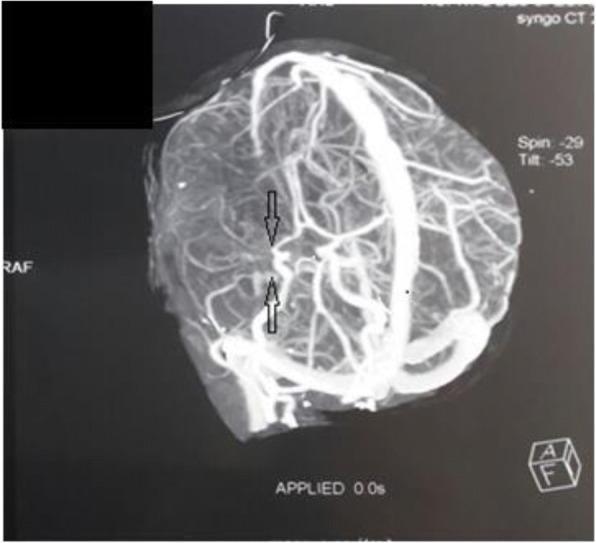
This diagnostic image is a cerebral angiogram (CT angiography) at the level of the circle of Willis, illustrating a classic presentation of Moyamoya disease. The visual highlights a proximal obliteration or severe stenosis of the right middle cerebral artery (MCA), marked by black arrows. Distal to the site of occlusion, there is a dense, hazy network of fine collateral vessels. This cluster of small, anastomotic vessels creates the pathognomonic 'puff of smoke' (moyamoya) appearance, which develops as a compensatory mechanism for the chronic, progressive narrowing of the major intracranial arteries. The image demonstrates significant vascular remodeling and diminished vessel caliber in the primary arterial territory. This finding is clinically significant for the evaluation of pediatric stroke and ischemic risk, representing a primary vasculopathy that affects the internal carotid arteries and their branches.
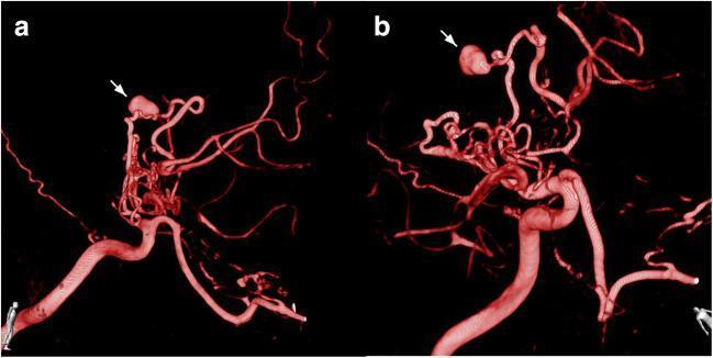
This diagnostic image shows a 3D reconstructed CT angiogram (CTA) of the cerebral vasculature in two views (a and b), highlighting a complex vascular network characteristic of Moyamoya disease (MMD). The reconstruction demonstrates significant stenosis of the distal internal carotid artery (ICA) and proximal segments of the anterior and middle cerebral arteries (ACA/MCA). A prominent, dense network of tortuous, 'puff-of-smoke' collateral vessels is visible in the basal regions, compensating for the occlusive disease. A distinctive white arrow in both views points to a large, saccular aneurysm (approximately 7mm) originating from a fragile collateral vessel near the terminal branches of the ACA. The aneurysm exhibits a wide neck and irregular morphology, appearing as a significant outpouching compared to the slender parent vessel. The images illustrate the increased hemodynamic stress placed on collateral pathways in MMD, leading to the formation and rapid growth of secondary aneurysms, which pose a high risk for intracerebral hemorrhage.
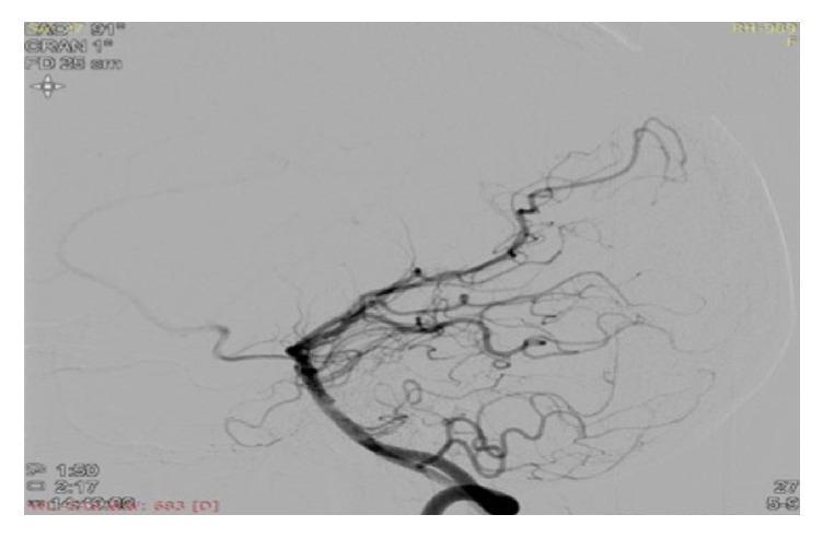
This diagnostic image is a cerebral angiogram (digital subtraction angiography) illustrating significant neurovascular pathology in an 18-year-old patient. The image shows the right internal carotid artery and middle cerebral artery (MCA) territory. There is a near-total to complete occlusion of the right M1 segment of the MCA, occurring just distal to its origin. Characteristic of Moyamoya disease or syndrome, a dense, hazy network of fine, thread-like collateral vessels—known as 'puff of smoke' or Moyamoya collaterals—is visible, attempting to reform the distal M1 segment through small perforating arteries. Comparison reveals a reduced caliber of the right internal carotid artery relative to the contralateral side, suggesting proximal stenosis. The peripheral vasculature appears as a complex, delicate web of compensatory vessels. This finding is clinically significant for explaining ischemic events and stroke in young patients, particularly in the context of hyperhomocysteinemia or idiopathic Moyamoya. The image serves as a classic educational example of intracranial arterial stenosis with resultant secondary collateralization.
moyamoya disease hemorrhagic stroke treatment revascularization
CT Cerebral Angiogram Report - Analysis
What the Report Shows
- Significantly reduced calibre of bilateral ICA throughout all intracranial segments
- Non-visualization of cavernous and supracavernous segments of bilateral ICA, bilateral MCA, and bilateral ACA
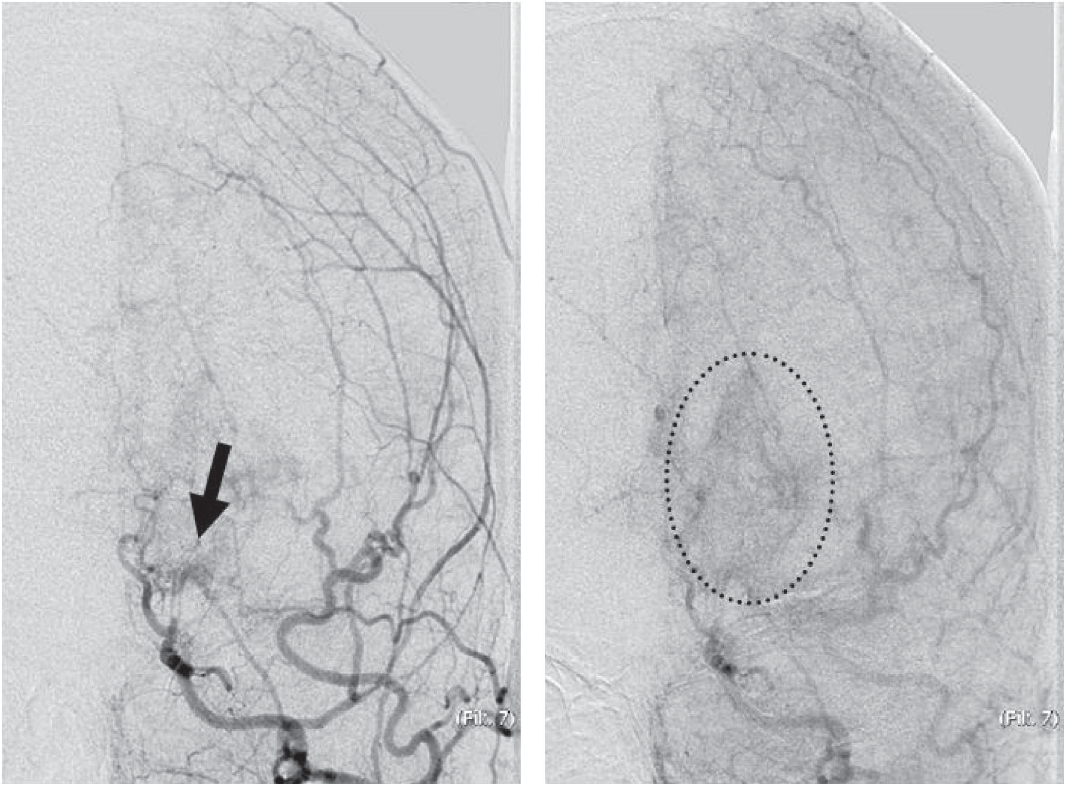
- Multiple pial-pial collateral circulation - this is the hallmark "puff of smoke" appearance
- Acute intraparenchymal hematoma in the left thalamus, 25 x 22 mm
- Mass effect: sulcal space effacement; no midline shift
- Intraventricular extension into bilateral lateral ventricles, 3rd and 4th ventricles
- Mild obstructive hydrocephalus with periventricular seepage (transependymal CSF flow)
Moyamoya Disease - Clinical Context
| Feature | This Patient | Moyamoya Characteristic |
|---|---|---|
| Age | 38F | Bimodal peaks: 1st and 4th decades; adult onset = hemorrhagic type |
| Bilateral ICA stenosis/occlusion | Yes - bilateral, supracavernous | Pathognomonic |
| MCA + ACA non-visualization | Yes, bilateral | Typical extension |
| Pial-pial collaterals | Yes | "Puff of smoke" vessels |
| Posterior circulation normal | Yes | Classic pattern |
| Thalamic hemorrhage | Yes | Adult Moyamoya bleeds in basal ganglia/thalamus |
| No aneurysm/AVM | Confirmed | Hemorrhage from fragile collateral rupture |
Why Thalamic Hemorrhage in Moyamoya?
Suzuki's Angiographic Staging (for reference)
- Carotid fork stenosis
- Appearance of moyamoya vessels at base of brain
- Intensification of moyamoya vessels
- Minimization of moyamoya vessels
- Reduction of moyamoya vessels
- Disappearance of moyamoya vessels (only ECA collaterals remain)
Immediate Management Priorities
- Hemorrhage management: Reverse any coagulopathy; target SBP <160 mmHg; INR correction if needed
- Hydrocephalus monitoring: Given IVH into all 4 ventricles + mild hydrocephalus + periventricular seepage - neurosurgical consult for possible EVD (external ventricular drain) if ICP rises or clinical deterioration occurs
- NO anticoagulation - contraindicated in hemorrhagic Moyamoya; also avoid aggressive antiplatelet agents acutely
- Seizure prophylaxis: Consider given thalamic location and mass effect
- ICU-level monitoring: GCS, pupillary responses, ICP signs
- Catheter (DSA) angiogram - rightly recommended by radiologist; gold standard for definitive Moyamoya diagnosis, Suzuki staging, and surgical planning
- MRI brain with DWI, FLAIR, T2* (GRE/SWI) to characterize hemorrhage extent and identify any ischemic territories
- Surgical revascularization is the mainstay for Moyamoya - STA-MCA (superficial temporal artery to MCA) direct bypass, indirect procedures (EDAS - encephaloduroarteriosynangiosis), or combined
- This reduces rebleeding risk by relieving hemodynamic stress on fragile collateral vessels
- According to a 2023 AHA/ASA Scientific Statement (Gonzalez et al., Stroke) and a 2025 review in Neurology (Sun et al.), revascularization is recommended for symptomatic Moyamoya; evidence for hemorrhagic type is less definitive but revascularization is generally favored
Key Clinical Red Flags in This Patient
- Bilateral disease (both hemispheres at risk)
- Young adult female - higher rebleeding risk if untreated
- IVH with hydrocephalus - risk of acute decompensation from CSF obstruction
- Posterior circulation is spared now but the basilar perforators that supply the thalamus arise from posterior circulation - the thalamic bleed is from Moyamoya collateral rupture, not basilar disease
- No AVM/aneurysm found - confirms Moyamoya as source
Search for latest AHA/ASA guidelines on Moyamoya hemorrhage and revascularization
moyamoya disease guidelines AHA ASA hemorrhage revascularization
AHA ASA 2023 Moyamoya disease guidelines hemorrhage revascularization recommendations
PMID: 37609846
PMID: 41195473
PMID: 40036714
https://www.ahajournals.org/doi/10.1161/STR.0000000000000443
moyamoya hemorrhagic stroke revascularization bypass outcome rebleeding
PMID: 37609846
Gonzalez 2023 AHA Moyamoya statement surgical revascularization hemorrhagic type recommendations summary
PMID: 37543198
PMID: 41573187
https://www.ncbi.nlm.nih.gov/books/NBK535455
AHA/ASA Guidelines on Moyamoya: Hemorrhage & Revascularization
The Key Guideline Document
I. Medical Management Recommendations
| Recommendation | Evidence Basis |
|---|---|
| Antiplatelet therapy (aspirin) - reasonable for prevention of ischemic events in both surgical and non-surgical patients with ischemic Moyamoya vasculopathy | Low level of evidence; may improve bypass patency perioperatively |
| Cilostazol (vasodilator antiplatelet) - may improve survival, cerebral blood flow, and cognition vs. other antiplatelets | Requires further validation |
| Avoid vasoconstricting migraine drugs (triptans, ergotamines) - migraine-like headaches are common in Moyamoya | Consensus |
| No anticoagulation - not useful and contraindicated given hemorrhagic risk | Standard |
| Control hypertension - hypertension is an independent risk factor for rebleeding in non-surgical patients | See Hirano et al. 2023 below |
II. Surgical Revascularization Recommendations (AHA/ASA 2023)
| Recommendation | Strength |
|---|---|
| Symptomatic MMD patients should be referred for surgical revascularization | Recommended |
| Endovascular treatment (stent/angioplasty) is NOT recommended for ischemic Moyamoya | Explicitly discouraged |
| Hemorrhagic aneurysms of Moyamoya vessels should be treated - either endovascularly OR with revascularization surgery | Recommended |
| For hemorrhagic MMD - revascularization is supported to reduce rebleeding by relieving hemodynamic stress on fragile collateral vessels | Supported; evidence evolving |
III. The Japan Adult Moyamoya (JAM) Trial - The Pivotal Evidence
- Direct bypass surgery significantly reduced rebleeding compared to conservative management in adult hemorrhagic MMD
- Posterior circulation hemorrhages had the highest risk of rebleeding and gained the most benefit from surgery
- Anterior hemorrhages also benefited (confirmed by Hirano et al. 2023 below)
IV. Latest Supporting Evidence (2023-2025)
- 41 adult patients, 45 hemispheres, 22-year follow-up
- Surgical group: rebleeding rate 1.3%/year vs non-surgical group: 5.3%/year (p=0.002)
- OR for rebleeding: 0.09 (surgical vs non-surgical) - dramatic benefit
- Anterior hemorrhage: surgery also protective (1.3% vs 5.1%/year, p=0.019)
- Hypertension significantly shortened time to rebleed in non-surgical patients (p=0.004)
- Takeaway for your patient: Even with anterior/thalamic hemorrhage, bypass is beneficial. Hypertension control is critical.
- 116 patients, 232 hemispheres, median follow-up 6.3 years
- Post-bypass rebleeding: 2.0%/year in periventricular anastomosis (PA)-positive vs 0.46%/year in PA-negative hemispheres
- Adjusted HR 4.11 for rebleeding if PA persists (especially lenticulostriate anastomosis)
- Takeaway: Post-bypass imaging surveillance is essential. If periventricular anastomosis persists on follow-up angiogram, rebleeding risk remains elevated.
- Confirms surgical revascularization as definitive treatment; perioperative ischemic events occur in 4-18% of cases
- Perioperative goals: normocarbia, normotension, normovolemia, normothermia, adequate oxygenation
- Long-term neuroimaging surveillance, neuropsychological evaluation, epilepsy/headache management emphasized
- Combined bypass surgery: low complication rate (6.3%) at a high-volume center (>500 patients)
- RNF213 gene mutation (especially R4810K variant) - key genetic factor; more severe/earlier in Asian populations
- Modern diagnostic evaluation: DSA + MRI + PET perfusion + cerebrovascular reactivity assessment
V. Types of Surgical Revascularization
| Type | Description | Notes |
|---|---|---|
| Direct | STA-MCA (superficial temporal artery to middle cerebral artery) anastomosis | Immediate perfusion augmentation; technically demanding |
| Indirect | EDAS (encephaloduroarteriosynangiosis), EMS, EMAS, burr hole surgery | Gradual neovascularization; preferred in children |
| Combined | Direct + indirect in same or staged surgery | Most widely practiced in adults; best long-term outcomes per Mertens 2025 |
VI. Application to Your Patient (38F, Thalamic Hemorrhage + Bilateral MMD)
- Acute phase now: No tPA, no anticoagulation; standard hemorrhagic stroke care; manage ICP from IVH/hydrocephalus
- Hypertension control is non-negotiable - independent predictor of rebleeding in non-surgical patients (Hirano 2023)
- Catheter DSA - essential next step for Suzuki staging and surgical planning
- Surgical referral to a Moyamoya-experienced center as soon as she is medically stable (typically 4-8 weeks after hemorrhage stabilization)
- Combined bypass is the preferred surgical strategy for adult hemorrhagic MMD
- Post-operative surveillance: DSA/MRI at 3-6 months to assess persistence of periventricular anastomosis - if present, close monitoring required (Hamano 2025)
- Long-term: Antiplatelet therapy post-revascularization; neuropsychological follow-up; epilepsy screening
Evidence gap to note: No large RCTs exist on optimal timing of surgery after hemorrhagic Moyamoya. The JAM trial and institutional series support surgery, but the AHA/ASA 2023 statement acknowledges this as an area needing stronger evidence.