
Explain blood supply of brain
Reading File
Searching sources...
This is a broad anatomical topic. Let me search the medical library for comprehensive coverage.
Finding Sources
Finding Sources
Reading File
Reading File
Reading File
Reading File
Reading File
Reading File
Reading File
Loading Image
Loading Image
Now let me get the venous drainage and cortical territory details:
Finding Sources
Loading Image
Finding Sources
Finding Sources
Reading File
Finding Sources
Reading File
Reading File
Loading Image
I now have comprehensive content from multiple authoritative textbooks. Let me compile the full answer.
Blood Supply of the Brain
The brain receives its arterial supply from two pairs of vessels - the internal carotid arteries (anterior circulation) and the vertebral arteries (posterior circulation). These four vessels are interconnected at the base of the brain to form the circle of Willis (cerebral arterial circle).
- Gray's Anatomy for Students, p. 1016
- Neuroanatomy through Clinical Cases 3rd Edition, p. 70
Arterial Supply - Overview
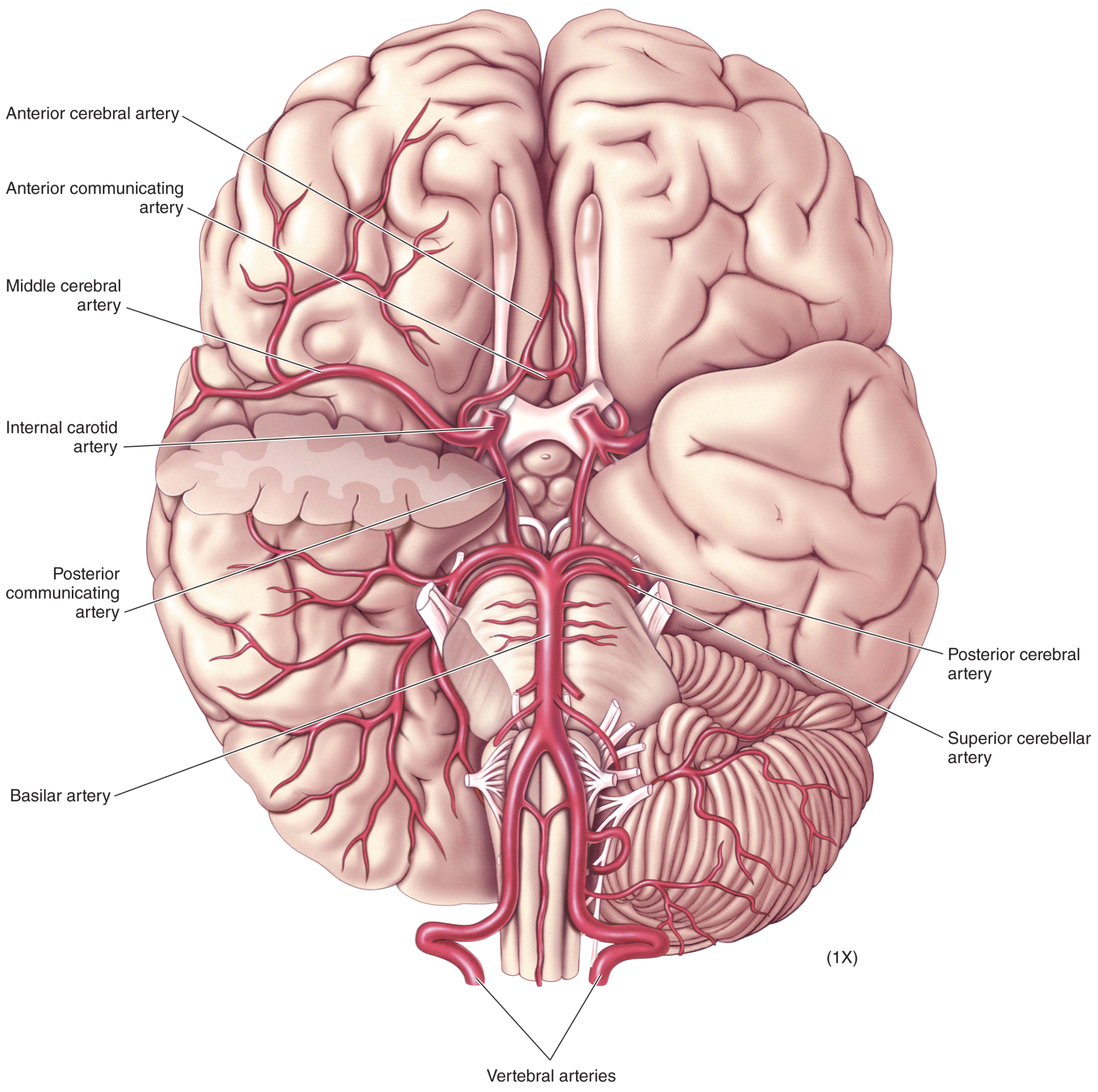
Ventral view: All major cerebral arteries - Neuroscience: Exploring the Brain, 5th Edition
1. Anterior (Carotid) Circulation
Internal Carotid Artery (ICA)
- Arises as a terminal branch of the common carotid artery in the neck
- Ascends to the base of the skull and enters the carotid canal
- On entering the cranial cavity, each ICA gives off four major branches:
- Ophthalmic artery - supplies the orbit and eye
- Posterior communicating artery (PComm) - connects to the posterior cerebral artery, completing the circle of Willis
- Middle cerebral artery (MCA)
- Anterior cerebral artery (ACA)
Middle Cerebral Artery (MCA)
- The largest branch of the ICA
- Travels through the lateral (Sylvian) fissure
- Supplies the entire lateral surface of the cerebral hemisphere - including the lateral frontal, parietal, and temporal lobes
- Gives off lenticulostriate arteries that supply the basal ganglia and internal capsule (deep perforating branches)
- MCA territory infarction causes contralateral hemiplegia, hemisensory loss, and aphasia (dominant side)
Anterior Cerebral Artery (ACA)
- Courses anteriorly and medially
- The two ACAs are connected by the anterior communicating artery (AComm)
- Supplies the medial surface of the frontal and parietal lobes (including the leg area of the motor cortex)
- ACA stroke characteristically causes contralateral leg weakness
- The recurrent artery of Heubner (a branch of ACA) supplies the head of the caudate nucleus and anterior limb of the internal capsule
2. Posterior (Vertebrobasilar) Circulation
Vertebral Arteries
- Each arises from the first part of the subclavian artery in the lower neck
- Pass superiorly through the foramina transversaria of the upper six cervical vertebrae
- Enter the cranial cavity through the foramen magnum
- Each gives off a small meningeal branch on entry
- Before joining, each gives off:
- Posterior inferior cerebellar artery (PICA) - supplies the posterior and inferior cerebellum and the lateral medulla
- Posterior spinal artery - descends on the posterior spinal cord (can also arise from PICA)
- Contribution to the anterior spinal artery (the two contributions join to form a single midline vessel)
- The two vertebral arteries fuse at the lower border of the pons to form the basilar artery
Basilar Artery
- Travels rostrally along the anterior surface of the pons
- Gives off (caudal to rostral):
- Anterior inferior cerebellar arteries (AICA) - supply the anterior inferior cerebellum and the pons/inner ear via the labyrinthine artery
- Pontine arteries - multiple small perforating branches to the pons
- Superior cerebellar arteries (SCA) - supply the superior cerebellum and dorsal pons/midbrain
- Terminates at the level of the midbrain by bifurcating into the two posterior cerebral arteries (PCA)
Posterior Cerebral Artery (PCA)
- Connected to the ICA via the posterior communicating artery
- Supplies the medial occipital lobe (primary visual cortex), the inferior temporal lobe, the thalamus (via thalamogeniculate and thalamoperforating branches), and the midbrain
- PCA infarction causes contralateral homonymous hemianopia (with macular sparing)
3. Circle of Willis (Cerebral Arterial Circle)
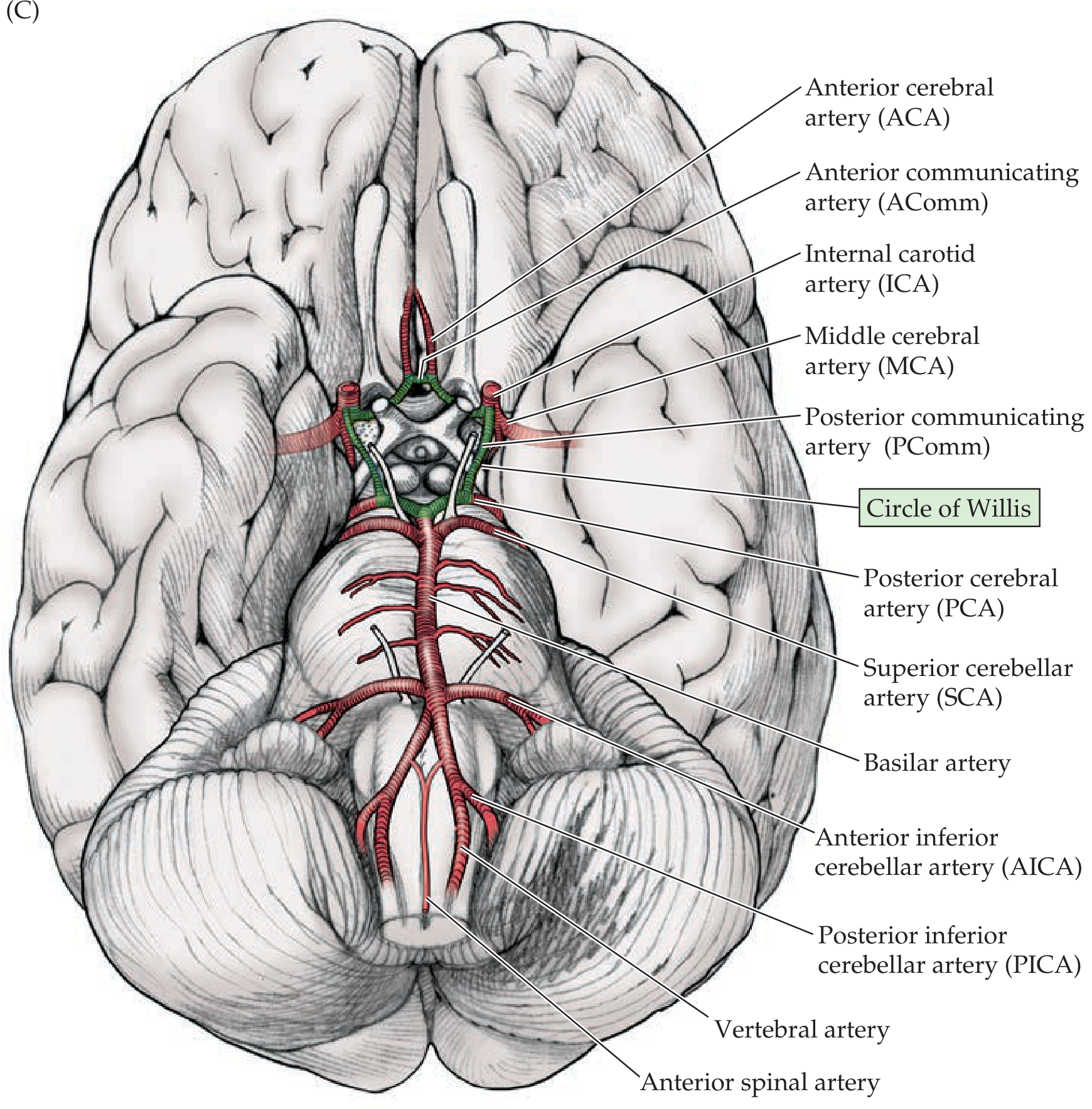
Circle of Willis - Neuroanatomy through Clinical Cases 3rd Edition, p. 71
The circle of Willis is a polygonal anastomotic ring at the base of the brain formed by:
| Component | Connects |
|---|---|
| Anterior communicating artery (AComm) | Left ACA ↔ Right ACA |
| Two posterior communicating arteries (PComm) | ICA ↔ PCA (each side) |
| Anterior cerebral arteries (×2) | Part of the ring |
| Internal carotid arteries (×2) | Part of the ring |
| Posterior cerebral arteries (×2) | Part of the ring |
Function: Provides collateral flow so that occlusion of one feeding vessel can be compensated by flow through the circle. However, in practice, the anastomoses are often insufficient during sudden occlusion.
Clinical significance: Saccular (berry) aneurysms most commonly form at the branch points of the circle - particularly at the AComm, PComm origin, and MCA bifurcation. Rupture causes subarachnoid hemorrhage presenting with a sudden thunderclap headache.
4. Cortical Territory Map
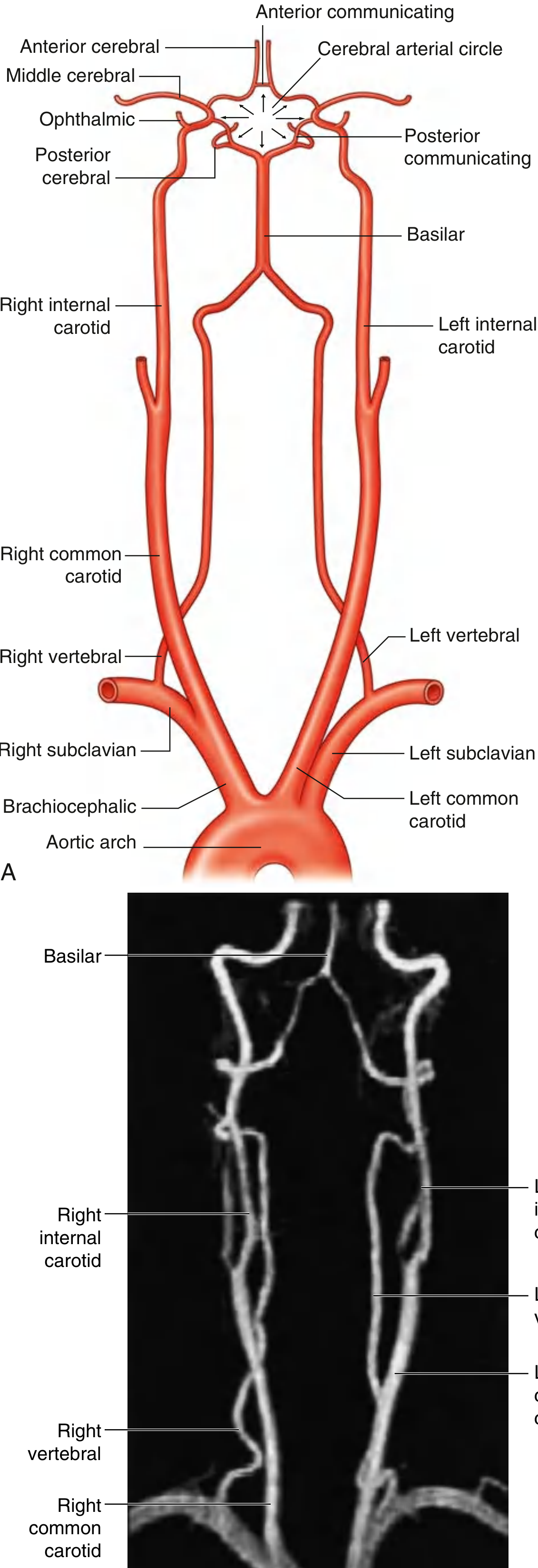
| Artery | Cortical Territory | Deep Structures |
|---|---|---|
| ACA | Medial frontal + parietal (leg area) | Anterior corpus callosum, septal region |
| MCA | Entire lateral hemisphere (face/arm motor cortex) | Basal ganglia, internal capsule (via lenticulostriates) |
| PCA | Medial occipital, inferior temporal | Thalamus, midbrain |
| PICA | Posterior inferior cerebellum | Lateral medulla |
| AICA | Anterior inferior cerebellum | Pontine tegmentum |
| SCA | Superior cerebellum | Dorsal pons, midbrain |
- Goldman-Cecil Medicine, p. 1932; Gray's Anatomy for Students, p. 1019
5. Venous Drainage
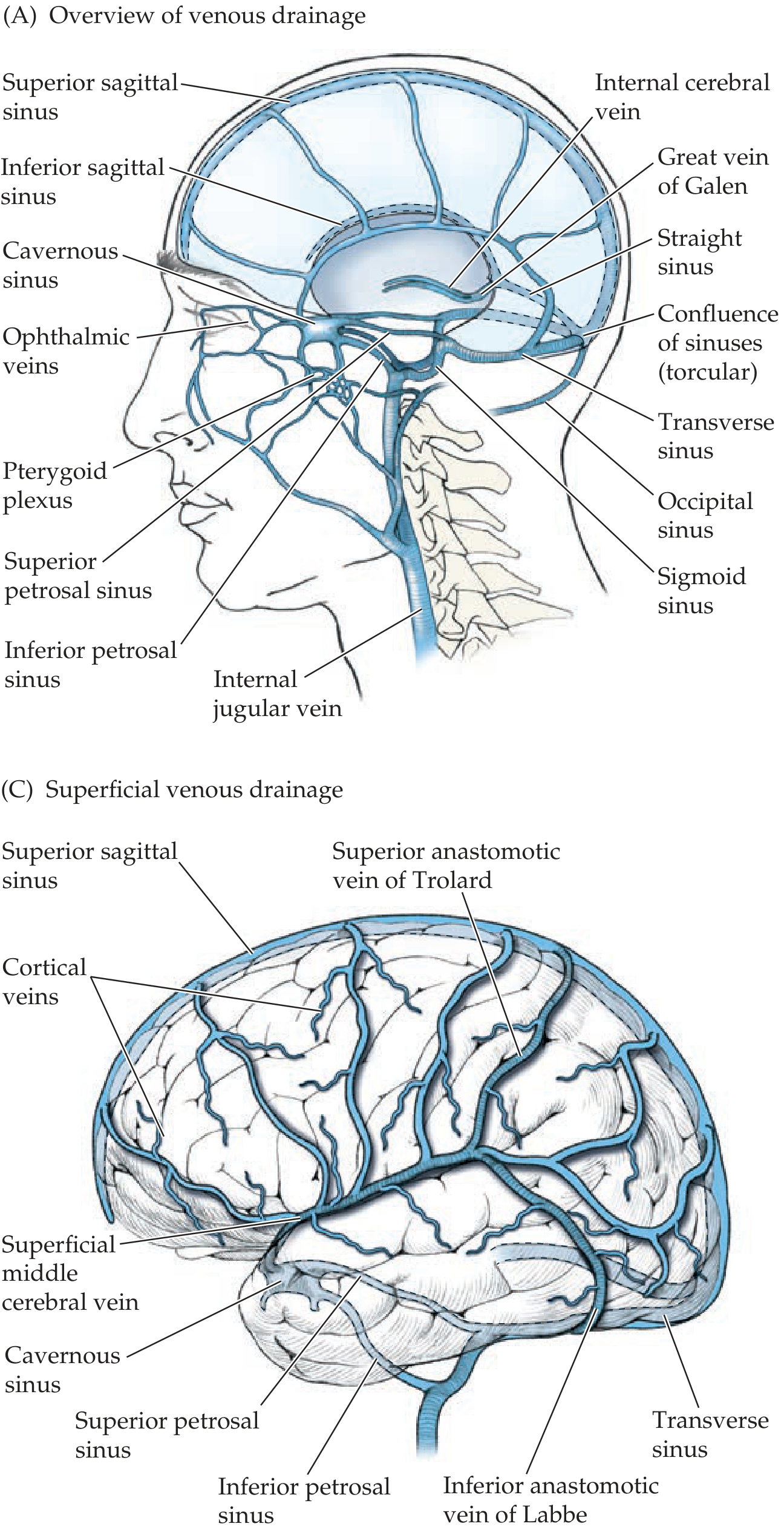
Venous drainage of cerebral hemispheres - Neuroanatomy through Clinical Cases 3rd Edition, p. 435
Venous drainage is divided into superficial and deep systems. Both ultimately drain into the internal jugular veins.
Superficial System
- Cortical veins drain into the superior sagittal sinus (runs along the superior border of the falx cerebri)
- The cavernous sinus (on either side of the sella turcica) receives blood from the face/orbit and drains via:
- Superior petrosal sinus → transverse sinus
- Inferior petrosal sinus → internal jugular vein
- Important anastomotic veins:
- Vein of Trolard (superior anastomotic vein) - drains into the superior sagittal sinus
- Vein of Labbe (inferior anastomotic vein) - drains into the transverse sinus
- Superficial middle cerebral vein - drains into the cavernous sinus
Deep System
- Deep white matter and basal ganglia drain via internal cerebral veins and basal veins of Rosenthal
- These join to form the great cerebral vein of Galen
- Vein of Galen joins the inferior sagittal sinus to form the straight sinus (sinus rectus)
Final Pathway
Superior sagittal sinus ──┐
Straight sinus ├──→ Confluence of sinuses (Torcular Herophili)
Occipital sinus ┘
↓
Transverse sinuses → Sigmoid sinuses → Internal jugular veins
The cavernous sinus is also clinically important - the ICA runs through it, and cranial nerves III, IV, V1, V2, and VI all pass through or in its walls (relevant in cavernous sinus thrombosis).
6. Clinical Correlates
| Condition | Vessel Involved | Key Features |
|---|---|---|
| Ischemic stroke | Usually MCA (most common) | Hemiplegia, aphasia, hemisensory loss |
| Berry aneurysm | Circle of Willis branch points | Thunderclap headache, SAH |
| Subdural hematoma | Bridging veins (cortical veins → sinuses) | Venous bleeding, delayed symptoms |
| Lateral medullary syndrome (Wallenberg) | PICA | Dysphagia, ipsilateral Horner, contralateral pain/temp loss |
| Weber syndrome | Posterior cerebral / basilar perforators | CN III palsy + contralateral hemiplegia |
| Cavernous sinus thrombosis | Cavernous sinus | CN III/IV/VI palsy, chemosis, proptosis |
- Gray's Anatomy for Students, p. 1020-1021; Neuroanatomy through Clinical Cases 3rd Edition, p. 70-71; Color Atlas of Human Anatomy, p. 392
This is a shared conversation. Sign in to Orris to start your own chat.