
How to put rules tube aspiration
Reading File
The query "rules tube aspiration" likely refers to the Ryle's tube (nasogastric tube) aspiration procedure - a common clinical skill. Let me search the medical library for detailed instructions.
Searching sources...
Finding Sources
Finding Sources
Reading File
Reading File
Now let me search for the step-by-step procedure for Ryle's tube insertion and aspiration from clinical procedure textbooks.
Finding Sources
Finding Sources
Reading File
Reading File
Reading File
Reading File
Reading File
Reading File
I now have comprehensive information from the textbooks. Let me fetch the key procedure image for a visual reference.
Loading Image
Excellent! I now have all the information needed for a complete, well-illustrated answer. Here is the full guide:
Ryle's Tube (Nasogastric Tube) Insertion and Aspiration
A Ryle's tube is a non-vented, single-lumen nasogastric (NG) tube used primarily for gastric decompression and aspiration. It is named after Dr. John Alfred Ryle. The vented equivalent is the Salem sump tube (double-lumen). Both pass through the nose into the stomach.
Indications
- Gastric decompression (bowel obstruction, ileus)
- Prevention of aspiration before/after surgery
- Monitoring and evaluation of upper GI bleeding
- Administration of medications or feeds when the patient cannot swallow
- Gastric lavage in poisoning
Contraindications
- Midface injury or basilar skull fracture (use orogastric route instead)
- Severe coagulopathy (relative)
- History of gastric bypass or lap banding
- Esophageal strictures or alkali injury
Equipment Needed
- Ryle's tube (appropriate size - 14-18 Fr for adults)
- Topical vasoconstrictor (phenylephrine 0.5% or oxymetazoline 0.05% spray)
- Topical anesthetic (2% lidocaine gel or nebulized 4-10% lidocaine)
- Lubricant (viscous lidocaine 2% or water-soluble gel)
- Cup of water with a straw
- Catheter-tipped syringe (20-60 mL)
- Tape for securing
- pH paper or stethoscope for confirmation
- Gloves, gown, kidney dish
Step-by-Step Procedure
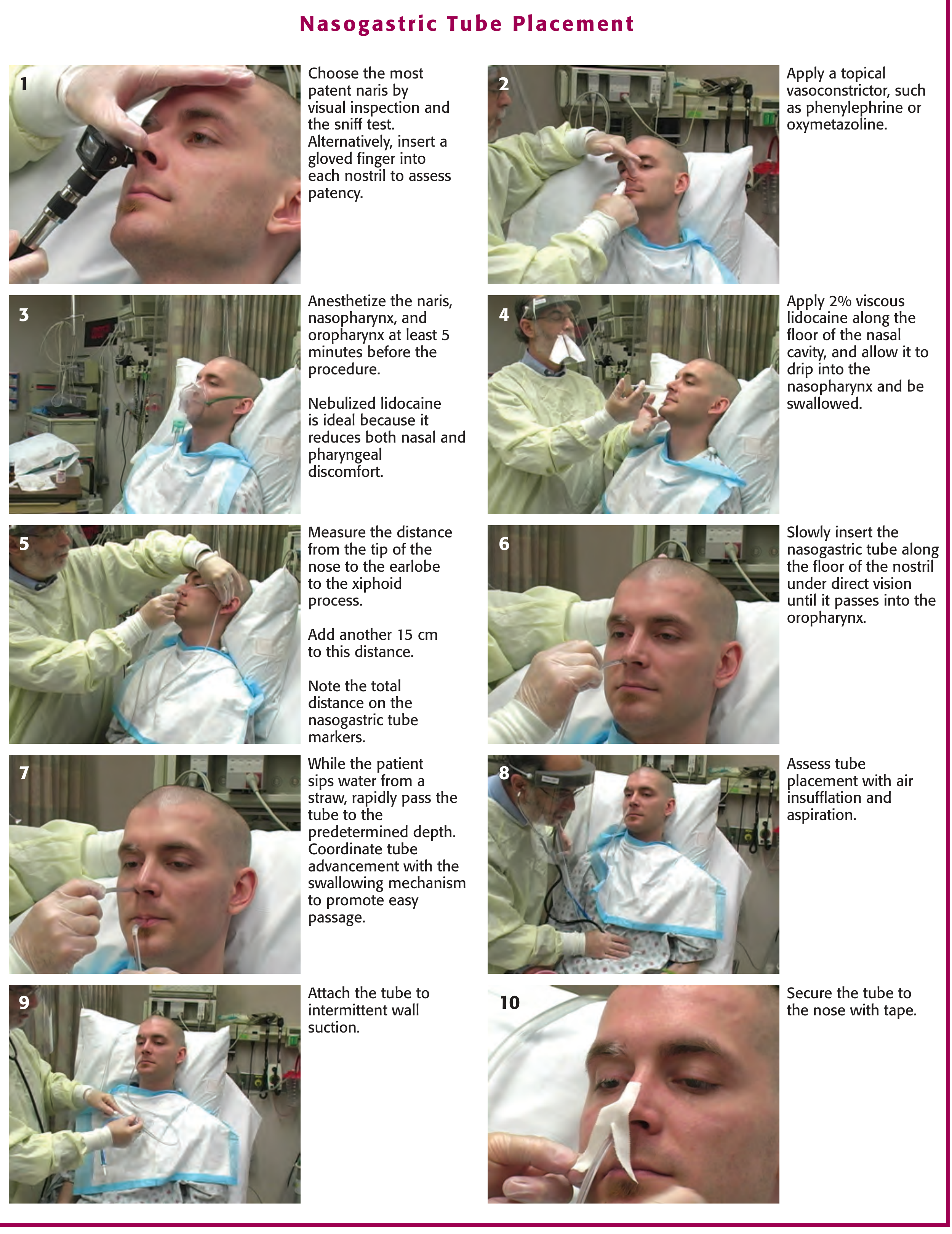
1. Patient Preparation
- Sit the patient upright at 45-90 degrees (semi-Fowler's or fully upright if possible)
- Explain the procedure to the patient
- Check for and remove dentures if present
- Place a kidney dish and tissue near the patient
2. Choose the Nostril (Step 1)
- Inspect both nostrils and ask the patient to sniff through each
- Choose the most patent naris - insert a gloved finger gently to assess patency
- Avoid any nostril with recent trauma or nasal polyps
3. Apply Vasoconstrictor (Step 2)
- Spray a topical vasoconstrictor (phenylephrine 0.5% or oxymetazoline) into both nares
- This reduces nasal mucosal congestion and minimizes bleeding
4. Anesthetize (Step 3 & 4)
- Anesthetize the naris, nasopharynx, and oropharynx at least 5 minutes before proceeding
- Nebulized lidocaine (4 mL of 10% solution) is the most effective method - reduces both nasal and pharyngeal discomfort
- Apply 2% viscous lidocaine along the floor of the nasal cavity and allow it to drip into the nasopharynx to be swallowed
5. Measure the Insertion Depth (Step 5)
- Measure from the tip of the nose → earlobe → xiphoid process
- Add an additional 15 cm to this distance
- Mark this total depth on the tube (Ryle's tubes have depth markings)
6. Lubricate the Tube
- Coat the first 15-20 cm of the tube liberally with 2% lidocaine gel or water-soluble lubricant
- Do not rely only on the lubricant for anesthesia - it must be pre-applied to the mucosa first
7. Insert the Tube (Step 6 & 7)
- With the patient's head in neutral or slightly flexed position, slowly insert the tube along the floor of the nostril (aim horizontally backward toward the ear, NOT upward)
- Pass it gently into the oropharynx under direct vision
- Once it reaches the oropharynx (~15 cm), ask the patient to sip water through a straw and swallow repeatedly
- Rapidly advance the tube to the predetermined depth while the patient is swallowing - this coordinates tube advancement with the swallowing mechanism
Key tip: If resistance is met, withdraw slightly, rotate the tube, and retry. If the patient coughs, chokes, or cannot speak - stop immediately and withdraw as the tube may be in the airway.
8. Confirm Placement (Step 8)
Use more than one method:
| Method | Detail |
|---|---|
| Air insufflation + auscultation | Inject 10-20 mL of air rapidly via syringe; auscultate over the epigastrium for a whooshing sound. Quick but not fully reliable |
| Aspiration + pH testing | Aspirate gastric contents; pH <4 = ~95% chance of correct gastric placement. More reliable |
| Patient speech | Ask the patient to speak - inability to speak suggests tracheal placement |
| Chest X-ray | Gold standard, especially in unconscious/obtunded patients. Tube should be seen passing below the diaphragm, tip at or below the gastroesophageal junction |
| Ultrasound | Highly reliable when successful |
9. Attach to Suction (Step 9)
- Connect the tube to intermittent wall suction (or leave on free drainage)
- For a Ryle's tube used for gastric aspiration: place on free drainage with 4-hourly aspiration or connect to continuous/intermittent suction as clinically needed
10. Secure the Tube (Step 10)
- Clean the nose and tube with tincture of benzoin (if available)
- Tape the tube to the nose using a butterfly/split-tape technique - ensure no pressure on the nostril margins to prevent necrosis
- Also secure the tube to the patient's gown to prevent accidental traction on the nose
Ongoing Aspiration Management
- For decompression (e.g., bowel obstruction): place on free drainage with 4-hourly aspiration or continuous suction
- Record the volume, color, and character of aspirate each time
- For the Salem sump tube: keep the blue pigtail (air vent) above the level of stomach fluid at all times to prevent siphoning
Confirming and Monitoring
- Inspect the aspirate: gastric contents are typically pale/yellow; bilious if the tube is past the pylorus; frank blood if upper GI bleeding
- Re-confirm position by X-ray if the patient's condition changes or placement is uncertain
- Replace the nose tape and reapply benzoin if the tape loosens
Complications to Watch For
| Complication | Watch for |
|---|---|
| Pulmonary misplacement | Coughing, cyanosis, inability to speak |
| Intracranial placement | Only in head trauma with basilar skull fracture |
| Epistaxis | Common - minimize with vasoconstrictor and gentle technique |
| Sinusitis | With prolonged tube placement |
| Aspiration pneumonia | If tube is misplaced or patient vomits around tube |
| Nasal necrosis | From poorly secured, tight-fitting tube |
Sources:
- Roberts and Hedges' Clinical Procedures in Emergency Medicine, Chapter 40 - Nasogastric Tube Placement (pp. 961-965)
- Current Surgical Therapy, 14th Edition - Nasogastric Tube chapter (p. 162)
- Bailey and Love's Short Practice of Surgery, 28th Edition - Supportive Management / Ryle's tube for bowel decompression (p. 1407)
This is a shared conversation. Sign in to Orris to start your own chat.