Physical examination for Down syndrome

Reading File
Finding Sources
Searching Images

Down syndrome physical features clinical examination

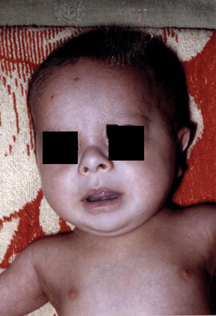
This clinical photograph shows the head and torso of a four-month-old infant, highlighting classic craniofacial dysmorphism associated with Trisomy 21 (Down Syndrome). Visible features include a prominent flat nasal bridge and a depressed nasal root, contributing to a flattened facial profile. The infant exhibits upslanting palpebral fissures (partially obscured for privacy) and a slightly protruding tongue (macroglossia or relative macroglossia). The head shape suggests a flat occiput, and the ears appear small and low-set. These phenotypic markers are key diagnostic indicators in pediatric genetics. The image serves as a clinical reference for identifying midface hypoplasia and other soft markers of chromosomal abnormalities during physical examination. The medical specialty is Pediatrics and Medical Genetics, intended for educational identification of syndromic facies.

This clinical photograph shows the head and torso of a four-month-old infant, highlighting classic craniofacial dysmorphism associated with Trisomy 21 (Down Syndrome). Visible features include a prominent flat nasal bridge and a depressed nasal root, contributing to a flattened facial profile. The infant exhibits upslanting palpebral fissures (partially obscured for privacy) and a slightly protruding tongue (macroglossia or relative macroglossia). The head shape suggests a flat occiput, and the ears appear small and low-set. These phenotypic markers are key diagnostic indicators in pediatric genetics. The image serves as a clinical reference for identifying midface hypoplasia and other soft markers of chromosomal abnormalities during physical examination. The medical specialty is Pediatrics and Medical Genetics, intended for educational identification of syndromic facies.

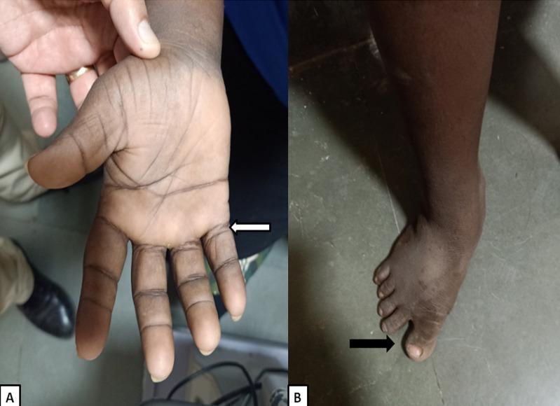
Clinical photographs showing characteristic dysmorphic features associated with Trisomy 21 (Down syndrome). Panel A illustrates the palmar aspect of the left hand, highlighting a short and broad hand morphology, a single transverse palmar crease (simian crease), and clinodactyly (incurving) of the shortened fifth finger. Panel B shows the patient's foot from a dorsal view, demonstrating a prominent 'sandal gap' deformity, which is a widened space between the first (hallux) and second toes. These physical findings are classic phenotypic markers used in the clinical diagnosis of genetic trisomy. The images serve as educational visual aids for identifying common musculoskeletal and dermatoglyphic manifestations of chromosomal abnormalities during a physical examination.

Clinical photographs showing characteristic dysmorphic features associated with Trisomy 21 (Down syndrome). Panel A illustrates the palmar aspect of the left hand, highlighting a short and broad hand morphology, a single transverse palmar crease (simian crease), and clinodactyly (incurving) of the shortened fifth finger. Panel B shows the patient's foot from a dorsal view, demonstrating a prominent 'sandal gap' deformity, which is a widened space between the first (hallux) and second toes. These physical findings are classic phenotypic markers used in the clinical diagnosis of genetic trisomy. The images serve as educational visual aids for identifying common musculoskeletal and dermatoglyphic manifestations of chromosomal abnormalities during a physical examination.

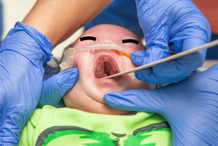
Clinical photograph of an infant in the supine position displaying a complete cleft palate. The image focuses on the open oral cavity, where a significant midline defect is visible, extending from the hard palate anteriorly to the soft palate posteriorly, creating a direct communication with the nasal cavity. The infant presents with dysmorphic facial features consistent with Trisomy 21 (Down syndrome), including a flattened nasal bridge and almond-shaped, upward-slanting palpebral fissures (partially obscured). Medical interventions are visible, including an orogastric tube secured to the left cheek with adhesive tape and a thin medical probe or tongue depressor used to facilitate the examination of the oral defect. Neonatal teeth or tooth buds are visible on the upper alveolar ridge. The surrounding clinical context includes gloved hands of healthcare providers performing the physical examination. This image is an educational example of congenital craniofacial anomalies and their association with chromosomal syndromes.

Clinical photograph of an infant in the supine position displaying a complete cleft palate. The image focuses on the open oral cavity, where a significant midline defect is visible, extending from the hard palate anteriorly to the soft palate posteriorly, creating a direct communication with the nasal cavity. The infant presents with dysmorphic facial features consistent with Trisomy 21 (Down syndrome), including a flattened nasal bridge and almond-shaped, upward-slanting palpebral fissures (partially obscured). Medical interventions are visible, including an orogastric tube secured to the left cheek with adhesive tape and a thin medical probe or tongue depressor used to facilitate the examination of the oral defect. Neonatal teeth or tooth buds are visible on the upper alveolar ridge. The surrounding clinical context includes gloved hands of healthcare providers performing the physical examination. This image is an educational example of congenital craniofacial anomalies and their association with chromosomal syndromes.

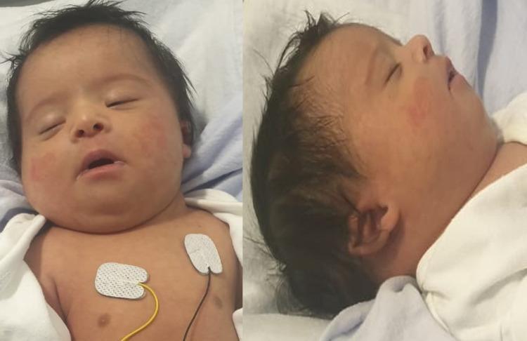
This clinical photograph consists of frontal and lateral views of a newborn infant displaying multiple dysmorphic facial features. Key clinical findings include a broad forehead, ocular hypertelorism (increased distance between the eyes), and upward slanting palpebral fissures. A visible infraorbital crease is present beneath the eyes. The nasal anatomy shows a depressed nasal bridge. Midface examination reveals a flat philtrum and micrognathia (small jaw). The lateral view highlights low-set, malformed ears with attached pinnae and a short, webbed neck. These features are characteristic of chromosomal abnormalities, specifically consistent with the phenotype of Trisomy 21 (Down syndrome). The infant is shown in a clinical setting with ECG monitoring leads visible on the chest. This image serves as an educational reference for neonatal physical examination and the identification of genetic dysmorphism in pediatrics and medical genetics.

This clinical photograph consists of frontal and lateral views of a newborn infant displaying multiple dysmorphic facial features. Key clinical findings include a broad forehead, ocular hypertelorism (increased distance between the eyes), and upward slanting palpebral fissures. A visible infraorbital crease is present beneath the eyes. The nasal anatomy shows a depressed nasal bridge. Midface examination reveals a flat philtrum and micrognathia (small jaw). The lateral view highlights low-set, malformed ears with attached pinnae and a short, webbed neck. These features are characteristic of chromosomal abnormalities, specifically consistent with the phenotype of Trisomy 21 (Down syndrome). The infant is shown in a clinical setting with ECG monitoring leads visible on the chest. This image serves as an educational reference for neonatal physical examination and the identification of genetic dysmorphism in pediatrics and medical genetics.

Reading File
Finding Sources
Reading File
Reading File
Reading File
Loading Image
Loading Image
Loading Image
I now have comprehensive information to write a detailed, well-organized answer. Let me compile the full physical examination findings for Down syndrome.

Physical Examination in Down Syndrome (Trisomy 21)

Down syndrome (trisomy 21) is the most common chromosomal disorder and the single most common genetic cause of moderate intellectual disability, with an incidence of approximately 1 in 700 live births. The diagnosis can usually be made clinically at birth or shortly thereafter from a characteristic - though variable - phenotype. Karyotyping remains essential for confirmation and genetic counseling.
  • Thompson & Thompson Genetics and Genomics in Medicine, 9th ed.

1. General Appearance

FeatureDetail
HypotoniaOften the first abnormality noticed in the newborn; generalized, "floppy baby"
Short staturePresent in most individuals
HabitusBroad, stocky build; obesity common in older children and adults

2. Head and Face

Infant with Down syndrome - facial features including flat nasal bridge and upslanting palpebral fissures
Newborn with Down syndrome - frontal and lateral views showing flat occiput, low-set ears, and upslanting palpebral fissures
RegionFindings
SkullBrachycephaly; flat occiput
FaceFlat facial profile; midface hypoplasia
FontanelleWide anterior fontanelle; delayed closure
Nasal bridgeFlat/depressed
EyesUpslanting palpebral fissures; epicanthal folds; Brushfield spots (speckled iris); strabismus common
EarsSmall, low-set, dysplastic ears; narrow ear canals
Mouth/tongueProtruding tongue (relative macroglossia); fissured tongue in older children; open mouth posture
PalateHigh-arched; narrow
NeckShort; loose skin on the nape of the neck (in neonates)

3. Hands and Feet

Down syndrome hands and feet - single palmar crease (simian crease) and sandal gap deformity
RegionFindings
HandsShort, broad hands; single transverse palmar crease (simian crease); fifth finger clinodactyly (incurving); brachydactyly
Feet"Sandal gap" - widened space between 1st and 2nd toes; plantar crease between 1st and 2nd toe
DermatoglyphicsIncreased ulnar loops; distal axial triradius

4. Cardiovascular Examination

Congenital heart disease is present in ~50% of all liveborn infants with Down syndrome. It is the most important associated anomaly and the most common cause of early death.
DefectNotes
Atrioventricular septal defect (AVSD)Most characteristic; also called endocardial cushion defect
Ventricular septal defect (VSD)Common
Atrial septal defect (ASD)Common
Patent ductus arteriosus (PDA)Present in a subset
Tetralogy of FallotLess common
Examine for: murmurs, signs of heart failure (tachycardia, hepatomegaly, poor feeding), cyanosis, and abnormal precordial impulse. An echocardiogram is mandatory in every newborn with Down syndrome.

5. Abdominal Examination

FindingNotes
Duodenal atresia"Double bubble" sign on imaging; presents with bilious vomiting in neonates - strongly associated with Down syndrome
Hirschsprung diseaseAbsent ganglion cells; constipation, abdominal distension
Tracheoesophageal fistulaCheck for polyhydramnios history, inability to pass NG tube
Umbilical herniaCommon due to hypotonia

6. Neurological / Musculoskeletal

FeatureNotes
HypotoniaGeneralized, central; hyperextensible joints
Atlantoaxial instabilityDue to ligamentous laxity at C1-C2; check for neck pain, limb weakness - important before any surgical procedure or contact sport
ReflexesReduced deep tendon reflexes in infancy due to hypotonia
Intellectual disabilityModerate (IQ typically 35-70); developmental delay evident by end of 1st year
Short stature / growth delayProgressive throughout childhood

7. Eyes (Ophthalmological)

FindingNotes
Brushfield spotsWhite/yellow spots on the iris periphery
Refractive errorsMyopia, hyperopia, astigmatism - very common
Strabismus~30-45%
NystagmusPresent in some
CataractsCongenital or early-onset

8. Ears, Nose, Throat (ENT)

Per Scott-Brown's Otorhinolaryngology:
  • Otitis media with effusion (OME) - extremely common; conductive hearing loss
  • Narrow, waxy ear canals
  • Sensorineural hearing loss with inner ear dysplasia
  • Increased incidence of cholesteatoma
  • Obstructive sleep apnoea (OSA) - due to relative adenotonsillar hypertrophy, midface/pharyngeal hypoplasia, and hypotonia
  • Subglottic stenosis; need for smaller-diameter ET tube than age would predict
  • Annual OSA screening recommended until age 3-5 years

9. Skin

FindingNotes
Loose skin on nape (neonates)Often one of the first clinical clues
Cutis marmorataMottled skin, especially in cold
Dry, rough skinIncreases with age
Increased risk of alopecia areataAutoimmune

10. Haematological / Oncological (Examination Implications)

  • 15-fold increase in risk of leukaemia (especially ALL and AML/transient myeloproliferative disorder in neonates)
  • Examine for pallor, petechiae, hepatosplenomegaly, lymphadenopathy

11. Alzheimer Disease / Dementia (Adults)

Nearly all individuals with Down syndrome develop Alzheimer-type neuropathology (cortical atrophy, neurofibrillary tangles) several decades earlier than the general population. On examination in adults: progressive cognitive decline, behavioral change, seizures.

Summary Mnemonic: "DOWN"

LetterFeatures
DDuodenal atresia; Dermatoglyphics (simian crease); Dementia (Alzheimer)
OOcciput flat; Oblique palpebral fissures (upslanting); Otitis media
WWide fontanelle; Wide sandal gap
NNeck short with loose skin; Nape laxity; Nystagmus

Key references:
  • Thompson & Thompson Genetics and Genomics in Medicine, 9th ed., pp. 102-103
  • Scott-Brown's Otorhinolaryngology Head & Neck Surgery, Vol. 2
  • Fuster & Hurst's The Heart, 15th ed., Table 2-4 (Genetic Syndromes and Cardiovascular Features)
  • Cummings Otolaryngology Head and Neck Surgery
This is a shared conversation. Sign in to Orris to start your own chat.