
make a case study for MRI BRAIN WITH CONTRAST for space occupying lession rule out a high grade glioma use the guide attached above
complete the above request
cd /tmp/workspace && python3 -c " import pdfplumber with pdfplumber.open('/tmp/workspace/attachments/8b5d69b7-2549-4130-8d4c-cca731810dc7/REVISED GUIDE TO CASE STUDY WRITING.pdf') as pdf: for i, page in enumerate(pdf.pages): text = page.extract_text() print(f'--- PAGE {i+1} ---') print(text) "
standardized brain tumor MRI protocol sequences glioma imaging T1 FLAIR DWI perfusion post-contrast
"high grade glioma" AND MRI diagnosis
glioblastoma high grade glioma MRI axial T1 post contrast ring enhancing lesion with necrosis and edema
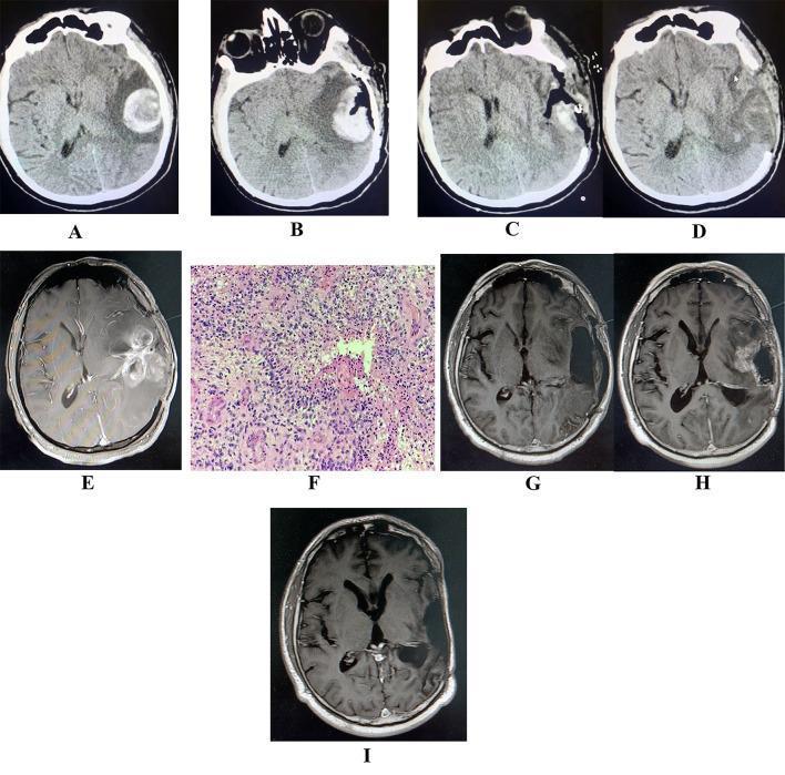
A longitudinal sequence of neuroradiological and histopathological images illustrating the progression and management of glioblastoma. (A) Initial non-contrast head CT shows an acute hyperdense left temporoparietal intraparenchymal hemorrhage with surrounding hypodense perihematomal edema. (B-D) Sequential CT scans following hematoma evacuation demonstrate post-surgical changes, including a decompressive craniectomy (bone flap removal) and persistent left-sided cerebral edema. (E) Axial T1-weighted contrast-enhanced MRI reveals a large, peripherally enhancing, ring-like space-occupying lesion with central necrosis in the same region, consistent with high-grade glioma. (F) H&E stained histopathology slide shows high-grade pleomorphic cells, hypercellularity, and microvascular proliferation, diagnostic of glioblastoma (WHO grade 4). (G) Post-resection MRI confirms gross total tumor removal. (H) Follow-up MRI at four months shows an enhancing mass in the surgical bed, indicating in-situ tumor recurrence. (I) Final MRI following targeted chemotherapeutic treatment with temozolomide and bevacizumab demonstrates a significant reduction in the recurrent tumor signal. This series highlights the clinical course of apoplectic glioma presentation, surgical intervention, and recurrence management.
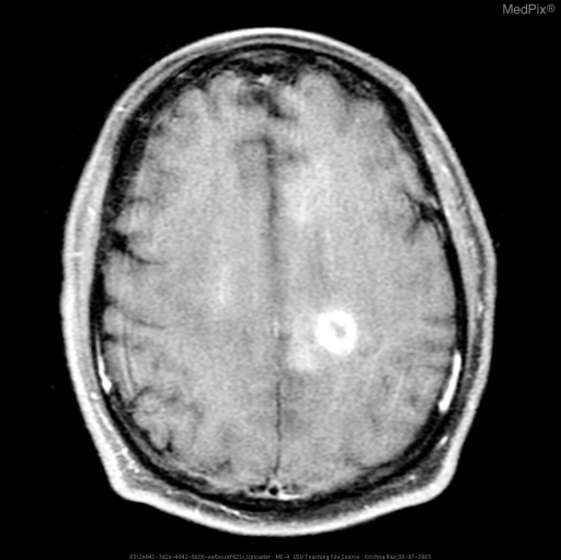
**Imaging Modality:** Axial T1-weighted post-contrast Magnetic Resonance Imaging (MRI) of the brain.
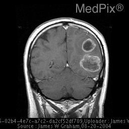
**Imaging Modality:** T1-weighted post-contrast Magnetic Resonance Imaging (MRI), coronal view.
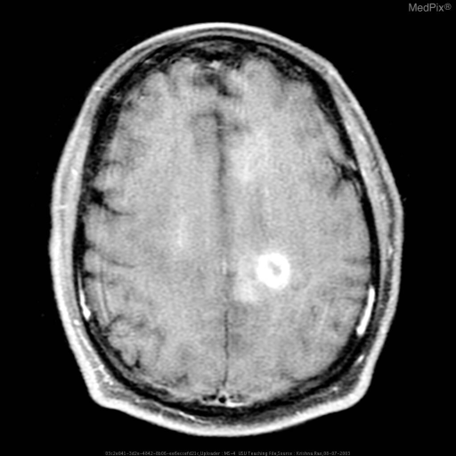
**Imaging Modality:** Axial T1-weighted post-contrast Magnetic Resonance Imaging (MRI).
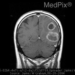
**Imaging Modality:** Coronal T1-weighted post-contrast Magnetic Resonance Imaging (MRI) of the brain.
~/case-study-mri-glioma/build_docx.py
#!/usr/bin/env python3
from docx import Document
from docx.shared import Pt, Inches, RGBColor, Cm
from docx.enum.text import WD_ALIGN_PARAGRAPH
from docx.enum.table import WD_TABLE_ALIGNMENT
from docx.oxml.ns import qn
import docx

doc = Document()

style = doc.styles["Normal"]
style.font.name = "Arial"
style.font.size = Pt(11)

def h1(text):
    p = doc.add_heading(text, level=1)
    return p

def h2(text):
    p = doc.add_heading(text, level=2)
    return p

def para(text, bold=False, italic=False, size=None, align=None):
    p = doc.add_paragraph()
    run = p.add_run(text)
    run.bold = bold
    run.italic = italic
    if size:
        run.font.size = Pt(size)
    if align:
        p.alignment = align
    return p

def bullet(text):
    p = doc.add_paragraph(text, style='List Bullet')
    return p

def numbered(text):
    p = doc.add_paragraph(text, style='List Number')
    return p

# ---------------- COVER PAGE ----------------
title = doc.add_paragraph()
title.alignment = WD_ALIGN_PARAGRAPH.CENTER
r = title.add_run("SCHOOL OF HEALTH SCIENCES")
r.bold = True
r.font.size = Pt(16)

sub = doc.add_paragraph()
sub.alignment = WD_ALIGN_PARAGRAPH.CENTER
r = sub.add_run("Department of Clinical Imaging")
r.bold = True
r.font.size = Pt(13)

sub2 = doc.add_paragraph()
sub2.alignment = WD_ALIGN_PARAGRAPH.CENTER
r = sub2.add_run("CASE STUDY")
r.bold = True
r.font.size = Pt(20)
r.font.color.rgb = RGBColor(0x1F, 0x4E, 0x79)

sub3 = doc.add_paragraph()
sub3.alignment = WD_ALIGN_PARAGRAPH.CENTER
r = sub3.add_run("MRI Brain with Contrast for a Space-Occupying Lesion:\nRule Out High-Grade Glioma")
r.bold = True
r.font.size = Pt(15)

doc.add_paragraph()
fields = [
    "Course code: ____________________________________________",
    "Name of student: ____________________________________________",
    "Registration number: ____________________________________________",
    "Name & signature of clinical instructor: ____________________________________________",
    "Examination assessed: MRI Brain with Contrast",
    "Date: ____________________________________________",
]
for f in fields:
    p = doc.add_paragraph(f)
    p.paragraph_format.space_after = Pt(10)

doc.add_page_break()

# ---------------- TABLE OF CONTENTS (manual) ----------------
h1("Table of Contents")
toc_items = [
    "1. Introduction",
    "2. Brief Gross Anatomy",
    "3. Patient History",
    "4. Discussion of Provisional Diagnosis and Aetiology",
    "5. Equipment and Accessories",
    "6. Patient Preparation for the Study",
    "7. Discussion of Technique Employed, Imaging Protocol/Procedure",
    "8. Patient Care - Before, During and After the Study",
    "9. Discussion of Findings/Pathology (Radiological Report)",
    "10. Further Role of the Imaging Department",
    "11. Patient Management",
    "12. Inter-professional Collaboration",
    "13. Reflection",
    "14. Conclusion",
    "15. References",
    "16. Appendices",
]
for t in toc_items:
    doc.add_paragraph(t)

doc.add_page_break()

# ---------------- 1. INTRODUCTION ----------------
h1("1. Introduction")
para(
    "A space-occupying lesion (SOL) of the brain is any focal abnormality that occupies space within "
    "the cranial cavity and disturbs the normal architecture or function of surrounding brain tissue. "
    "Common causes include primary brain tumours (gliomas, meningiomas, lymphoma), metastatic disease, "
    "abscesses, and less commonly vascular malformations or granulomas. Magnetic Resonance Imaging (MRI) "
    "of the brain with intravenous gadolinium-based contrast is the imaging investigation of choice for the "
    "detection, characterisation, and pre-treatment work-up of a suspected SOL, because of its superior soft "
    "tissue contrast resolution, multiplanar capability, and absence of ionising radiation."
)
para(
    "This case study follows a patient referred for MRI Brain with Contrast to investigate a clinically and "
    "radiologically suspected space-occupying lesion, with the provisional differential of a high-grade glioma "
    "(WHO Grade III/IV astrocytic tumour, including glioblastoma). The purpose of this case study is to "
    "correlate the clinical presentation, relevant gross anatomy, imaging technique, and radiological findings "
    "with the underlying pathology, and to discuss the multidisciplinary management pathway for a patient with "
    "a suspected high-grade glioma."
)

# ---------------- 2. GROSS ANATOMY ----------------
h1("2. Brief Gross Anatomy")
para("Relevant surface and gross anatomy of the brain includes the following structures:", bold=True)
bullet("Cerebral hemispheres: Paired structures separated by the longitudinal (interhemispheric) fissure, each divided into frontal, parietal, temporal and occipital lobes, demarcated by the central sulcus (Rolando) and lateral sulcus (Sylvian fissure).")
bullet("Cortex and white matter: An outer six-layered grey matter cortex (gyri and sulci) surrounds the internal white matter, which condenses into the corona radiata and internal capsule (anterior limb, genu, posterior limb) connecting the cortex to deep grey nuclei, brainstem and spinal cord.")
bullet("Deep grey matter: Basal ganglia (caudate, putamen, globus pallidus), thalamus - frequently displaced or infiltrated by deep-seated gliomas.")
bullet("Corpus callosum: The major white matter commissure connecting the two hemispheres (rostrum, genu, body, splenium); a classic site of infiltration for a 'butterfly glioma' crossing the midline.")
bullet("Ventricular system: Paired lateral ventricles, third ventricle and fourth ventricle, connected by the interventricular foramina (of Monro) and cerebral aqueduct, filled with cerebrospinal fluid (CSF) produced mainly by the choroid plexus. Mass lesions may cause ventricular effacement, midline shift or obstructive hydrocephalus.")
bullet("Meninges: Dura mater (periosteal and meningeal layers, forming the falx cerebri and tentorium cerebelli), arachnoid mater, and pia mater, enclosing the subarachnoid space through which CSF and cerebral vessels travel.")
bullet("Vascular supply: The anterior and middle cerebral arteries (from the internal carotid arteries) and posterior cerebral arteries (from the vertebrobasilar system), joined at the Circle of Willis; venous drainage via cortical veins into the superior sagittal and other dural venous sinuses. Tumours frequently show neovascularity and are related to major vascular territories used to plan safe surgical access.")
bullet("Cranial vault and skull base: The neurocranium (frontal, parietal, temporal, occipital, sphenoid, ethmoid bones) forms a rigid, closed compartment - the basis for raised intracranial pressure when any space-occupying lesion increases intracranial volume (Monro-Kellie doctrine), potentially progressing to cerebral herniation/'coning' through the foramen magnum. (Gray's Anatomy for Students, p. 1302; p. 1032)")

# ---------------- 3. PATIENT HISTORY ----------------
h1("3. Patient History")
para("Patient demographics (for illustrative/teaching purposes):", bold=True)
bullet("Age/Sex: 54-year-old male")
bullet("Presenting complaint: Progressively worsening early-morning headache for 6 weeks, associated with nausea and one episode of vomiting")
bullet("Associated symptoms: New-onset expressive dysphasia and subtle right-sided weakness noticed by family over the preceding 2 weeks; one witnessed focal seizure with secondary generalisation 3 days prior to presentation")
bullet("Past medical history: Hypertension, well controlled on medication; no known malignancy, no prior cranial surgery or radiation")
bullet("Social/family history: Non-smoker, occasional alcohol use; no significant family history of CNS tumours")
bullet("Clinical examination: Glasgow Coma Scale 15/15, mild right upper-limb pronator drift and expressive dysphasia; fundoscopy shows early bilateral papilloedema; no other focal neurological deficit")
bullet("Referral reason: General Practitioner/Neurology referral for MRI Brain with Contrast to investigate a suspected intracranial space-occupying lesion following an outpatient non-contrast CT brain that demonstrated a left frontal hypodense mass with surrounding oedema and mass effect")
para(
    "This history of progressive focal neurological deficit, new-onset seizure, and signs of raised intracranial "
    "pressure (early-morning headache, vomiting, papilloedema) in a middle-aged adult is a classic presentation "
    "for a primary intra-axial brain tumour and warrants urgent contrast-enhanced MRI."
)

# ---------------- 4. DISCUSSION OF PROVISIONAL DIAGNOSIS AND AETIOLOGY ----------------
h1("4. Discussion of Provisional Diagnosis and Aetiology")
h2("4.1 Provisional Diagnosis")
para(
    "Based on the clinical presentation and prior CT findings, the provisional diagnosis is an intracranial "
    "space-occupying lesion, most likely a primary high-grade glioma (WHO Grade 3 anaplastic astrocytoma or "
    "WHO Grade 4 glioblastoma, IDH-wildtype). The key differential diagnoses for a solitary, oedema-associated, "
    "enhancing cerebral mass in an adult include:"
)
bullet("High-grade glioma (anaplastic astrocytoma / glioblastoma) - most likely given the infiltrative margin and progressive focal deficit")
bullet("Solitary metastasis - especially from lung, breast, melanoma, renal or colorectal primary; typically well-circumscribed with disproportionate vasogenic oedema")
bullet("Primary CNS lymphoma - typically periventricular, homogeneously enhancing, restricted diffusion")
bullet("Cerebral abscess - clinical infective features, thin smooth rim enhancement, marked diffusion restriction of the pus cavity")
bullet("Low-grade glioma or meningioma - generally less aggressive imaging behaviour, meningioma is extra-axial and dural-based")
bullet("Demyelinating (tumefactive) lesion or subacute infarct - can mimic tumour but usually has different clinical course and enhancement pattern")

h2("4.2 Aetiology and Pathogenesis of High-Grade Glioma")
para(
    "Gliomas arise from glial precursor cells (astrocytes, oligodendrocytes) and are classified by the WHO "
    "2021 CNS tumour classification using both histology and molecular markers - principally IDH mutation "
    "status, ATRX loss, and 1p/19q co-deletion. Glioblastoma, IDH-wildtype (WHO Grade 4) is the most common "
    "primary malignant brain tumour in adults, accounting for approximately half of all primary malignant "
    "brain tumours. More than 90% are primary (de novo) tumours lacking IDH mutation; a smaller proportion "
    "(secondary glioblastoma) evolve from a pre-existing lower-grade IDH-mutant astrocytoma (Robbins & Kumar "
    "Basic Pathology, p. 858; Grainger & Allison's Diagnostic Radiology, p. 1406)."
)
para("Key molecular/genetic drivers include:", bold=True)
bullet("Telomerase reverse transcriptase (TERT) promoter mutations or alternative telomere lengthening - evasion of cellular senescence")
bullet("CDKN2A biallelic deletion - loss of normal growth control (p16 pathway)")
bullet("EGFR and PDGFR gene amplification - activation of growth-factor signalling")
bullet("TP53 mutation - resistance to apoptosis")
bullet("MGMT gene promoter methylation - silences a DNA repair enzyme; methylated tumours are more sensitive to temozolomide chemotherapy and carry a better prognosis")
para(
    "Established risk factors are limited: prior therapeutic ionising radiation to the head and certain rare "
    "hereditary syndromes (e.g. Li-Fraumeni syndrome, neurofibromatosis, Turcot syndrome) increase risk; most "
    "cases, however, are sporadic with no identifiable cause. Pathologically, glioblastoma is characterised by "
    "a densely cellular, infiltrative astrocytic tumour with marked nuclear pleomorphism, mitotic activity, "
    "microvascular proliferation, and pseudopalisading necrosis - features that correspond directly with the "
    "central necrosis and avid rim enhancement seen on contrast MRI (Robbins & Kumar Basic Pathology, p. 858)."
)

# ---------------- 5. EQUIPMENT AND ACCESSORIES ----------------
h1("5. Equipment and Accessories")
para("The following equipment and accessories are required for MRI Brain with Contrast:", bold=True)
bullet("MRI scanner: Superconducting magnet, ideally 1.5T or 3T field strength for optimal signal-to-noise ratio and spatial resolution in neuro-oncology imaging")
bullet("Dedicated head/neurovascular receive coil (multi-channel phased-array head coil, e.g. 16/32-channel) to maximise signal in the brain")
bullet("Patient table with head cradle/immobilisation foam pads and straps to minimise motion artefact")
bullet("MR-safe/MR-conditional power injector for a controlled, timed bolus injection of gadolinium-based contrast agent (for perfusion-weighted and dynamic sequences)")
bullet("MR-compatible intravenous cannula (20-22G), extension tubing, and non-ferromagnetic infusion line")
bullet("Gadolinium-based contrast agent (e.g. gadoterate meglumine, gadobutrol) and saline flush")
bullet("Pulse oximeter/MR-compatible physiological monitoring equipment, hearing protection (earplugs/headphones), and emergency call bell")
bullet("Resuscitation trolley with anaphylaxis kit kept immediately outside the scan room in accordance with contrast safety protocols")
bullet("Ferromagnetic detector/screening wand and MRI safety checklist for pre-scan screening")

# ---------------- 6. PATIENT PREPARATION ----------------
h1("6. Patient Preparation for the Study")
bullet("Verify the request form, clinical history and indication; confirm patient identity using at least two identifiers")
bullet("MRI safety screening questionnaire: exclude absolute/relative contraindications - non-MRI-conditional pacemakers/implants, aneurysm clips, cochlear implants, metallic foreign bodies, claustrophobia")
bullet("Screen renal function (eGFR/serum creatinine) prior to gadolinium administration, especially in patients with known renal impairment, to reduce the risk of nephrogenic systemic fibrosis")
bullet("Take a focused history of prior contrast reactions or allergies")
bullet("Explain the procedure, duration (typically 30-45 minutes), the need to remain still, and the sensation of the contrast injection; obtain informed written consent for IV contrast administration")
bullet("Ask the patient to remove all metallic objects, jewellery, hairpins, dentures, hearing aids and change into an MRI-safe gown")
bullet("Insert an intravenous cannula (usually in the antecubital fossa) prior to the scan for contrast administration")
bullet("Provide hearing protection (earplugs or headphones) due to acoustic noise, and an emergency call bell")
bullet("Position the patient supine, head centred in the head coil, using foam pads for immobilisation and comfort; offer a blanket and reassurance for anxious or claustrophobic patients (sedation may rarely be required)")
bullet("Confirm no pregnancy where relevant, and follow local departmental policy regarding contrast use in pregnancy and breastfeeding")

# ---------------- 7. TECHNIQUE / IMAGING PROTOCOL ----------------
h1("7. Discussion of Technique Employed, Imaging Protocol/Procedure")
para(
    "The patient is positioned supine on the table, head placed within the dedicated multi-channel head coil, "
    "immobilised with foam padding, and centred to the isocentre of the magnet at the level of the glabella. "
    "Localiser (scout) images are first acquired in three planes, followed by a standardised brain tumour "
    "imaging protocol combining anatomical, diffusion-weighted and post-contrast sequences, broadly consistent "
    "with international standardised Brain Tumor Imaging Protocol (BTIP) recommendations:"
)

table = doc.add_table(rows=1, cols=3)
table.style = 'Light Grid Accent 1'
hdr = table.rows[0].cells
hdr[0].text = 'Sequence'
hdr[1].text = 'Plane'
hdr[2].text = 'Clinical purpose'
rows_data = [
    ("Axial T1-weighted (pre-contrast)", "Axial", "Baseline anatomy; identifies haemorrhage, fat, and pre-contrast T1 hyperintensities for comparison with post-contrast images"),
    ("Axial/Sagittal T2-weighted", "Axial/Sagittal", "Demonstrates lesion signal characteristics, cystic/necrotic change, and surrounding oedema"),
    ("Axial FLAIR", "Axial", "Highly sensitive for vasogenic oedema and non-enhancing infiltrative tumour margins by suppressing CSF signal"),
    ("Diffusion-weighted imaging (DWI) + ADC map", "Axial", "Assesses tumour cellularity; areas of reduced diffusion (low ADC) suggest densely cellular, higher-grade tumour or abscess"),
    ("Susceptibility-weighted imaging (SWI) / GRE", "Axial", "Detects intratumoural haemorrhage, calcification and microvascularity"),
    ("3D T1-weighted post-contrast (IR-GRE, e.g. MPRAGE/BRAVO)", "3D (reformatted in all planes)", "Core diagnostic sequence - demonstrates the pattern, thickness and nodularity of contrast enhancement; enables thin-slice reformatting for surgical/biopsy planning"),
    ("Axial/Coronal T1 post-contrast (2D spin echo)", "Axial/Coronal", "Confirms and further characterises enhancement pattern"),
    ("Dynamic susceptibility contrast (DSC) perfusion MRI", "Axial", "Measures relative cerebral blood volume (rCBV) - elevated rCBV supports high-grade glioma and helps differentiate tumour from pseudo-progression or radiation necrosis"),
    ("MR spectroscopy (MRS) (where available)", "Voxel/multi-voxel", "Elevated choline, choline/creatine and choline/NAA ratios, reduced NAA, and presence of lactate/lipid support high-grade neoplasm"),
]
for seq, plane, purpose in rows_data:
    row_cells = table.add_row().cells
    row_cells[0].text = seq
    row_cells[1].text = plane
    row_cells[2].text = purpose

doc.add_paragraph()
para("Contrast administration:", bold=True)
bullet("A weight-based dose of gadolinium-based contrast agent (typically 0.1 mmol/kg body weight) is administered intravenously via the pre-sited cannula, either as a manual bolus or using a power injector, followed by a saline flush")
bullet("Post-contrast 3D T1-weighted imaging is acquired after an appropriate delay (typically 3-5 minutes) to allow adequate contrast distribution and equilibration, maximising conspicuity of enhancing tumour components")
bullet("If perfusion imaging is included, a rapid bolus injection with a power injector and simultaneous dynamic axial T2*-weighted acquisition through the lesion is performed before the diagnostic post-contrast T1 sequence")
para(
    "Field of view typically covers the whole brain (approximately 220-240 mm), with 3-5 mm slice thickness for "
    "2D sequences and sub-millimetre isotropic voxels for the 3D T1 post-contrast sequence, enabling multiplanar "
    "reformatting for surgical navigation and stereotactic biopsy planning. Throughout acquisition the "
    "radiographer monitors the patient via intercom and observation window, checking for motion artefact and "
    "patient comfort between sequences."
)

# ---------------- 8. PATIENT CARE ----------------
h1("8. Patient Care - Before, During and After the Study")
h2("Before")
bullet("Full explanation of the procedure and contrast injection to reduce anxiety; address claustrophobia")
bullet("Completed safety screening and consent; cannulation performed with aseptic technique")
bullet("Confirm the patient has emptied their bladder and is comfortable for the duration of the scan")
h2("During")
bullet("Continuous visual and intercom communication; use of an emergency call bell placed in the patient's hand")
bullet("Observation for adverse contrast reactions (flushing, nausea, urticaria, anaphylaxis) immediately after injection")
bullet("Monitoring for signs of distress, seizure activity (relevant given this patient's seizure history) or deterioration in conscious level")
h2("After")
bullet("Observe the patient for 15-30 minutes post-contrast for delayed hypersensitivity reactions before discharge or transfer back to the ward")
bullet("Remove the IV cannula using standard aseptic and sharps-safety precautions")
bullet("Advise on adequate oral hydration to aid contrast excretion")
bullet("Provide clear information on when and how results/follow-up will be communicated, and ensure timely, secure transfer of images to PACS and the reporting radiologist")
bullet("Given the patient's known seizure and raised intracranial pressure risk, ensure safe transfer with escort/monitoring back to the referring ward or clinic")

# ---------------- 9. FINDINGS / PATHOLOGY ----------------
h1("9. Discussion of Findings/Pathology (Radiological Report)")
h2("9.1 Typical Imaging Findings of High-Grade Glioma")
para(
    "The classic MRI appearance of glioblastoma (WHO Grade 4) is a centrally necrotic mass with avid, often "
    "thick and nodular, irregular rim enhancement, surrounded by non-enhancing T2/FLAIR hyperintense signal "
    "representing vasogenic oedema. This oedema is typically infiltrated by tumour cells beyond what is visible "
    "on conventional imaging. Areas of reduced diffusion (low ADC signal) may be present within solid tumour "
    "components, reflecting high cellularity, though this is not entirely specific. Mass effect with sulcal "
    "effacement, ventricular compression and midline shift is common, and Dynamic Susceptibility Contrast "
    "perfusion typically shows markedly elevated relative cerebral blood volume (rCBV) due to neovascularity "
    "and microvascular proliferation (Grainger & Allison's Diagnostic Radiology, p. 1406)."
)
h2("9.2 Illustrative Radiological Report")
para("REFERRING CLINICIAN: Neurology / Neurosurgery      EXAMINATION: MRI Brain with Contrast", italic=True)
para("CLINICAL HISTORY: 54-year-old male with progressive headache, focal seizure, expressive dysphasia and right-sided weakness. CT showed a left frontal hypodense mass. Rule out high-grade glioma.", italic=True)
para("FINDINGS:", bold=True)
bullet("There is an irregular, heterogeneous mass lesion measuring approximately 4.2 x 3.6 x 3.8 cm centred in the left frontal lobe, extending toward the genu of the corpus callosum.")
bullet("The lesion demonstrates a large central area of T1 hypointensity/T2 hyperintensity in keeping with necrosis, with thick, irregular, nodular peripheral enhancement following contrast administration.")
bullet("Extensive surrounding T2/FLAIR hyperintense signal change consistent with vasogenic oedema extends into the adjacent frontal white matter.")
bullet("Areas of restricted diffusion (reduced ADC) are noted within the solid enhancing component, in keeping with a densely cellular tumour.")
bullet("Mass effect with effacement of the frontal horn of the left lateral ventricle and 6 mm rightward midline shift.")
bullet("DSC perfusion imaging demonstrates markedly elevated relative cerebral blood volume (rCBV) within the enhancing component.")
bullet("No significant blooming on SWI to suggest substantial haemorrhage; no abnormal leptomeningeal enhancement.")
bullet("No other intracranial lesion identified. Ventricular system otherwise unremarkable in configuration apart from the described compression.")
para("IMPRESSION: Appearances are highly suspicious for a high-grade glial neoplasm (WHO Grade 3/4 astrocytic tumour, glioblastoma most likely), with mass effect and midline shift. Neurosurgical referral for biopsy/resection and molecular (IDH, MGMT) characterisation is recommended.", bold=True)

# ---------------- 10. FURTHER ROLE OF IMAGING ----------------
h1("10. Further Role of the Imaging Department")
bullet("Stereotactic/frameless neuro-navigation MRI: thin-slice 3D T1 post-contrast datasets are used intraoperatively for surgical/biopsy guidance")
bullet("Functional MRI (fMRI) and Diffusion Tensor Imaging (DTI) tractography: pre-operative mapping of eloquent cortex (e.g. speech, motor areas) and white matter tracts to minimise post-surgical deficit, particularly relevant given this patient's dysphasia and weakness")
bullet("MR perfusion and MR spectroscopy: further molecular/physiological characterisation and grading of the lesion, and differentiation of tumour from treatment-related change")
bullet("CT Brain: may be used as an initial or complementary modality, particularly in the acute setting or where MRI is contraindicated")
bullet("PET imaging (FDG or amino-acid tracers such as 18F-FET): can help distinguish tumour recurrence from radiation necrosis and delineate metabolically active tumour extent")
bullet("Image-guided biopsy: CT- or MRI-guided stereotactic biopsy for histological and molecular diagnosis when resection is not feasible")
bullet("Post-treatment surveillance imaging: early postoperative MRI (ideally within 48 hours of surgery) to assess extent of resection before postoperative enhancement obscures residual tumour, followed by serial contrast MRI during and after chemoradiotherapy to assess treatment response using standardised (RANO) criteria")
bullet("Radiotherapy planning imaging: fused MRI-CT datasets for accurate delineation of the gross tumour volume and organs at risk")

# ---------------- 11. PATIENT MANAGEMENT ----------------
h1("11. Patient Management")
para(
    "Once high-grade glioma is confirmed or strongly suspected on imaging, management is coordinated through a "
    "neuro-oncology multidisciplinary team (MDT) meeting. The standard treatment paradigm for glioblastoma "
    "comprises:"
)
numbered("Maximal safe surgical resection (or stereotactic biopsy if the lesion is unresectable/eloquent), guided by neuro-navigation and, where appropriate, awake craniotomy with intraoperative mapping")
numbered("Concurrent radiotherapy with daily oral temozolomide chemotherapy over approximately six weeks")
numbered("Adjuvant (maintenance) temozolomide for a further six cycles")
numbered("Corticosteroids (e.g. dexamethasone) to reduce peritumoural oedema and control symptoms of raised intracranial pressure; anti-epileptic medication for seizure control")
numbered("Molecular testing (IDH mutation status, MGMT promoter methylation) to refine prognosis and guide chemotherapy responsiveness - MGMT promoter methylation predicts better response to temozolomide")
numbered("Consideration of tumour-treating fields therapy and enrolment into clinical trials where available")
numbered("Supportive/palliative care input given the guarded prognosis (median survival of primary IDH-wildtype glioblastoma is approximately 12-14 months even with maximal treatment), including psychological support, physiotherapy/speech and language therapy for the patient's dysphasia and weakness, and end-of-life planning discussions as appropriate")

# ---------------- 12. INTER-PROFESSIONAL COLLABORATION ----------------
h1("12. Inter-professional Collaboration")
para("Optimal care of a patient with a suspected high-grade glioma requires close collaboration across multiple disciplines:", bold=True)
bullet("Radiographer: performs safe, high-quality MRI acquisition, patient screening, contrast administration and immediate patient monitoring")
bullet("Radiologist: interprets and reports the MRI findings, communicates urgent/critical findings to the referring team, and may perform image-guided biopsy")
bullet("Neurosurgeon: performs biopsy/resection, informed by neuro-navigation imaging and functional mapping")
bullet("Neurologist: manages seizures, initial diagnostic work-up and neurological follow-up")
bullet("Histopathologist/neuropathologist: provides histological grading and molecular profiling (IDH, MGMT, ATRX, 1p/19q) essential for WHO classification and treatment planning")
bullet("Clinical/radiation oncologist: plans and delivers chemoradiotherapy")
bullet("Specialist neuro-oncology nurse/clinical nurse specialist: coordinates care, patient education and psychosocial support")
bullet("Pharmacist: advises on chemotherapy dosing, drug interactions and monitoring")
bullet("Physiotherapist, speech and language therapist, occupational therapist: address motor deficit, dysphasia and functional rehabilitation")
bullet("Palliative care team and general practitioner: involved in symptom management, holistic support and long-term/end-of-life care")
para("This multidisciplinary team approach, coordinated through regular neuro-oncology MDT meetings, ensures that imaging findings are translated efficiently into a safe, evidence-based, patient-centred management plan.")

# ---------------- 13. REFLECTION ----------------
h1("13. Reflection")
para(
    "This case reinforced the central role of contrast-enhanced MRI in the diagnostic pathway of suspected "
    "high-grade glioma and highlighted the importance of accurate patient preparation, safety screening and "
    "vigilant monitoring, particularly for a patient with a seizure history and features of raised intracranial "
    "pressure. It highlighted how imaging findings - the pattern of enhancement, necrosis, oedema and perfusion "
    "characteristics - map directly onto the underlying tumour pathology and molecular biology, and how this "
    "informs surgical planning, chemoradiotherapy and prognosis. It also reinforced that a single scan does not "
    "exist in isolation: safe, effective care depends on inter-professional communication between the imaging "
    "department, neurosurgical, oncology, nursing and allied health teams. Areas for continued learning include "
    "developing greater familiarity with advanced sequences (perfusion, spectroscopy, DTI tractography) and how "
    "these are integrated into surgical navigation systems, as well as maintaining up-to-date knowledge of the "
    "evolving WHO molecular classification of gliomas."
)

# ---------------- 14. CONCLUSION ----------------
h1("14. Conclusion")
para(
    "MRI Brain with Contrast remains the definitive non-invasive investigation for characterising a suspected "
    "intracranial space-occupying lesion and differentiating high-grade glioma from its key differentials. A "
    "structured, standardised imaging protocol combining anatomical, diffusion, perfusion and post-contrast "
    "sequences, delivered with meticulous patient preparation and care, allows accurate diagnosis, appropriate "
    "grading, and safe surgical and oncological planning, ultimately guiding the multidisciplinary management "
    "of this aggressive disease."
)

# ---------------- 15. REFERENCES ----------------
h1("15. References")
refs = [
    "Adam, A., Dixon, A.K., Gillard, J.H. and Schaefer-Prokop, C.M., 2021. Grainger & Allison's Diagnostic Radiology: A Textbook of Medical Imaging. 7th ed. Amsterdam: Elsevier. (Glioblastoma WHO IV, p. 1406-1407).",
    "Kumar, V., Abbas, A.K. and Aster, J.C., 2023. Robbins & Kumar Basic Pathology. 11th ed. Philadelphia: Elsevier. (Glioblastoma, IDH-Wild-Type, p. 857-858).",
    "Standring, S., 2023. Gray's Anatomy for Students. 5th ed. Philadelphia: Elsevier. (Cerebral Hemispheres, p. 1302; Increased Intracranial Pressure and Coning, p. 1032).",
    "Jankovic, J., Mazziotta, J.C., Pomeroy, S.L. and Newman, N.J., 2021. Bradley and Daroff's Neurology in Clinical Practice. 8th ed. Philadelphia: Elsevier. (Glioblastoma WHO Grade IV).",
    "Loscalzo, J., Fauci, A., Kasper, D., Hauser, S., Longo, D. and Jameson, J.L., 2025. Harrison's Principles of Internal Medicine. 22nd ed. New York: McGraw Hill Medical. (Alkylating agents / temozolomide, high-grade glioma).",
    "Wolters Kluwer, 2023. The Washington Manual of Medical Therapeutics. 37th ed. Philadelphia: Wolters Kluwer. (Treatment of glioblastoma).",
    "Loevner, L.A. and Kim, J., 2023. 'Consensus recommendations for a standardized Brain Tumor Imaging Protocol in clinical trials.' Neuro-Oncology / PMC4588759. Available at: https://pmc.ncbi.nlm.nih.gov/articles/PMC4588759",
    "PMC10751345, 2023. 'Standardized brain tumor imaging protocols for clinical trials: current recommendations and tips for integration.' Available at: https://pmc.ncbi.nlm.nih.gov/articles/PMC10751345",
    "De Maria, L., Ponzio, F., Cho, H.H., et al., 2024. 'The Current Diagnostic Performance of MRI-Based Radiomics for Glioma Grading: A Meta-Analysis.' Journal of Integrative Neuroscience, 23(5). PMID: 38812383.",
]
for r in refs:
    doc.add_paragraph(r, style='List Bullet')

# ---------------- 16. APPENDICES ----------------
h1("16. Appendices")
para("Appendix A: Sample MRI Brain with Contrast Request Form (fields to be completed by referring clinician)", bold=True)
bullet("Patient name / ID / DOB")
bullet("Referring clinician and contact details")
bullet("Clinical history and indication: r/o high-grade glioma / space-occupying lesion")
bullet("Relevant renal function (eGFR), allergy history, pregnancy status")
bullet("MRI safety screening checklist (implants, pacemaker, claustrophobia)")
bullet("Signature and date")
para("Appendix B: Sample structured radiological report (see Section 9.2 above)", bold=True)
para("Appendix C: Placeholder for annotated MRI images / radiographs illustrating the ring-enhancing necrotic left frontal mass, associated vasogenic oedema, and mass effect (to be inserted from the departmental PACS with patient consent).", bold=True)

doc.save('/tmp/workspace/case-study-mri-glioma/MRI_Brain_Contrast_Case_Study_HighGradeGlioma.docx')
print("Saved.")
mkdir -p /tmp/workspace/case-study-mri-glioma && cd /tmp/workspace/case-study-mri-glioma && python3 build_docx.py
MRI Brain Contrast Case Study HighGradeGlioma
Word Document · DOCX
- Introduction - rationale for MRI Brain with contrast in SOL work-up
- Brief Gross Anatomy - cerebral hemispheres, ventricular system, meninges, vascular supply, cranial vault (Monro-Kellie doctrine)
- Patient History - illustrative 54-year-old male with headache, seizure, dysphasia, right-sided weakness
- Provisional Diagnosis & Aetiology - differential diagnosis (glioma vs metastasis, lymphoma, abscess, meningioma) plus glioma molecular pathogenesis (IDH, TERT, MGMT, TP53, EGFR)
- Equipment and Accessories - scanner, head coil, power injector, contrast agents, safety kit
- Patient Preparation - screening, consent, renal function check, cannulation
- Technique/Imaging Protocol - full sequence table (T1, T2, FLAIR, DWI/ADC, SWI, 3D post-contrast T1, DSC perfusion, MRS) with contrast timing
- Patient Care - before/during/after
- Findings/Pathology - typical glioblastoma imaging appearance + a sample structured radiological report
- Further Role of Imaging - fMRI/DTI tractography, PET, image-guided biopsy, RANO follow-up
- Patient Management - resection, chemoradiotherapy (temozolomide), MGMT-guided therapy, palliative input
- Inter-professional Collaboration - full MDT breakdown
- Reflection
- Conclusion
- References - textbook citations with page numbers plus recent literature (including a 2024 meta-analysis on MRI radiomics for glioma grading, PMID 38812383)
- Appendices - sample request form, report, image placeholder
make a case study on gastric outlet obstruction with the use of barium meal use the above format
barium meal technique protocol for gastric outlet obstruction radiography positioning
"gastric outlet obstruction" diagnosis
barium meal upper GI series showing gastric outlet obstruction dilated stomach with delayed emptying
gastric outlet obstruction dilated stomach radiograph
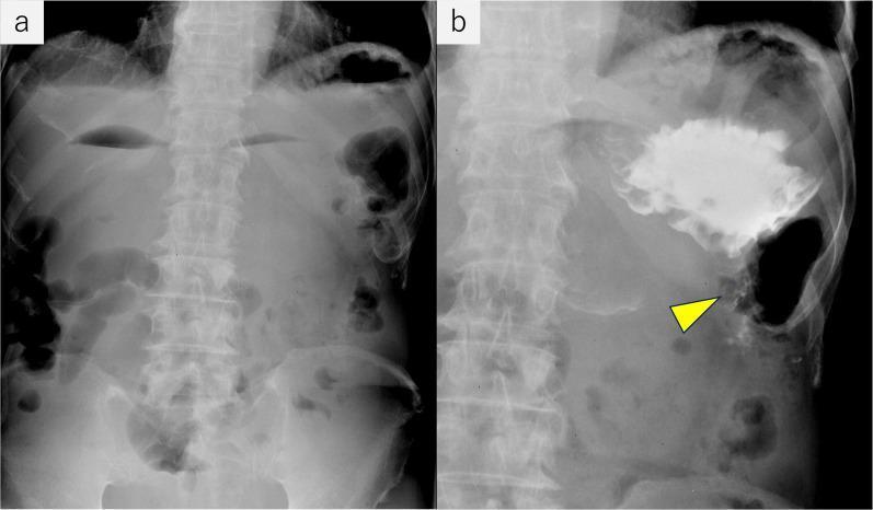
This dual-panel medical visual displays upper gastrointestinal fluoroscopy and abdominal radiography of a 60-year-old male with signs of high intestinal obstruction. Panel (a) is a plain abdominal radiograph showing multiple horizontal air-fluid levels (niveau formations) in the upper quadrant, corresponding to a dilated stomach, duodenum, and proximal jejunum. Panel (b) shows a contrast-enhanced fluoroscopic image following a gastrojejunostomy. A large volume of radiopaque contrast medium is retained within a significantly dilated stomach, indicating delayed gastric emptying. A yellow arrowhead points to a minimal trace of contrast passing into the efferent loop of the gastrojejunostomy, demonstrating a severe functional or mechanical stricture at the anastomosis site. The images illustrate gastric outlet-type obstruction secondary to external compression from metastatic renal cancer, highlighting the characteristic radiological signs of proximal bowel distension and impaired contrast transit in a postoperative setting.
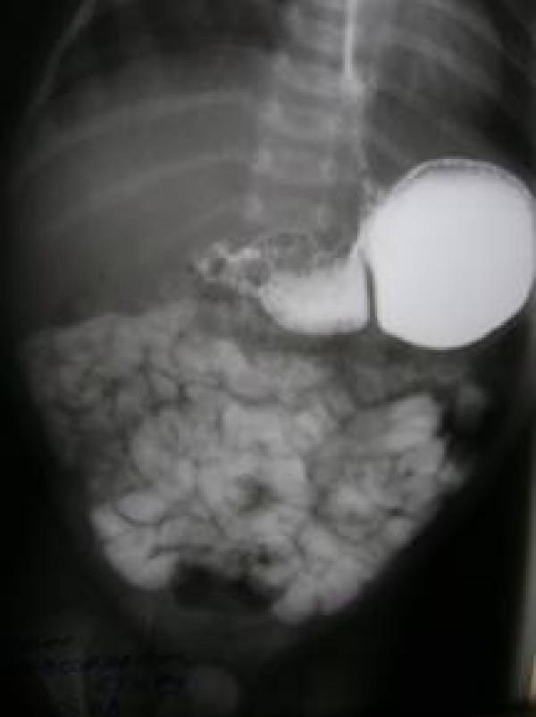
This diagnostic image is an abdominal X-ray from an upper gastrointestinal (GI) contrast study. The radiograph reveals a significantly dilated stomach filled with radiopaque contrast material, exhibiting a 'double loop' appearance or marked gastric distension. A focal, high-grade constriction is visible at the prepyloric region, characterizing a prepyloric stricture. Distal to the site of obstruction, a small amount of contrast has passed into the small bowel, which appears in a clustered pattern. The imaging findings are consistent with gastric outlet obstruction, specifically prepyloric stenosis or stricture, causing delayed gastric emptying. The study highlights the anatomical distortion and proximal dilatation typical of obstructive pathology in the pediatric or adult upper GI tract.
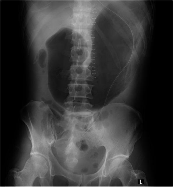
This abdominal radiograph (X-ray) demonstrates significant gastric distention in a postoperative patient. The most prominent feature is a large, radiolucent (air-filled) stomach bubble occupying a substantial portion of the upper abdominal cavity, extending across the midline and down toward the pelvis. A radiopaque nasogastric (NG) tube is visible, entering from the upper aspect of the image and descending along the patient's left side. However, the distal end of the tube appears malpositioned or ineffective, failing to decompress the markedly dilated stomach. Surgical staples are visible along the spine, consistent with recent thoracic or abdominal surgery. The large bowel and small intestinal loops are visible in the lower abdomen but appear non-dilated compared to the massive gastric air. This image illustrates gastric outlet obstruction or severe aerophagia in a clinical setting, highlighting the importance of verifying NG tube positioning and functionality for gastric decompression.
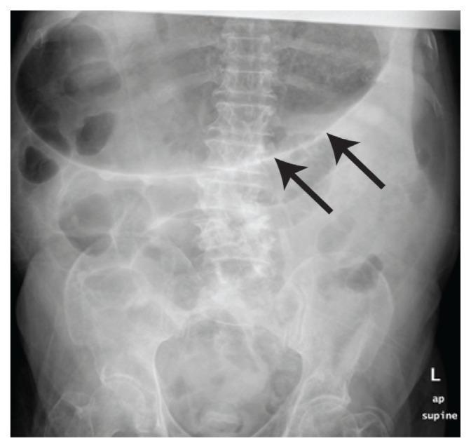
Diagnostic Image: Anteroposterior (AP) supine abdominal radiograph of an adult patient. The primary finding is a grossly distended stomach, visualized as a large, radiolucent (gas-filled) structure occupying the epigastrium and extending into the mid-abdomen (indicated by black arrows). The gastric wall appears relatively thin. There is a lack of multiple dilated small bowel loops or 'step-ladder' air-fluid levels, suggesting the absence of distal small bowel obstruction. No subdiaphragmatic free air is visible, excluding pneumoperitoneum in this view. The anatomical context shows the lumbar spine and pelvic girdle within normal radiological limits. This image is significant for evaluating acute abdominal distention, specifically highlighting gastric dilatation as a potential indicator of gastric outlet obstruction or gastroparesis.
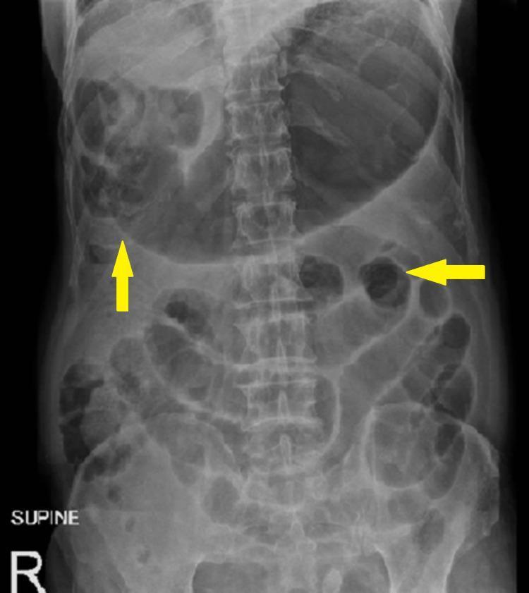
This diagnostic image is a supine plain abdominal radiograph (X-ray) illustrating significant gastrointestinal pathology. The primary finding is marked gastric distension, characterized by a large, gas-filled stomach shadow occupying the upper left quadrant and crossing the midline. Additionally, multiple dilated small bowel loops are visible throughout the central and lower abdominal regions, demonstrating pathologically increased luminal diameter. Yellow arrows highlight specific areas of gaseous distension and prominent bowel loops. The valvulae conniventes are visible within the dilated loops, identifying them as small bowel. There is a notable absence of significant colonic gas or pneumoperitoneum in this projection. These radiographic signs are highly suggestive of an intestinal obstruction or generalized ileus. The image serves as a clinical example of mechanical bowel obstruction or gastric outlet obstruction for medical education, emphasizing the radiographic appearance of dilated hollow viscera and the importance of supine imaging in initial diagnostic workups for acute abdominal pain.
~/case-study-goo-barium/build_docx.py
#!/usr/bin/env python3
from docx import Document
from docx.shared import Pt, RGBColor
from docx.enum.text import WD_ALIGN_PARAGRAPH

doc = Document()

style = doc.styles["Normal"]
style.font.name = "Arial"
style.font.size = Pt(11)

def h1(text):
    doc.add_heading(text, level=1)

def h2(text):
    doc.add_heading(text, level=2)

def para(text, bold=False, italic=False, size=None):
    p = doc.add_paragraph()
    run = p.add_run(text)
    run.bold = bold
    run.italic = italic
    if size:
        run.font.size = Pt(size)
    return p

def bullet(text):
    doc.add_paragraph(text, style='List Bullet')

def numbered(text):
    doc.add_paragraph(text, style='List Number')

# ---------------- COVER PAGE ----------------
title = doc.add_paragraph()
title.alignment = WD_ALIGN_PARAGRAPH.CENTER
r = title.add_run("SCHOOL OF HEALTH SCIENCES")
r.bold = True
r.font.size = Pt(16)

sub = doc.add_paragraph()
sub.alignment = WD_ALIGN_PARAGRAPH.CENTER
r = sub.add_run("Department of Clinical Imaging")
r.bold = True
r.font.size = Pt(13)

sub2 = doc.add_paragraph()
sub2.alignment = WD_ALIGN_PARAGRAPH.CENTER
r = sub2.add_run("CASE STUDY")
r.bold = True
r.font.size = Pt(20)
r.font.color.rgb = RGBColor(0x1F, 0x4E, 0x79)

sub3 = doc.add_paragraph()
sub3.alignment = WD_ALIGN_PARAGRAPH.CENTER
r = sub3.add_run("Barium Meal Examination in the Investigation of\nGastric Outlet Obstruction")
r.bold = True
r.font.size = Pt(15)

doc.add_paragraph()
fields = [
    "Course code: ____________________________________________",
    "Name of student: ____________________________________________",
    "Registration number: ____________________________________________",
    "Name & signature of clinical instructor: ____________________________________________",
    "Examination assessed: Barium Meal",
    "Date: ____________________________________________",
]
for f in fields:
    p = doc.add_paragraph(f)
    p.paragraph_format.space_after = Pt(10)

doc.add_page_break()

# ---------------- TABLE OF CONTENTS ----------------
h1("Table of Contents")
toc_items = [
    "1. Introduction",
    "2. Brief Gross Anatomy",
    "3. Patient History",
    "4. Discussion of Provisional Diagnosis and Aetiology",
    "5. Equipment and Accessories",
    "6. Patient Preparation for the Study",
    "7. Discussion of Technique Employed, Imaging Protocol/Procedure",
    "8. Patient Care - Before, During and After the Study",
    "9. Discussion of Findings/Pathology (Radiological Report)",
    "10. Further Role of the Imaging Department",
    "11. Patient Management",
    "12. Inter-professional Collaboration",
    "13. Reflection",
    "14. Conclusion",
    "15. References",
    "16. Appendices",
]
for t in toc_items:
    doc.add_paragraph(t)

doc.add_page_break()

# ---------------- 1. INTRODUCTION ----------------
h1("1. Introduction")
para(
    "Gastric outlet obstruction (GOO) is a clinical syndrome resulting from mechanical impedance to the "
    "emptying of gastric contents through the pyloric channel or proximal duodenum. It presents with "
    "postprandial vomiting, early satiety, abdominal distension and weight loss, and if untreated leads to "
    "progressive dehydration and metabolic derangement. Historically, peptic ulcer disease (PUD) was the "
    "leading cause; with the widespread use of proton pump inhibitors and Helicobacter pylori eradication, "
    "malignancy (gastric or periampullary cancer) has become an increasingly common cause and GOO should be "
    "regarded as malignant until proven otherwise."
)
para(
    "A barium meal (upper gastrointestinal contrast study) is a fluoroscopic examination using an ingested "
    "barium sulfate suspension, with or without gas-forming granules, to outline the mucosal contour and "
    "assess the calibre, motility and emptying of the stomach and duodenum. Although endoscopy is now the "
    "first-line investigation for suspected GOO, the barium meal remains a valuable, widely available "
    "technique for demonstrating the site, length and functional severity of an obstructing lesion, and for "
    "assessing gastric emptying dynamically under fluoroscopic control. This case study discusses the use of "
    "barium meal examination in a patient presenting with clinical features of gastric outlet obstruction, "
    "correlating anatomy, technique, radiological findings and multidisciplinary management."
)

# ---------------- 2. GROSS ANATOMY ----------------
h1("2. Brief Gross Anatomy")
para("Relevant gross anatomy for interpretation of a barium meal study of the gastric outlet includes:", bold=True)
bullet("Stomach: A distensible J-shaped sac situated in the epigastrium/left hypochondrium, immediately inferior to the diaphragm. It is divided into the cardia (adjoining the gastro-oesophageal junction), fundus (superior dome), body, antrum and pylorus. Gastric capacity ranges from a few hundred millilitres when empty to over 2 litres when fully distended (Yamada's Textbook of Gastroenterology, p. 138).")
bullet("Pylorus and pyloric canal: The narrow, muscular distal segment of the stomach, guarded by the pyloric sphincter, which regulates the rate of gastric emptying into the duodenum. This is the classic site of obstruction from peptic scarring, hypertrophy, or infiltrating tumour.")
bullet("Duodenum: The first part of the small bowel, comprising the duodenal bulb (cap), and a C-shaped loop (descending, horizontal/transverse, and ascending parts) that curves around the head of the pancreas before becoming the jejunum at the duodenojejunal flexure; largely retroperitoneal.")
bullet("Relations: The stomach abuts the diaphragm superiorly, the liver and biliary tree to the right, the spleen to the left, the pancreas posteriorly (via the lesser sac), and the transverse colon inferiorly. The proximity of the pancreatic head to the duodenal C-loop explains why periampullary and pancreatic head tumours commonly cause extrinsic duodenal/gastric outlet obstruction (Yamada's Textbook of Gastroenterology, p. 138).")
bullet("Mucosal folds (rugae): Longitudinal gastric mucosal folds, most prominent along the greater curvature, which flatten with gastric distension - visualised as areae gastricae on double-contrast studies.")
bullet("Peritoneal attachments: The gastrohepatic and gastrocolic ligaments (lesser and greater omentum) anchor the stomach and permit its mobility while transmitting the vascular supply (coeliac trunk branches: left gastric, splenic, common hepatic arteries).")

# ---------------- 3. PATIENT HISTORY ----------------
h1("3. Patient History")
para("Patient demographics (for illustrative/teaching purposes):", bold=True)
bullet("Age/Sex: 58-year-old male")
bullet("Presenting complaint: 3-month history of recurrent, large-volume vomiting of undigested food, occurring 30-60 minutes after meals, with no bile staining")
bullet("Associated symptoms: Early satiety, progressive epigastric fullness and bloating, 8 kg unintentional weight loss, and generalised weakness/lethargy")
bullet("Past medical history: Long-standing dyspepsia treated intermittently with over-the-counter antacids; no formal H. pylori testing; heavy NSAID use for chronic knee pain")
bullet("Social history: Smoker (20 cigarettes/day for 30 years), moderate alcohol intake")
bullet("Clinical examination: Visibly wasted, dehydrated; epigastric distension with a positive succussion splash on abdominal auscultation/shaking; no palpable abdominal mass; mild epigastric tenderness")
bullet("Basic investigations: Blood gas/electrolytes show hypochloraemic, hypokalaemic metabolic alkalosis; urea and creatinine elevated in keeping with dehydration")
bullet("Referral reason: Referred for a barium meal (and subsequent upper GI endoscopy) to confirm and characterise a suspected gastric outlet obstruction")
para(
    "This history of non-bilious, undigested-food vomiting, weight loss, a succussion splash and hypochloraemic "
    "hypokalaemic alkalosis is the classic clinical and biochemical picture of gastric outlet obstruction, and "
    "in a patient of this age with chronic NSAID use and smoking history, both benign (peptic) and malignant "
    "aetiologies must be actively considered."
)

# ---------------- 4. PROVISIONAL DIAGNOSIS AND AETIOLOGY ----------------
h1("4. Discussion of Provisional Diagnosis and Aetiology")
h2("4.1 Provisional Diagnosis")
para(
    "The provisional diagnosis is gastric outlet obstruction, most likely secondary to either chronic peptic "
    "ulcer disease with pyloric scarring/stenosis or an underlying gastric/periampullary malignancy. As gastric "
    "cancer and pyloric stenosis secondary to peptic ulceration are the two most common causes, and PUD-related "
    "GOO has become less common with modern acid-suppression therapy, obstruction in this age group should be "
    "considered malignant until proven otherwise (Bailey and Love's Short Practice of Surgery, p. 1189-1190)."
)
h2("4.2 Aetiology - Benign Causes")
bullet("Peptic ulcer disease: Chronic duodenal or pyloric channel ulceration causing oedema (acute, reversible) or fibrotic scarring/stenosis (chronic, fixed) of the pylorus - the classic benign cause")
bullet("Adult hypertrophic pyloric stenosis: A rare acquired thickening of the pyloric muscle, anatomically identical to the infantile form, presenting with progressive gastric outlet symptoms in adulthood")
bullet("Caustic/corrosive ingestion strictures, and post-surgical anastomotic strictures")
bullet("Crohn's disease involving the gastric antrum/duodenum")
bullet("Gastric polyps, bezoars, or foreign bodies causing mechanical obstruction")
bullet("Gastric volvulus and diaphragmatic hernia (uncommon but important causes of relapsing obstructive symptoms)")
h2("4.3 Aetiology - Malignant Causes")
bullet("Gastric adenocarcinoma involving the antrum/pylorus - the leading malignant cause")
bullet("Pancreatic head or periampullary carcinoma causing extrinsic duodenal compression/invasion")
bullet("Duodenal carcinoma, gastric lymphoma, and metastatic disease or peritoneal carcinomatosis encasing the antro-pyloric region")
h2("4.4 Pathophysiology")
para(
    "Mechanical narrowing of the pyloric channel or proximal duodenum leads to progressive gastric stasis and "
    "dilatation. Repeated vomiting of hydrochloric acid-rich gastric contents (without loss of duodenal "
    "bicarbonate-rich bile, since vomitus is non-bilious) produces a hypochloraemic, hypokalaemic metabolic "
    "alkalosis: initially the kidney excretes bicarbonate-rich, low-chloride urine; as dehydration progresses, "
    "sodium is preferentially retained at the expense of potassium and hydrogen ion excretion, producing a "
    "paradoxically acidic urine and worsening hypokalaemia. Alkalosis further lowers ionised calcium and can "
    "precipitate tetany (Bailey and Love's Short Practice of Surgery, p. 1190)."
)

# ---------------- 5. EQUIPMENT AND ACCESSORIES ----------------
h1("5. Equipment and Accessories")
bullet("Fluoroscopy (screening) unit with an image intensifier or flat-panel digital detector, tilting table, and spot-film/digital acquisition capability")
bullet("Barium sulfate suspension (appropriate concentration for a single- or double-contrast study)")
bullet("Effervescent gas-forming granules/carbon dioxide-producing agent for double-contrast technique")
bullet("Smooth-muscle relaxant/hypotonic agent - intravenous or intramuscular hyoscine butylbromide (Buscopan) or glucagon, to reduce gastric peristalsis and spasm during the examination")
bullet("Wide-bore nasogastric tube and suction apparatus, available for pre-procedure gastric decompression in cases of gross gastric stasis/retained food")
bullet("Disposable cups/straws for barium ingestion, paper towels, emesis bags")
bullet("Lead aprons/thyroid shields and radiation dosimetry badges for staff; gonad shielding for the patient where practicable")
bullet("Compression paddle for spot compression views")
bullet("Automatic exposure control and image-recording/PACS workstation for permanent record and reporting")
bullet("Resuscitation equipment readily available given the risk of aspiration in patients with gross gastric outlet obstruction")

# ---------------- 6. PATIENT PREPARATION ----------------
h1("6. Patient Preparation for the Study")
bullet("Verify the request form, clinical indication and identity of the patient using at least two identifiers")
bullet("Fasting for 6-8 hours prior to the examination (no food or fluids) to ensure an empty stomach and adequate mucosal coating; in known/suspected GOO with gross gastric stasis, a period of nasogastric aspiration/decompression the night before may be required to empty retained food and fluid residue")
bullet("Discontinue antispasmodic and prokinetic medication as per departmental protocol prior to the study, since these affect gastric motility and pyloric tone")
bullet("Explain the procedure fully, including the taste/texture of the barium suspension, the need to swallow on command, and the positional changes (rotation, tilting table) required during screening; obtain verbal/informed consent")
bullet("Exclude contraindications to barium: suspected perforation or complete high-grade obstruction with risk of aspiration - in these situations a water-soluble contrast agent is substituted for barium")
bullet("Screen for pregnancy in women of childbearing age, given the use of ionising radiation")
bullet("Remove any radio-opaque objects/clothing from the imaging field")
bullet("Ensure intravenous access is available if a smooth-muscle relaxant is to be given, and check for contraindications (e.g. glaucoma, prostatic hypertrophy, cardiac arrhythmia for hyoscine)")

# ---------------- 7. TECHNIQUE ----------------
h1("7. Discussion of Technique Employed, Imaging Protocol/Procedure")
para(
    "Barium meal examination can be performed as a single-contrast (barium suspension alone, filling the "
    "lumen) or double-contrast (barium plus gas-distension) study; the double-contrast technique provides "
    "superior mucosal detail and is preferred where mucosal disease (ulceration, early neoplasm) is also "
    "suspected. The technique proceeds as follows:"
)
numbered("The patient is positioned erect or supine on the tilting fluoroscopy table. An intravenous smooth-muscle relaxant (hyoscine butylbromide or glucagon) may be given to reduce peristalsis and gastric spasm and improve mucosal coating.")
numbered("For a double-contrast study, the patient first swallows effervescent gas-forming granules, followed by a mouthful of high-density barium suspension, timed to fluoroscopic observation of the pharynx and oesophagus.")
numbered("The patient is rotated from supine through a full 360-degree roll, coming to rest in a right anterior oblique (RAO) position; this manoeuvre coats the gastric mucosa evenly with barium and distributes the gas to distend the fundus and body.")
numbered("Spot fluoroscopic images (and permanent digital acquisitions) are obtained systematically in the following projections to fully evaluate the stomach and pyloro-duodenal region: AP, RAO, left posterior oblique (LPO), lateral, and left anterior oblique (LAO), with the table semi-erect to double-contrast the fundus and cardia.")
numbered("Compression views over the antrum and pylorus (using a compression paddle or manual palpation under fluoroscopic guidance) are used to separate overlapping bowel loops and assess distensibility and peristalsis through the pyloric channel.")
numbered("The passage of the barium bolus through the pylorus into the duodenal bulb and C-loop is observed continuously under fluoroscopy, noting the calibre, contour and rate of transit; delayed spot films are taken to record the site and character of any narrowing.")
numbered("Where gastric outlet obstruction is clinically suspected, the study is extended with delayed erect and supine images at intervals (e.g. 30 minutes, 1, 2, 4, and up to 6 hours) to assess the gastric emptying time and the pattern/degree of barium retention, since delayed emptying beyond 4-6 hours is a key functional indicator of obstruction.")
numbered("All relevant images are reviewed on the workstation, archived to PACS, and a structured report is issued.")

# ---------------- 8. PATIENT CARE ----------------
h1("8. Patient Care - Before, During and After the Study")
h2("Before")
bullet("Full explanation of the procedure, including sensations associated with the gas-forming granules and smooth-muscle relaxant, to reduce anxiety")
bullet("Confirm fasting status and, where indicated, prior nasogastric decompression has been performed")
bullet("Assess hydration and electrolyte status; correct significant dehydration or hypokalaemia before the study where clinically safe to do so")
h2("During")
bullet("Continuous observation for signs of aspiration, distress, or vasovagal reaction during positional changes and swallowing")
bullet("Monitor for side effects of hyoscine butylbromide (dry mouth, blurred vision, tachycardia) or glucagon (nausea)")
bullet("Provide clear verbal instructions throughout positional changes; assist frail or unstable patients with rotation on the table to prevent falls")
h2("After")
bullet("Advise on increased oral fluid intake to aid barium passage and prevent constipation/faecal impaction")
bullet("Warn the patient that stools will appear white/pale for 1-3 days")
bullet("Advise on mild laxative use if barium retention or constipation is anticipated, particularly relevant here given delayed gastric and possibly bowel transit")
bullet("Provide information on when results will be available and arrange prompt referral to gastroenterology/surgery given the suspicion of gastric outlet obstruction")
bullet("Monitor for continued vomiting or aspiration risk after the study and ensure safe transfer back to the ward with appropriate nursing handover")

# ---------------- 9. FINDINGS / PATHOLOGY ----------------
h1("9. Discussion of Findings/Pathology (Radiological Report)")
h2("9.1 Typical Barium Meal Findings in Gastric Outlet Obstruction")
para(
    "Gastric outlet obstruction produces a combination of morphological and functional radiological signs:"
)
bullet("A grossly dilated, atonic stomach, often containing residual fluid and food debris visible even before contrast administration as a mottled, granular appearance within the barium column")
bullet("Poor or absent peristaltic activity through the antro-pyloric region on fluoroscopy")
bullet("A narrowed, irregular or smoothly tapered pyloric channel - a long, smooth, tapering 'beak-like' narrowing favours a benign peptic stricture, whereas an abrupt, irregular, shouldered narrowing with mucosal destruction favours malignancy")
bullet("A thin trickle or delayed, intermittent passage of barium through the stenotic segment into the duodenum")
bullet("Markedly delayed gastric emptying, with substantial barium retained in the stomach at delayed imaging (typically beyond 4-6 hours), in contrast to the normal stomach which should be largely empty of contrast by around 4 hours")
bullet("In adult hypertrophic pyloric stenosis, an elongated, narrow pyloric channel producing the classic 'string sign'")
bullet("In malignant obstruction, an irregular filling defect, mucosal destruction, rigidity of the gastric wall ('leather bottle' stomach in diffuse infiltrative carcinoma), or an extrinsic mass effect narrowing/displacing the duodenal C-loop (e.g. from a pancreatic head tumour)")

h2("9.2 Illustrative Radiological Report")
para("EXAMINATION: Barium Meal (double contrast)", italic=True)
para("CLINICAL HISTORY: 58-year-old male with non-bilious vomiting of undigested food, weight loss, succussion splash, hypochloraemic hypokalaemic alkalosis. Rule out gastric outlet obstruction.", italic=True)
para("FINDINGS:", bold=True)
bullet("The stomach is grossly dilated and atonic, containing a moderate volume of retained fluid and food debris, producing a mottled appearance within the barium pool prior to full contrast administration.")
bullet("There is a smooth, tapering narrowing of the pyloric channel and proximal duodenal bulb measuring approximately 3 cm in length, through which only a thin trickle of barium passes into the duodenum.")
bullet("Peristaltic activity across the antro-pyloric segment is markedly reduced.")
bullet("Delayed erect films at 4 hours demonstrate significant retention of barium within the stomach, in keeping with markedly delayed gastric emptying.")
bullet("No discrete irregular filling defect or mucosal destruction is identified at the pylorus on this study; the duodenal C-loop is otherwise unremarkable in contour.")
para("IMPRESSION: Findings confirm gastric outlet obstruction with a smooth, tapering pyloric stricture and markedly delayed gastric emptying. Appearances are more suggestive of a benign (peptic) stricture; however, mucosal detail is a limitation of contrast studies and upper GI endoscopy with biopsy is recommended to exclude an underlying malignant cause.", bold=True)

# ---------------- 10. FURTHER ROLE OF IMAGING ----------------
h1("10. Further Role of the Imaging Department")
bullet("Upper GI endoscopy (gastroscopy): now the first-line investigation, allowing direct visualisation, biopsy for histology and H. pylori testing, and therapeutic endoscopic balloon dilation of benign strictures")
bullet("CT abdomen with contrast: assesses gastric wall thickening, locoregional invasion, lymphadenopathy and metastatic disease for staging when malignancy is suspected or confirmed, and can identify pancreatic head masses causing extrinsic obstruction")
bullet("Endoscopic ultrasound (EUS): further local staging of gastric/periampullary tumours and EUS-guided tissue sampling")
bullet("Water-soluble contrast studies: used instead of barium if perforation is suspected or in the acute unstable patient")
bullet("Interventional radiology/endoscopy: fluoroscopically or endoscopically guided balloon dilation of benign strictures; placement of self-expanding metal stents (SEMS) or EUS-guided gastroenterostomy for palliation of malignant, unresectable gastric outlet obstruction")
bullet("Follow-up barium studies or repeat endoscopy to assess response to dilation/stenting or resolution of oedema-related obstruction with medical therapy")
para(
    "Recent evidence note: pooled analyses and network meta-analyses (2023-2026) comparing EUS-guided "
    "gastroenterostomy against enteral stenting and surgical gastrojejunostomy suggest EUS-guided "
    "gastroenterostomy offers durable patency with a favourable safety profile for both benign and malignant "
    "gastric outlet obstruction, and is increasingly used as a minimally invasive alternative where expertise "
    "is available (Canakis et al. 2026, PMID 39761155; Tran et al. 2024, PMID 38641337). This does not replace "
    "the diagnostic role of barium meal/endoscopy but is relevant to the further management pathway."
)

# ---------------- 11. PATIENT MANAGEMENT ----------------
h1("11. Patient Management")
para("Management of confirmed gastric outlet obstruction addresses both the metabolic derangement and the underlying mechanical cause:", bold=True)
numbered("Resuscitation: intravenous rehydration with isotonic saline and potassium supplementation to correct the hypochloraemic, hypokalaemic metabolic alkalosis and dehydration")
numbered("Gastric decompression with a wide-bore nasogastric tube to rest the stomach, reduce vomiting/aspiration risk, and allow gastric oedema to settle")
numbered("Correction of nutritional deficits, with dietetic input and consideration of nasoenteric or parenteral feeding if prolonged obstruction is anticipated")
numbered("Endoscopy with biopsy to obtain a definitive histological diagnosis and exclude malignancy, and H. pylori testing/eradication where relevant")
numbered("Benign disease: aggressive acid-suppression therapy (high-dose proton pump inhibitors), cessation of NSAIDs/smoking, and endoscopic balloon dilation of the stricture; surgery (pyloroplasty or gastrojejunostomy) is reserved for cases refractory to medical/endoscopic therapy")
numbered("Malignant disease: staging (CT/EUS +/- PET) to determine resectability; potentially curative gastrectomy with lymphadenectomy for resectable disease, or palliative measures (endoscopic stenting, EUS-guided gastroenterostomy, or surgical bypass) for unresectable/metastatic disease, combined with oncological therapy as appropriate")
numbered("Ongoing multidisciplinary follow-up, including nutritional support, symptom control and, where relevant, palliative care input")

# ---------------- 12. INTER-PROFESSIONAL COLLABORATION ----------------
h1("12. Inter-professional Collaboration")
bullet("Radiographer: performs the barium meal examination safely, ensures correct patient preparation, positioning and radiation protection")
bullet("Radiologist: supervises/performs fluoroscopic screening, interprets and reports findings, and may perform image-guided interventions")
bullet("Gastroenterologist: performs diagnostic and therapeutic endoscopy, biopsy, and endoscopic dilation/stenting")
bullet("General/upper GI surgeon: undertakes surgical management (pyloroplasty, gastrojejunostomy, gastrectomy) where indicated")
bullet("Oncologist: coordinates chemotherapy/radiotherapy for malignant causes")
bullet("Histopathologist: provides tissue diagnosis from endoscopic or surgical biopsy specimens")
bullet("Dietitian: manages nutritional support and dietary modification during recovery")
bullet("Nursing staff: manage nasogastric tube care, fluid balance monitoring, and patient education")
bullet("Pharmacist: advises on acid-suppression therapy, electrolyte replacement and drug interactions")
para("Effective communication between these professionals, coordinated through gastrointestinal multidisciplinary team meetings, ensures timely diagnosis and an individualised, safe management plan.")

# ---------------- 13. REFLECTION ----------------
h1("13. Reflection")
para(
    "This case demonstrated how a structured barium meal technique, combined with careful attention to patient "
    "preparation and positioning, can clearly demonstrate both the morphological site of an obstructing lesion "
    "and its functional impact on gastric emptying. It highlighted the importance of correlating the clinical "
    "presentation (non-bilious vomiting, weight loss, succussion splash) and biochemical picture (hypochloraemic "
    "hypokalaemic alkalosis) with imaging findings, and reinforced that while barium studies are excellent at "
    "demonstrating the site and length of a stricture and quantifying delayed emptying, they cannot reliably "
    "exclude malignancy on morphology alone - endoscopic biopsy remains essential. It also highlighted the "
    "practical importance of pre-procedure gastric decompression in patients with gross gastric stasis, both "
    "for diagnostic image quality and patient safety (aspiration risk). Continued learning points include "
    "developing familiarity with the evolving role of minimally invasive endoscopic/EUS-guided techniques in "
    "managing both benign and malignant gastric outlet obstruction."
)

# ---------------- 14. CONCLUSION ----------------
h1("14. Conclusion")
para(
    "Gastric outlet obstruction is a mechanical and functional syndrome with important benign (peptic) and "
    "malignant (gastric/periampullary cancer) causes, producing a characteristic clinical and metabolic "
    "picture. The barium meal remains a useful, accessible fluoroscopic technique for demonstrating the site, "
    "length and severity of an obstructing lesion and for objectively assessing gastric emptying, complementing "
    "endoscopy in the diagnostic pathway. Prompt correction of fluid and electrolyte disturbance, accurate "
    "diagnosis of the underlying cause, and coordinated multidisciplinary management are essential to good "
    "patient outcomes."
)

# ---------------- 15. REFERENCES ----------------
h1("15. References")
refs = [
    "Williams, N.S., O'Connell, P.R. and McCaskie, A.W., 2023. Bailey and Love's Short Practice of Surgery. 28th ed. Boca Raton: CRC Press. (Gastric Outlet Obstruction, p. 1189-1190).",
    "Feldman, M., Friedman, L.S. and Brandt, L.J., 2020. Sleisenger and Fordtran's Gastrointestinal and Liver Disease. 11th ed. Philadelphia: Elsevier. (Gastric Outlet Obstruction, p. 213; Adult Hypertrophic Pyloric Stenosis, p. 731).",
    "Podolsky, D.K., Camilleri, M., Fitz, J.G., Kalloo, A.N., Shanahan, F. and Wang, T.C., 2022. Yamada's Textbook of Gastroenterology. 7th ed. Hoboken: Wiley-Blackwell. (Gross Anatomy of the Stomach and Duodenum, p. 138; Contrast Studies).",
    "Cameron, J.L. and Cameron, A.M., 2019. Current Surgical Therapy. 14th ed. Philadelphia: Elsevier. (Gastric Outlet Obstruction).",
    "Adam, A., Dixon, A.K., Gillard, J.H. and Schaefer-Prokop, C.M., 2021. Grainger & Allison's Diagnostic Radiology: A Textbook of Medical Imaging. 7th ed. Amsterdam: Elsevier. (Fluoroscopy/double-contrast barium technique).",
    "Sabiston, D.C. and Townsend, C.M., 2022. Sabiston Textbook of Surgery: The Biological Basis of Modern Surgical Practice. 21st ed. Philadelphia: Elsevier. (Barium Swallow/Upper GI Series).",
    "Canakis, A., Tugarinov, N., Bapaye, J., et al., 2026. 'EUS-Guided Gastroenterostomy for Benign Gastric Outlet Obstruction: A Systematic Review and Meta-analysis.' Journal of Clinical Gastroenterology. PMID: 39761155.",
    "Tran, K.V., Vo, N.P., Nguyen, H.S., et al., 2024. 'Palliative procedures for malignant gastric outlet obstruction: a network meta-analysis.' Endoscopy. PMID: 38641337.",
]
for r in refs:
    doc.add_paragraph(r, style='List Bullet')

# ---------------- 16. APPENDICES ----------------
h1("16. Appendices")
para("Appendix A: Sample Barium Meal Request Form (fields to be completed by referring clinician)", bold=True)
bullet("Patient name / ID / DOB")
bullet("Referring clinician and contact details")
bullet("Clinical history and indication: r/o gastric outlet obstruction")
bullet("Relevant history: vomiting pattern, weight loss, prior PUD, NSAID/smoking history")
bullet("Pregnancy status; contraindications to barium (suspected perforation)")
bullet("Signature and date")
para("Appendix B: Sample structured radiological report (see Section 9.2 above)", bold=True)
para(
    "Appendix C: Placeholder for annotated barium meal radiographs illustrating gastric dilatation, the "
    "tapering pyloric stricture, delayed emptying on the delayed film, and (where applicable) mucosal "
    "irregularity of a malignant stricture (to be inserted from the departmental PACS with patient consent).",
    bold=True,
)

doc.save('/tmp/workspace/case-study-goo-barium/Barium_Meal_Case_Study_Gastric_Outlet_Obstruction.docx')
print("Saved.")
mkdir -p /tmp/workspace/case-study-goo-barium && cd /tmp/workspace/case-study-goo-barium && python3 build_docx.py
Barium Meal Case Study Gastric Outlet Obstruction
Word Document · DOCX
- Introduction - GOO overview and rationale for barium meal
- Brief Gross Anatomy - stomach regions, pylorus, duodenum, adjacent organ relations
- Patient History - illustrative 58-year-old male with non-bilious vomiting, weight loss, succussion splash
- Provisional Diagnosis & Aetiology - benign (PUD, adult hypertrophic pyloric stenosis, Crohn's, corrosive stricture) vs malignant (gastric/periampullary cancer) causes, plus the hypochloraemic hypokalaemic alkalosis pathophysiology
- Equipment and Accessories - fluoroscopy unit, barium, effervescent granules, Buscopan/glucagon, NG tube/suction
- Patient Preparation - fasting, decompression if grossly dilated stomach, contraindication screening (perforation → water-soluble contrast instead)
- Technique/Protocol - single vs double contrast, positioning sequence (AP/RAO/LPO/lateral/LAO), compression views, delayed emptying films at 30 min–6 hr
- Patient Care - before/during/after
- Findings/Pathology - typical radiological signs (dilated atonic stomach, tapering "beak" vs irregular malignant narrowing, string sign, delayed emptying) plus a sample structured report
- Further Role of Imaging - endoscopy, CT staging, EUS, SEMS/EUS-guided gastroenterostomy, with a note flagging recent meta-analyses (PMID 39761155, 38641337) on EUS-guided gastroenterostomy outcomes
- Patient Management - resuscitation, decompression, benign vs malignant treatment pathways
- Inter-professional Collaboration - full MDT breakdown
- Reflection
- Conclusion
- References - textbook citations with page numbers plus recent PubMed evidence
- Appendices - sample request form, report, image placeholder
make ppt for both presentations
/tmp/workspace
~/case-study-mri-glioma
~/case-study-mri-glioma/build_pptx.py
#!/usr/bin/env python3
"""Build MRI Brain with Contrast - High Grade Glioma case study presentation."""
from pptx import Presentation
from pptx.util import Inches, Pt, Emu
from pptx.dml.color import RGBColor
from pptx.enum.text import PP_ALIGN, MSO_ANCHOR
from pptx.enum.shapes import MSO_SHAPE

NAVY = RGBColor(0x1F, 0x4E, 0x79)
ACCENT = RGBColor(0x2E, 0x86, 0xAB)
LIGHT_BG = RGBColor(0xF4, 0xF7, 0xFA)
DARK_TEXT = RGBColor(0x2B, 0x2B, 0x2B)
WHITE = RGBColor(0xFF, 0xFF, 0xFF)

prs = Presentation()
prs.slide_width = Inches(13.333)
prs.slide_height = Inches(7.5)
SW, SH = prs.slide_width, prs.slide_height
BLANK = prs.slide_layouts[6]


def add_background(slide, color=WHITE):
    rect = slide.shapes.add_shape(MSO_SHAPE.RECTANGLE, 0, 0, SW, SH)
    rect.fill.solid()
    rect.fill.fore_color.rgb = color
    rect.line.fill.background()
    rect.shadow.inherit = False
    slide.shapes._spTree.remove(rect._element)
    slide.shapes._spTree.insert(2, rect._element)
    return rect


def add_header_bar(slide, title, kicker=None):
    bar = slide.shapes.add_shape(MSO_SHAPE.RECTANGLE, 0, 0, SW, Inches(1.15))
    bar.fill.solid()
    bar.fill.fore_color.rgb = NAVY
    bar.line.fill.background()
    bar.shadow.inherit = False
    tb = slide.shapes.add_textbox(Inches(0.6), Inches(0.12), SW - Inches(1.2), Inches(0.9))
    tf = tb.text_frame
    tf.word_wrap = True
    tf.vertical_anchor = MSO_ANCHOR.MIDDLE
    p = tf.paragraphs[0]
    p.text = title
    r = p.runs[0]
    r.font.size = Pt(28)
    r.font.bold = True
    r.font.color.rgb = WHITE
    r.font.name = "Arial"
    if kicker:
        kb = slide.shapes.add_textbox(Inches(0.6), Inches(1.2), Inches(6), Inches(0.35))
        ktf = kb.text_frame
        kp = ktf.paragraphs[0]
        kp.text = kicker
        kr = kp.runs[0]
        kr.font.size = Pt(13)
        kr.font.italic = True
        kr.font.color.rgb = ACCENT
    accent_line = slide.shapes.add_shape(MSO_SHAPE.RECTANGLE, 0, Inches(1.15), SW, Pt(4))
    accent_line.fill.solid()
    accent_line.fill.fore_color.rgb = ACCENT
    accent_line.line.fill.background()
    accent_line.shadow.inherit = False


def add_footer(slide, num):
    tb = slide.shapes.add_textbox(SW - Inches(1.2), SH - Inches(0.45), Inches(0.9), Inches(0.35))
    tf = tb.text_frame
    p = tf.paragraphs[0]
    p.text = str(num)
    r = p.runs[0]
    r.font.size = Pt(11)
    r.font.color.rgb = RGBColor(0x99, 0x99, 0x99)
    tb2 = slide.shapes.add_textbox(Inches(0.6), SH - Inches(0.45), Inches(6), Inches(0.35))
    tf2 = tb2.text_frame
    p2 = tf2.paragraphs[0]
    p2.text = "MRI Brain with Contrast | High-Grade Glioma Case Study"
    r2 = p2.runs[0]
    r2.font.size = Pt(10)
    r2.font.color.rgb = RGBColor(0xAA, 0xAA, 0xAA)


def bullet_slide(title, bullets, kicker=None, num=None, two_col=False):
    slide = prs.slides.add_slide(BLANK)
    add_background(slide)
    add_header_bar(slide, title, kicker)
    top = Inches(1.55)
    if two_col:
        half = (SW - Inches(1.6)) / 2
        cols = [bullets[:len(bullets)//2 + len(bullets) % 2], bullets[len(bullets)//2 + len(bullets) % 2:]]
        for i, col in enumerate(cols):
            box = slide.shapes.add_textbox(Inches(0.6) + i * (half + Inches(0.4)), top, half, SH - top - Inches(0.6))
            tf = box.text_frame
            tf.word_wrap = True
            _fill_bullets(tf, col)
    else:
        box = slide.shapes.add_textbox(Inches(0.6), top, SW - Inches(1.2), SH - top - Inches(0.6))
        tf = box.text_frame
        tf.word_wrap = True
        _fill_bullets(tf, bullets)
    if num is not None:
        add_footer(slide, num)
    return slide


def _fill_bullets(tf, bullets):
    first = True
    for item in bullets:
        if isinstance(item, tuple):
            text, level = item
        else:
            text, level = item, 0
        p = tf.paragraphs[0] if first else tf.add_paragraph()
        first = False
        p.text = "●  " + text if level == 0 else "–  " + text
        p.level = 0
        r = p.runs[0]
        r.font.size = Pt(18) if level == 0 else Pt(15)
        r.font.color.rgb = DARK_TEXT if level == 0 else RGBColor(0x55, 0x55, 0x55)
        r.font.bold = (level == 0)
        r.font.name = "Arial"
        p.space_after = Pt(10)


def title_slide(main_title, subtitle, footer_text):
    slide = prs.slides.add_slide(BLANK)
    add_background(slide, NAVY)
    band = slide.shapes.add_shape(MSO_SHAPE.RECTANGLE, 0, Inches(4.6), SW, Pt(4))
    band.fill.solid(); band.fill.fore_color.rgb = ACCENT; band.line.fill.background(); band.shadow.inherit = False
    tb = slide.shapes.add_textbox(Inches(1), Inches(2.6), SW - Inches(2), Inches(1.8))
    tf = tb.text_frame; tf.word_wrap = True
    p = tf.paragraphs[0]; p.text = main_title; p.alignment = PP_ALIGN.CENTER
    r = p.runs[0]; r.font.size = Pt(40); r.font.bold = True; r.font.color.rgb = WHITE; r.font.name = "Arial"
    tb2 = slide.shapes.add_textbox(Inches(1), Inches(4.75), SW - Inches(2), Inches(1.0))
    tf2 = tb2.text_frame; tf2.word_wrap = True
    p2 = tf2.paragraphs[0]; p2.text = subtitle; p2.alignment = PP_ALIGN.CENTER
    r2 = p2.runs[0]; r2.font.size = Pt(18); r2.font.color.rgb = RGBColor(0xCF, 0xE3, 0xF0); r2.font.name = "Arial"
    tb3 = slide.shapes.add_textbox(Inches(1), SH - Inches(1.0), SW - Inches(2), Inches(0.6))
    tf3 = tb3.text_frame
    p3 = tf3.paragraphs[0]; p3.text = footer_text; p3.alignment = PP_ALIGN.CENTER
    r3 = p3.runs[0]; r3.font.size = Pt(13); r3.font.italic = True; r3.font.color.rgb = RGBColor(0xAA, 0xC6, 0xDE)
    return slide


def table_slide(title, headers, rows, kicker=None, num=None, col_widths=None):
    slide = prs.slides.add_slide(BLANK)
    add_background(slide)
    add_header_bar(slide, title, kicker)
    top = Inches(1.65)
    n_rows = len(rows) + 1
    n_cols = len(headers)
    tbl_w = SW - Inches(1.2)
    tbl_h = SH - top - Inches(0.6)
    gtable = slide.shapes.add_table(n_rows, n_cols, Inches(0.6), top, tbl_w, tbl_h).table
    if col_widths:
        for i, w in enumerate(col_widths):
            gtable.columns[i].width = Inches(w)
    for c, h in enumerate(headers):
        cell = gtable.cell(0, c)
        cell.text = h
        cell.fill.solid(); cell.fill.fore_color.rgb = NAVY
        p = cell.text_frame.paragraphs[0]
        p.runs[0].font.bold = True
        p.runs[0].font.color.rgb = WHITE
        p.runs[0].font.size = Pt(13)
    for r_i, row in enumerate(rows, start=1):
        for c_i, val in enumerate(row):
            cell = gtable.cell(r_i, c_i)
            cell.text = val
            cell.fill.solid()
            cell.fill.fore_color.rgb = LIGHT_BG if r_i % 2 == 0 else WHITE
            p = cell.text_frame.paragraphs[0]
            p.runs[0].font.size = Pt(11.5)
            p.runs[0].font.color.rgb = DARK_TEXT
    if num is not None:
        add_footer(slide, num)
    return slide


def section_divider(title, num=None):
    slide = prs.slides.add_slide(BLANK)
    add_background(slide, ACCENT)
    tb = slide.shapes.add_textbox(Inches(1), Inches(3.1), SW - Inches(2), Inches(1.3))
    tf = tb.text_frame; tf.word_wrap = True
    p = tf.paragraphs[0]; p.text = title; p.alignment = PP_ALIGN.CENTER
    r = p.runs[0]; r.font.size = Pt(34); r.font.bold = True; r.font.color.rgb = WHITE
    if num is not None:
        add_footer(slide, num)
    return slide


# ============================================================
# CONTENT
# ============================================================

n = 0
title_slide(
    "MRI Brain with Contrast",
    "Space-Occupying Lesion - Rule Out High-Grade Glioma\nA Radiography Case Study",
    "Department of Clinical Imaging | Case Study Presentation",
)

n += 1
bullet_slide("Presentation Outline", [
    "Introduction", "Brief Gross Anatomy", "Patient History",
    "Provisional Diagnosis & Aetiology", "Equipment and Accessories",
    "Patient Preparation", "Imaging Technique / Protocol", "Patient Care",
    "Findings / Radiological Report", "Further Role of Imaging",
    "Patient Management", "Inter-professional Collaboration",
    "Reflection & Conclusion", "References",
], num=n, two_col=True)

n += 1
bullet_slide("1. Introduction", [
    "A space-occupying lesion (SOL) is any focal abnormality that occupies space within the cranial cavity and disturbs normal brain architecture/function.",
    "Causes include primary brain tumours (gliomas, meningioma, lymphoma), metastases, abscesses, and vascular malformations.",
    "MRI Brain with IV gadolinium contrast is the investigation of choice: superior soft-tissue contrast, multiplanar capability, no ionising radiation.",
    "This case follows a patient referred for MRI Brain with Contrast for a suspected SOL, provisional diagnosis: high-grade glioma (WHO Grade III/IV).",
], num=n)

n += 1
bullet_slide("2. Brief Gross Anatomy", [
    "Cerebral hemispheres: frontal, parietal, temporal, occipital lobes - separated by central sulcus and lateral (Sylvian) sulcus.",
    "White matter pathways: corona radiata, internal capsule, corpus callosum (classic site for 'butterfly glioma').",
    "Deep grey matter: basal ganglia, thalamus - frequently displaced/infiltrated by deep gliomas.",
    "Ventricular system: lateral, third and fourth ventricles - mass lesions cause effacement, midline shift, hydrocephalus.",
    "Meninges: dura (falx cerebri, tentorium cerebelli), arachnoid, pia - enclose CSF and vessels.",
    "Vascular supply: ACA, MCA, PCA joined at the Circle of Willis; venous drainage via dural sinuses.",
    "Rigid cranial vault -> Monro-Kellie doctrine: any SOL raises ICP, risking cerebral herniation ('coning').",
], num=n)

n += 1
bullet_slide("3. Patient History", [
    "54-year-old male; 6-week history of worsening early-morning headache, nausea, one episode of vomiting.",
    "New expressive dysphasia and subtle right-sided weakness over 2 weeks; one focal-to-generalised seizure 3 days prior.",
    "PMH: hypertension (controlled); no prior malignancy, cranial surgery or radiation.",
    "Non-smoker; occasional alcohol; no significant family history of CNS tumours.",
    "Examination: GCS 15/15, mild right pronator drift, expressive dysphasia, early bilateral papilloedema.",
    "Referral: outpatient non-contrast CT showed a left frontal hypodense mass with oedema and mass effect -> referred for MRI Brain with Contrast.",
], num=n)

n += 1
bullet_slide("4. Provisional Diagnosis - Key Differentials", [
    "High-grade glioma (anaplastic astrocytoma / glioblastoma) - most likely given infiltrative margin and progressive deficit",
    "Solitary metastasis - well-circumscribed, disproportionate vasogenic oedema (lung, breast, melanoma, renal, colorectal primary)",
    "Primary CNS lymphoma - periventricular, homogeneous enhancement, restricted diffusion",
    "Cerebral abscess - infective features, thin smooth rim enhancement, marked diffusion restriction of pus cavity",
    "Low-grade glioma or meningioma - less aggressive behaviour; meningioma is extra-axial/dural-based",
    "Demyelination (tumefactive) or subacute infarct - can mimic tumour, different clinical course",
], num=n)

n += 1
bullet_slide("4. Aetiology & Pathogenesis of High-Grade Glioma", [
    "Arises from glial precursor cells; WHO 2021 classification uses histology + molecular markers (IDH status, ATRX, 1p/19q).",
    "Glioblastoma IDH-wildtype (Grade 4) = most common primary malignant brain tumour in adults (~50% of primary malignant brain tumours).",
    "Key molecular drivers:",
    ("TERT mutation / alternative telomere lengthening - evades senescence", 1),
    ("CDKN2A deletion - loss of growth control (p16 pathway)", 1),
    ("EGFR / PDGFR amplification - growth-factor signalling activation", 1),
    ("TP53 mutation - resistance to apoptosis", 1),
    ("MGMT promoter methylation - predicts better response to temozolomide", 1),
    "Risk factors: prior cranial radiation, rare hereditary syndromes (Li-Fraumeni, NF, Turcot); most cases sporadic.",
    "Pathology: densely cellular infiltrative tumour, nuclear pleomorphism, microvascular proliferation, pseudopalisading necrosis - corresponds to imaging necrosis/rim enhancement.",
], num=n)

n += 1
bullet_slide("5. Equipment and Accessories", [
    "MRI scanner: 1.5T or 3T superconducting magnet for optimal SNR/resolution in neuro-oncology imaging",
    "Dedicated multi-channel phased-array head coil (16/32-channel)",
    "Head cradle, foam immobilisation pads and straps to reduce motion artefact",
    "MR-safe power injector for controlled, timed gadolinium bolus (perfusion/dynamic sequences)",
    "MR-compatible IV cannula (20-22G), extension tubing, saline flush",
    "Gadolinium-based contrast agent (e.g. gadoterate meglumine, gadobutrol)",
    "Pulse oximeter, hearing protection, emergency call bell",
    "Resuscitation trolley with anaphylaxis kit outside scan room",
    "Ferromagnetic screening wand and MRI safety checklist",
], num=n, two_col=True)

n += 1
bullet_slide("6. Patient Preparation", [
    "Verify request form, clinical history and patient identity (2 identifiers)",
    "MRI safety screening: exclude pacemakers/implants, aneurysm clips, cochlear implants, metallic foreign bodies, claustrophobia",
    "Check renal function (eGFR/creatinine) before gadolinium - risk of nephrogenic systemic fibrosis",
    "History of prior contrast reaction/allergy",
    "Explain procedure (30-45 min), obtain informed consent for IV contrast",
    "Remove metallic objects; change into MRI-safe gown",
    "Insert IV cannula (antecubital fossa) before scanning",
    "Provide hearing protection and emergency call bell",
    "Position supine, head centred in coil, foam immobilisation; reassurance for anxious/claustrophobic patients",
    "Confirm pregnancy status per local policy",
], num=n, two_col=True)

n += 1
table_slide("7. Imaging Technique / Protocol", ["Sequence", "Plane", "Clinical Purpose"], [
    ["Axial T1 (pre-contrast)", "Axial", "Baseline anatomy; haemorrhage/fat; comparison with post-contrast"],
    ["Axial/Sagittal T2", "Axial/Sag", "Signal characteristics, cystic/necrotic change, oedema"],
    ["Axial FLAIR", "Axial", "Sensitive for vasogenic oedema & infiltrative margins"],
    ["DWI + ADC map", "Axial", "Tumour cellularity; low ADC = higher-grade/densely cellular"],
    ["SWI / GRE", "Axial", "Haemorrhage, calcification, microvascularity"],
    ["3D T1 post-contrast (IR-GRE)", "3D/reformats", "Core diagnostic sequence; enhancement pattern; surgical planning"],
    ["DSC Perfusion MRI", "Axial", "Elevated rCBV supports high-grade glioma"],
    ["MR Spectroscopy (MRS)", "Voxel", "High choline/Cr, low NAA, lactate/lipid = malignancy"],
], kicker="Standardised Brain Tumour Imaging Protocol (BTIP)", num=n, col_widths=[3.6, 2.0, 6.5])

n += 1
bullet_slide("7. Contrast Administration & Positioning", [
    "Patient supine, head in dedicated head coil, immobilised, centred at glabella",
    "3-plane localiser (scout) acquired first",
    "Weight-based gadolinium dose (~0.1 mmol/kg) IV via power injector or manual bolus + saline flush",
    "Post-contrast 3D T1 acquired after 3-5 minute delay for adequate distribution/equilibration",
    "Perfusion imaging (if performed): rapid bolus with power injector + dynamic T2* acquisition before diagnostic post-contrast T1",
    "FOV ~220-240mm; 3-5mm slice thickness (2D); sub-mm isotropic voxels for 3D T1 (surgical navigation)",
    "Continuous radiographer monitoring via intercom/observation window for motion and comfort",
], num=n)

n += 1
bullet_slide("8. Patient Care - Before, During, After", [
    ("Before:", 0),
    ("Explain procedure and contrast injection; address claustrophobia", 1),
    ("Safety screening + consent complete; aseptic cannulation", 1),
    ("Before scan, patient comfortable, bladder emptied", 1),
    ("During:", 0),
    ("Continuous visual/intercom communication; emergency call bell in hand", 1),
    ("Observe for adverse contrast reaction (flushing, nausea, urticaria, anaphylaxis)", 1),
    ("Monitor for seizure activity or deterioration (relevant given history)", 1),
    ("After:", 0),
    ("Observe 15-30 min post-contrast for delayed hypersensitivity", 1),
    ("Remove cannula (aseptic/sharps-safe); encourage hydration", 1),
    ("Timely PACS transfer to reporting radiologist; safe escorted transfer back to ward", 1),
], num=n)

n += 1
bullet_slide("9. Typical Imaging Findings - High-Grade Glioma", [
    "Centrally necrotic mass with avid, thick, nodular, irregular rim enhancement",
    "Surrounding non-enhancing T2/FLAIR hyperintense vasogenic oedema (often infiltrated by tumour cells beyond visible margin)",
    "Areas of reduced diffusion (low ADC) within solid components - reflects high cellularity",
    "Mass effect: sulcal effacement, ventricular compression, midline shift",
    "DSC perfusion: markedly elevated relative cerebral blood volume (rCBV) from neovascularity",
], num=n)

n += 1
bullet_slide("9. Sample Radiological Report - Impression", [
    "4.2 x 3.6 x 3.8 cm irregular, heterogeneous left frontal mass extending to genu of corpus callosum",
    "Large central necrosis; thick, irregular, nodular peripheral enhancement",
    "Extensive T2/FLAIR vasogenic oedema into adjacent frontal white matter",
    "Restricted diffusion within enhancing component",
    "6mm rightward midline shift; effacement of left frontal horn",
    "Markedly elevated rCBV on DSC perfusion",
    "IMPRESSION: Highly suspicious for high-grade glial neoplasm (glioblastoma most likely). Neurosurgical referral for biopsy/resection and molecular (IDH, MGMT) work-up recommended.",
], num=n)

n += 1
bullet_slide("10. Further Role of the Imaging Department", [
    "Stereotactic/neuro-navigation MRI - intraoperative surgical/biopsy guidance",
    "Functional MRI (fMRI) and DTI tractography - map eloquent cortex and white matter tracts pre-operatively",
    "MR perfusion and spectroscopy - further grading, distinguish tumour from treatment change",
    "CT Brain - acute setting or where MRI contraindicated",
    "PET (FDG or amino-acid tracers) - distinguish recurrence from radiation necrosis",
    "Image-guided stereotactic biopsy when resection not feasible",
    "Early postoperative MRI (within 48 hrs) to assess extent of resection",
    "Serial contrast MRI surveillance using RANO criteria",
    "Radiotherapy planning - fused MRI-CT for tumour volume delineation",
], num=n, two_col=True)

n += 1
bullet_slide("11. Patient Management", [
    "Maximal safe surgical resection or stereotactic biopsy, guided by neuro-navigation +/- awake craniotomy with mapping",
    "Concurrent radiotherapy with daily oral temozolomide (~6 weeks)",
    "Adjuvant (maintenance) temozolomide for 6 further cycles",
    "Corticosteroids for oedema/raised ICP; anti-epileptics for seizure control",
    "Molecular testing (IDH, MGMT) - guides prognosis and chemo-responsiveness",
    "Consider tumour-treating fields and clinical trial enrolment",
    "Supportive/palliative care: median survival ~12-14 months even with maximal treatment; rehabilitation for dysphasia/weakness",
], num=n)

n += 1
bullet_slide("12. Inter-professional Collaboration", [
    "Radiographer - safe, high-quality MRI acquisition, screening, contrast administration",
    "Radiologist - interpretation, reporting, urgent-finding communication, image-guided biopsy",
    "Neurosurgeon - biopsy/resection with neuro-navigation and functional mapping",
    "Neurologist - seizure management, neurological follow-up",
    "Neuropathologist - histological grading, molecular profiling (IDH, MGMT, ATRX, 1p/19q)",
    "Oncologist - chemoradiotherapy planning and delivery",
    "Nurse specialist, pharmacist, physiotherapist, SALT, OT - coordination, drug safety, rehabilitation",
    "Palliative care team and GP - holistic and long-term support",
], num=n, two_col=True)

n += 1
bullet_slide("13-14. Reflection & Conclusion", [
    "Reinforced the central role of contrast MRI in diagnosing suspected high-grade glioma",
    "Highlighted importance of safety screening and vigilant monitoring, especially with seizure risk and raised ICP",
    "Imaging findings (enhancement, necrosis, oedema, perfusion) map directly onto tumour pathology/biology",
    "Safe, effective care depends on inter-professional communication across imaging, surgical, oncology and allied health teams",
    "Conclusion: MRI Brain with Contrast remains the definitive non-invasive investigation for characterising a suspected SOL and differentiating high-grade glioma from key differentials, guiding accurate diagnosis, grading, and safe multidisciplinary management.",
], num=n)

n += 1
bullet_slide("References", [
    "Adam, A. et al., 2021. Grainger & Allison's Diagnostic Radiology. 7th ed. Elsevier. (Glioblastoma WHO IV, p.1406-1407)",
    "Kumar, V., Abbas, A.K. and Aster, J.C., 2023. Robbins & Kumar Basic Pathology. 11th ed. Elsevier. (p.857-858)",
    "Standring, S., 2023. Gray's Anatomy for Students. 5th ed. Elsevier. (p.1302; p.1032)",
    "Jankovic, J. et al., 2021. Bradley and Daroff's Neurology in Clinical Practice. 8th ed. Elsevier.",
    "Loscalzo, J. et al., 2025. Harrison's Principles of Internal Medicine. 22nd ed. McGraw Hill.",
    "Consensus recommendations for a standardized Brain Tumor Imaging Protocol. PMC4588759.",
    "De Maria, L. et al., 2024. MRI-Based Radiomics for Glioma Grading: A Meta-Analysis. PMID 38812383.",
], num=n)

section_divider("Thank You\nQuestions & Discussion", num=n+1)

prs.save('/tmp/workspace/case-study-mri-glioma/MRI_Brain_Glioma_Presentation.pptx')
print("Saved MRI deck.")
MRI Brain Contrast Case Study HighGradeGlioma
Word Document · DOCX
- Introduction - rationale for MRI Brain with contrast in SOL work-up
- Brief Gross Anatomy - cerebral hemispheres, ventricular system, meninges, vascular supply, cranial vault (Monro-Kellie doctrine)
- Patient History - illustrative 54-year-old male with headache, seizure, dysphasia, right-sided weakness
- Provisional Diagnosis & Aetiology - differential diagnosis (glioma vs metastasis, lymphoma, abscess, meningioma) plus glioma molecular pathogenesis (IDH, TERT, MGMT, TP53, EGFR)
- Equipment and Accessories - scanner, head coil, power injector, contrast agents, safety kit
- Patient Preparation - screening, consent, renal function check, cannulation
- Technique/Imaging Protocol - full sequence table (T1, T2, FLAIR, DWI/ADC, SWI, 3D post-contrast T1, DSC perfusion, MRS) with contrast timing
- Patient Care - before/during/after
- Findings/Pathology - typical glioblastoma imaging appearance + a sample structured radiological report
- Further Role of Imaging - fMRI/DTI tractography, PET, image-guided biopsy, RANO follow-up
- Patient Management - resection, chemoradiotherapy (temozolomide), MGMT-guided therapy, palliative input
- Inter-professional Collaboration - full MDT breakdown
- Reflection
- Conclusion
- References - textbook citations with page numbers plus recent literature (including a 2024 meta-analysis on MRI radiomics for glioma grading, PMID 38812383)
- Appendices - sample request form, report, image placeholder
Barium Meal Case Study Gastric Outlet Obstruction
Word Document · DOCX
- Introduction - GOO overview and rationale for barium meal
- Brief Gross Anatomy - stomach regions, pylorus, duodenum, adjacent organ relations
- Patient History - illustrative 58-year-old male with non-bilious vomiting, weight loss, succussion splash
- Provisional Diagnosis & Aetiology - benign (PUD, adult hypertrophic pyloric stenosis, Crohn's, corrosive stricture) vs malignant (gastric/periampullary cancer) causes, plus the hypochloraemic hypokalaemic alkalosis pathophysiology
- Equipment and Accessories - fluoroscopy unit, barium, effervescent granules, Buscopan/glucagon, NG tube/suction
- Patient Preparation - fasting, decompression if grossly dilated stomach, contraindication screening (perforation → water-soluble contrast instead)
- Technique/Protocol - single vs double contrast, positioning sequence (AP/RAO/LPO/lateral/LAO), compression views, delayed emptying films at 30 min–6 hr
- Patient Care - before/during/after
- Findings/Pathology - typical radiological signs (dilated atonic stomach, tapering "beak" vs irregular malignant narrowing, string sign, delayed emptying) plus a sample structured report
- Further Role of Imaging - endoscopy, CT staging, EUS, SEMS/EUS-guided gastroenterostomy, with a note flagging recent meta-analyses (PMID 39761155, 38641337) on EUS-guided gastroenterostomy outcomes
- Patient Management - resuscitation, decompression, benign vs malignant treatment pathways
- Inter-professional Collaboration - full MDT breakdown
- Reflection
- Conclusion
- References - textbook citations with page numbers plus recent PubMed evidence
- Appendices - sample request form, report, image placeholder
An error occurred (ValidationException) when calling the InvokeCodeInterpreter operation: Code interpreter session 01M0FD1KW9QNFQJYS31PCN8KHX is not active
We encountered an error while processing your request. Please try again or reach out to us for help.
An error occurred (ValidationException) when calling the InvokeCodeInterpreter operation: Code interpreter session 01M0FD1KW9QNFQJYS31PCN8KHX is not active
We encountered an error while processing your request. Please try again or reach out to us for help.
make ppt for barium swallow above
/tmp/workspace
~/case-study-goo-barium
~/case-study-mri-glioma
~/pptx_helpers.py
#!/usr/bin/env python3
"""Shared helpers for building styled case-study presentations with python-pptx."""
from pptx import Presentation
from pptx.util import Inches, Pt
from pptx.dml.color import RGBColor
from pptx.enum.text import PP_ALIGN, MSO_ANCHOR
from pptx.enum.shapes import MSO_SHAPE

NAVY = RGBColor(0x1F, 0x4E, 0x79)
ACCENT = RGBColor(0x2E, 0x86, 0xAB)
LIGHT_BG = RGBColor(0xF4, 0xF7, 0xFA)
DARK_TEXT = RGBColor(0x2B, 0x2B, 0x2B)
WHITE = RGBColor(0xFF, 0xFF, 0xFF)


def new_presentation():
    prs = Presentation()
    prs.slide_width = Inches(13.333)
    prs.slide_height = Inches(7.5)
    return prs


def add_background(slide, prs, color=WHITE):
    SW, SH = prs.slide_width, prs.slide_height
    rect = slide.shapes.add_shape(MSO_SHAPE.RECTANGLE, 0, 0, SW, SH)
    rect.fill.solid()
    rect.fill.fore_color.rgb = color
    rect.line.fill.background()
    rect.shadow.inherit = False
    slide.shapes._spTree.remove(rect._element)
    slide.shapes._spTree.insert(2, rect._element)
    return rect


def add_header_bar(slide, prs, title, kicker=None):
    SW = prs.slide_width
    bar = slide.shapes.add_shape(MSO_SHAPE.RECTANGLE, 0, 0, SW, Inches(1.15))
    bar.fill.solid(); bar.fill.fore_color.rgb = NAVY; bar.line.fill.background(); bar.shadow.inherit = False
    tb = slide.shapes.add_textbox(Inches(0.6), Inches(0.12), SW - Inches(1.2), Inches(0.9))
    tf = tb.text_frame; tf.word_wrap = True; tf.vertical_anchor = MSO_ANCHOR.MIDDLE
    p = tf.paragraphs[0]; p.text = title
    r = p.runs[0]; r.font.size = Pt(28); r.font.bold = True; r.font.color.rgb = WHITE; r.font.name = "Arial"
    if kicker:
        kb = slide.shapes.add_textbox(Inches(0.6), Inches(1.2), Inches(8), Inches(0.35))
        ktf = kb.text_frame
        kp = ktf.paragraphs[0]; kp.text = kicker
        kr = kp.runs[0]; kr.font.size = Pt(13); kr.font.italic = True; kr.font.color.rgb = ACCENT
    accent_line = slide.shapes.add_shape(MSO_SHAPE.RECTANGLE, 0, Inches(1.15), SW, Pt(4))
    accent_line.fill.solid(); accent_line.fill.fore_color.rgb = ACCENT
    accent_line.line.fill.background(); accent_line.shadow.inherit = False


def add_footer(slide, prs, num, footer_text):
    SW, SH = prs.slide_width, prs.slide_height
    tb = slide.shapes.add_textbox(SW - Inches(1.2), SH - Inches(0.45), Inches(0.9), Inches(0.35))
    tf = tb.text_frame; p = tf.paragraphs[0]; p.text = str(num)
    r = p.runs[0]; r.font.size = Pt(11); r.font.color.rgb = RGBColor(0x99, 0x99, 0x99)
    tb2 = slide.shapes.add_textbox(Inches(0.6), SH - Inches(0.45), Inches(8), Inches(0.35))
    tf2 = tb2.text_frame; p2 = tf2.paragraphs[0]; p2.text = footer_text
    r2 = p2.runs[0]; r2.font.size = Pt(10); r2.font.color.rgb = RGBColor(0xAA, 0xAA, 0xAA)


def _fill_bullets(tf, bullets):
    first = True
    for item in bullets:
        if isinstance(item, tuple):
            text, level = item
        else:
            text, level = item, 0
        p = tf.paragraphs[0] if first else tf.add_paragraph()
        first = False
        p.text = ("●  " + text) if level == 0 else ("–  " + text)
        r = p.runs[0]
        r.font.size = Pt(18) if level == 0 else Pt(15)
        r.font.color.rgb = DARK_TEXT if level == 0 else RGBColor(0x55, 0x55, 0x55)
        r.font.bold = (level == 0)
        r.font.name = "Arial"
        p.space_after = Pt(10)


def bullet_slide(prs, title, bullets, kicker=None, num=None, footer_text="", two_col=False):
    BLANK = prs.slide_layouts[6]
    SW, SH = prs.slide_width, prs.slide_height
    slide = prs.slides.add_slide(BLANK)
    add_background(slide, prs)
    add_header_bar(slide, prs, title, kicker)
    top = Inches(1.55)
    if two_col:
        half = (SW - Inches(1.6)) / 2
        mid = len(bullets)//2 + len(bullets) % 2
        cols = [bullets[:mid], bullets[mid:]]
        for i, col in enumerate(cols):
            box = slide.shapes.add_textbox(Inches(0.6) + i * (half + Inches(0.4)), top, half, SH - top - Inches(0.6))
            tf = box.text_frame; tf.word_wrap = True
            _fill_bullets(tf, col)
    else:
        box = slide.shapes.add_textbox(Inches(0.6), top, SW - Inches(1.2), SH - top - Inches(0.6))
        tf = box.text_frame; tf.word_wrap = True
        _fill_bullets(tf, bullets)
    if num is not None:
        add_footer(slide, prs, num, footer_text)
    return slide


def title_slide(prs, main_title, subtitle, footer_text):
    BLANK = prs.slide_layouts[6]
    SW, SH = prs.slide_width, prs.slide_height
    slide = prs.slides.add_slide(BLANK)
    add_background(slide, prs, NAVY)
    band = slide.shapes.add_shape(MSO_SHAPE.RECTANGLE, 0, Inches(4.6), SW, Pt(4))
    band.fill.solid(); band.fill.fore_color.rgb = ACCENT; band.line.fill.background(); band.shadow.inherit = False
    tb = slide.shapes.add_textbox(Inches(1), Inches(2.4), SW - Inches(2), Inches(1.8))
    tf = tb.text_frame; tf.word_wrap = True
    p = tf.paragraphs[0]; p.text = main_title; p.alignment = PP_ALIGN.CENTER
    r = p.runs[0]; r.font.size = Pt(40); r.font.bold = True; r.font.color.rgb = WHITE; r.font.name = "Arial"
    tb2 = slide.shapes.add_textbox(Inches(1), Inches(4.75), SW - Inches(2), Inches(1.3))
    tf2 = tb2.text_frame; tf2.word_wrap = True
    lines = subtitle.split("\n")
    p2 = tf2.paragraphs[0]; p2.text = lines[0]; p2.alignment = PP_ALIGN.CENTER
    r2 = p2.runs[0]; r2.font.size = Pt(18); r2.font.color.rgb = RGBColor(0xCF, 0xE3, 0xF0); r2.font.name = "Arial"
    for extra in lines[1:]:
        p2b = tf2.add_paragraph(); p2b.text = extra; p2b.alignment = PP_ALIGN.CENTER
        r2b = p2b.runs[0]; r2b.font.size = Pt(18); r2b.font.color.rgb = RGBColor(0xCF, 0xE3, 0xF0)
    tb3 = slide.shapes.add_textbox(Inches(1), SH - Inches(1.0), SW - Inches(2), Inches(0.6))
    tf3 = tb3.text_frame
    p3 = tf3.paragraphs[0]; p3.text = footer_text; p3.alignment = PP_ALIGN.CENTER
    r3 = p3.runs[0]; r3.font.size = Pt(13); r3.font.italic = True; r3.font.color.rgb = RGBColor(0xAA, 0xC6, 0xDE)
    return slide


def table_slide(prs, title, headers, rows, kicker=None, num=None, footer_text="", col_widths=None):
    BLANK = prs.slide_layouts[6]
    SW, SH = prs.slide_width, prs.slide_height
    slide = prs.slides.add_slide(BLANK)
    add_background(slide, prs)
    add_header_bar(slide, prs, title, kicker)
    top = Inches(1.65)
    n_rows = len(rows) + 1
    n_cols = len(headers)
    tbl_w = SW - Inches(1.2)
    tbl_h = SH - top - Inches(0.6)
    gtable = slide.shapes.add_table(n_rows, n_cols, Inches(0.6), top, tbl_w, tbl_h).table
    if col_widths:
        for i, w in enumerate(col_widths):
            gtable.columns[i].width = Inches(w)
    for c, h in enumerate(headers):
        cell = gtable.cell(0, c)
        cell.text = h
        cell.fill.solid(); cell.fill.fore_color.rgb = NAVY
        p = cell.text_frame.paragraphs[0]
        p.runs[0].font.bold = True; p.runs[0].font.color.rgb = WHITE; p.runs[0].font.size = Pt(13)
    for r_i, row in enumerate(rows, start=1):
        for c_i, val in enumerate(row):
            cell = gtable.cell(r_i, c_i)
            cell.text = val
            cell.fill.solid()
            cell.fill.fore_color.rgb = LIGHT_BG if r_i % 2 == 0 else WHITE
            p = cell.text_frame.paragraphs[0]
            p.runs[0].font.size = Pt(11.5); p.runs[0].font.color.rgb = DARK_TEXT
    if num is not None:
        add_footer(slide, prs, num, footer_text)
    return slide


def section_divider(prs, title, num=None, footer_text=""):
    BLANK = prs.slide_layouts[6]
    SW, SH = prs.slide_width, prs.slide_height
    slide = prs.slides.add_slide(BLANK)
    add_background(slide, prs, ACCENT)
    tb = slide.shapes.add_textbox(Inches(1), Inches(3.1), SW - Inches(2), Inches(1.3))
    tf = tb.text_frame; tf.word_wrap = True
    lines = title.split("\n")
    p = tf.paragraphs[0]; p.text = lines[0]; p.alignment = PP_ALIGN.CENTER
    r = p.runs[0]; r.font.size = Pt(34); r.font.bold = True; r.font.color.rgb = WHITE
    for extra in lines[1:]:
        pb = tf.add_paragraph(); pb.text = extra; pb.alignment = PP_ALIGN.CENTER
        rb = pb.runs[0]; rb.font.size = Pt(22); rb.font.color.rgb = WHITE
    if num is not None:
        add_footer(slide, prs, num, footer_text)
    return slide
~/case-study-goo-barium/build_pptx.py
#!/usr/bin/env python3
import sys
sys.path.insert(0, '/tmp/workspace')
from pptx_helpers import new_presentation, title_slide, bullet_slide, table_slide, section_divider

FOOTER = "Barium Meal | Gastric Outlet Obstruction Case Study"
prs = new_presentation()
n = 0

title_slide(
    prs,
    "Barium Meal Examination",
    "In the Investigation of Gastric Outlet Obstruction\nA Radiography Case Study",
    "Department of Clinical Imaging | Case Study Presentation",
)

n += 1
bullet_slide(prs, "Presentation Outline", [
    "Introduction", "Brief Gross Anatomy", "Patient History",
    "Provisional Diagnosis & Aetiology", "Equipment and Accessories",
    "Patient Preparation", "Imaging Technique / Protocol", "Patient Care",
    "Findings / Radiological Report", "Further Role of Imaging",
    "Patient Management", "Inter-professional Collaboration",
    "Reflection & Conclusion", "References",
], num=n, two_col=True, footer_text=FOOTER)

n += 1
bullet_slide(prs, "1. Introduction", [
    "Gastric outlet obstruction (GOO): mechanical impedance to gastric emptying through the pyloric channel or proximal duodenum.",
    "Presents with postprandial vomiting, early satiety, distension, weight loss; untreated leads to dehydration and metabolic derangement.",
    "Historically peptic ulcer disease (PUD) was the leading cause; with PPI/H. pylori therapy, malignancy is now increasingly common - GOO should be considered malignant until proven otherwise.",
    "Barium meal: fluoroscopic study using ingested barium (+/- gas) to outline mucosal contour and assess calibre, motility and emptying of the stomach/duodenum.",
    "Endoscopy is now first-line, but barium meal remains valuable for demonstrating site/length of obstruction and functional emptying dynamics.",
], num=n, footer_text=FOOTER)

n += 1
bullet_slide(prs, "2. Brief Gross Anatomy", [
    "Stomach: J-shaped sac in epigastrium/left hypochondrium - cardia, fundus, body, antrum, pylorus. Capacity: few hundred mL empty to >2L distended.",
    "Pylorus / pyloric canal: narrow muscular distal segment with pyloric sphincter regulating emptying - classic obstruction site.",
    "Duodenum: bulb (cap) + C-loop (descending, horizontal, ascending parts) curving around the pancreatic head; largely retroperitoneal.",
    "Relations: diaphragm superiorly, liver/biliary tree right, spleen left, pancreas posteriorly, transverse colon inferiorly.",
    "Pancreatic head proximity explains why periampullary/pancreatic tumours cause extrinsic duodenal/gastric outlet obstruction.",
    "Mucosal folds (rugae) - most prominent along greater curvature; seen as areae gastricae on double-contrast studies.",
    "Peritoneal attachments: gastrohepatic and gastrocolic ligaments anchor the stomach and carry vascular supply (coeliac trunk branches).",
], num=n, footer_text=FOOTER)

n += 1
bullet_slide(prs, "3. Patient History", [
    "58-year-old male; 3-month history of recurrent, large-volume vomiting of undigested food, 30-60 min post-meal, non-bilious",
    "Early satiety, progressive epigastric fullness/bloating, 8 kg unintentional weight loss, generalised weakness",
    "PMH: long-standing dyspepsia (OTC antacids only, no H. pylori testing); heavy NSAID use for chronic knee pain",
    "Smoker (20/day x 30 years); moderate alcohol intake",
    "Examination: wasted, dehydrated, epigastric distension, positive succussion splash, mild epigastric tenderness, no palpable mass",
    "Bloods: hypochloraemic, hypokalaemic metabolic alkalosis; raised urea/creatinine (dehydration)",
    "Referred for barium meal (and subsequent endoscopy) to confirm/characterise suspected GOO",
], num=n, footer_text=FOOTER)

n += 1
bullet_slide(prs, "4. Provisional Diagnosis & Aetiology - Benign Causes", [
    "Peptic ulcer disease - chronic duodenal/pyloric channel ulceration causing oedema (acute) or fibrotic stenosis (chronic) - classic benign cause",
    "Adult hypertrophic pyloric stenosis - rare acquired pyloric muscle thickening, identical anatomy to infantile form",
    "Caustic/corrosive strictures; post-surgical anastomotic strictures",
    "Crohn's disease involving antrum/duodenum",
    "Gastric polyps, bezoars, foreign bodies",
    "Gastric volvulus and diaphragmatic hernia (relapsing symptoms)",
], num=n, footer_text=FOOTER)

n += 1
bullet_slide(prs, "4. Provisional Diagnosis & Aetiology - Malignant Causes / Pathophysiology", [
    "Gastric adenocarcinoma of antrum/pylorus - leading malignant cause",
    "Pancreatic head / periampullary carcinoma - extrinsic duodenal compression/invasion",
    "Duodenal carcinoma, gastric lymphoma, metastatic disease/peritoneal carcinomatosis",
    "Pathophysiology: mechanical narrowing -> gastric stasis/dilatation -> repeated vomiting of HCl-rich, non-bilious contents",
    "Result: hypochloraemic, hypokalaemic metabolic alkalosis - Na retained preferentially, H+/K+ excreted, paradoxically acidic urine",
    "Alkalosis lowers ionised calcium -> can precipitate tetany",
], num=n, footer_text=FOOTER)

n += 1
bullet_slide(prs, "5. Equipment and Accessories", [
    "Fluoroscopy unit: image intensifier/flat-panel detector, tilting table, spot-film/digital acquisition",
    "Barium sulfate suspension (single- or double-contrast concentration)",
    "Effervescent gas-forming granules (CO2) for double-contrast technique",
    "Smooth-muscle relaxant: IV/IM hyoscine butylbromide (Buscopan) or glucagon",
    "Wide-bore NG tube and suction apparatus for pre-procedure decompression if needed",
    "Disposable cups/straws, paper towels, emesis bags",
    "Lead aprons/thyroid shields, dosimetry badges, gonad shielding where practicable",
    "Compression paddle for spot compression views",
    "PACS workstation for image recording and reporting",
    "Resuscitation equipment available (aspiration risk in gross GOO)",
], num=n, footer_text=FOOTER, two_col=True)

n += 1
bullet_slide(prs, "6. Patient Preparation", [
    "Verify request form, clinical indication, patient identity (2 identifiers)",
    "Fasting 6-8 hours prior; NG aspiration/decompression overnight if gross gastric stasis is present",
    "Discontinue antispasmodic/prokinetic medication per protocol before the study",
    "Explain procedure fully: barium taste/texture, swallowing on command, positional/table changes; obtain consent",
    "Exclude contraindications: suspected perforation or complete high-grade obstruction -> use water-soluble contrast instead",
    "Screen for pregnancy in women of childbearing age",
    "Remove radio-opaque objects/clothing from imaging field",
    "Ensure IV access if smooth-muscle relaxant given; check contraindications (glaucoma, prostatic hypertrophy, arrhythmia)",
], num=n, footer_text=FOOTER, two_col=True)

n += 1
bullet_slide(prs, "7. Imaging Technique / Protocol (Part 1)", [
    "Single- vs double-contrast: double-contrast (barium + gas) gives superior mucosal detail - preferred if mucosal disease also suspected",
    "IV smooth-muscle relaxant (hyoscine or glucagon) reduces peristalsis/spasm, improves mucosal coating",
    "Double-contrast: patient swallows effervescent granules then a mouthful of high-density barium, timed to fluoroscopic observation of pharynx/oesophagus",
    "Patient rolled supine through a full 360-degree turn, resting in RAO position - coats mucosa evenly, distributes gas to distend fundus/body",
    "Spot images in AP, RAO, LPO, lateral, LAO projections; table semi-erect to double-contrast fundus/cardia",
], num=n, footer_text=FOOTER)

n += 1
bullet_slide(prs, "7. Imaging Technique / Protocol (Part 2)", [
    "Compression views over antrum/pylorus separate overlapping loops, assess distensibility and peristalsis",
    "Continuous fluoroscopic observation of barium bolus passage through pylorus into duodenal bulb/C-loop - note calibre, contour, transit rate",
    "Delayed spot films record site/character of any narrowing",
    "If GOO suspected: extend study with delayed erect/supine images at 30 min, 1, 2, 4, up to 6 hours",
    "Delayed emptying beyond 4-6 hours is a key functional indicator of obstruction",
    "All images reviewed, archived to PACS, structured report issued",
], num=n, footer_text=FOOTER)

n += 1
bullet_slide(prs, "8. Patient Care - Before, During, After", [
    ("Before:", 0),
    ("Explain procedure, sensations of granules/relaxant, reduce anxiety", 1),
    ("Confirm fasting status and prior NG decompression if indicated", 1),
    ("Assess hydration/electrolytes; correct significant derangement where safe", 1),
    ("During:", 0),
    ("Observe for aspiration, distress, vasovagal reaction during positional change/swallowing", 1),
    ("Monitor for hyoscine (dry mouth, blurred vision, tachycardia) or glucagon (nausea) side effects", 1),
    ("Assist frail patients with table rotation to prevent falls", 1),
    ("After:", 0),
    ("Encourage oral fluids to aid barium passage; warn stools will be pale 1-3 days", 1),
    ("Mild laxative if retention/constipation anticipated", 1),
    ("Prompt referral to gastroenterology/surgery given suspected GOO; safe ward transfer", 1),
], num=n, footer_text=FOOTER)

n += 1
bullet_slide(prs, "9. Typical Barium Meal Findings in GOO", [
    "Grossly dilated, atonic stomach - mottled/granular appearance from residual food/fluid even before contrast",
    "Poor or absent peristaltic activity through antro-pyloric region",
    "Narrowed pyloric channel: smooth, tapering 'beak-like' narrowing favours benign peptic stricture",
    "Abrupt, irregular, shouldered narrowing with mucosal destruction favours malignancy",
    "Thin trickle / delayed, intermittent passage of barium through the stenotic segment",
    "Markedly delayed gastric emptying - substantial retained barium beyond 4-6 hours",
    "Adult hypertrophic pyloric stenosis: elongated narrow channel - classic 'string sign'",
    "Malignant obstruction: irregular filling defect, wall rigidity ('leather bottle' stomach), or extrinsic mass displacing duodenal C-loop",
], num=n, footer_text=FOOTER, two_col=True)

n += 1
bullet_slide(prs, "9. Sample Radiological Report - Impression", [
    "Grossly dilated, atonic stomach with retained fluid/food debris (mottled appearance)",
    "Smooth, tapering ~3cm narrowing of pyloric channel/proximal duodenal bulb; only thin trickle of barium passes",
    "Markedly reduced peristaltic activity across antro-pyloric segment",
    "Delayed 4-hour erect film: significant barium retention - markedly delayed gastric emptying",
    "No discrete irregular filling defect or mucosal destruction seen; duodenal C-loop otherwise unremarkable",
    "IMPRESSION: Confirms GOO with smooth, tapering pyloric stricture and delayed emptying - more suggestive of benign (peptic) stricture; endoscopy with biopsy recommended to exclude malignancy.",
], num=n, footer_text=FOOTER)

n += 1
bullet_slide(prs, "10. Further Role of the Imaging Department", [
    "Upper GI endoscopy (gastroscopy) - first-line: direct visualisation, biopsy, H. pylori testing, therapeutic balloon dilation",
    "CT abdomen with contrast - staging, wall thickening, invasion, lymphadenopathy, metastases; identifies pancreatic head masses",
    "Endoscopic ultrasound (EUS) - local staging, EUS-guided tissue sampling",
    "Water-soluble contrast studies - if perforation suspected or acute unstable patient",
    "Interventional radiology/endoscopy - balloon dilation of benign strictures; SEMS or EUS-guided gastroenterostomy for palliation of malignant, unresectable GOO",
    "Follow-up barium studies/endoscopy to assess response to dilation, stenting, or medical therapy",
], num=n, footer_text=FOOTER)

n += 1
bullet_slide(prs, "10. Recent Evidence Note", [
    "Pooled analyses/network meta-analyses (2023-2026) compared EUS-guided gastroenterostomy vs enteral stenting vs surgical gastrojejunostomy",
    "EUS-guided gastroenterostomy shows durable patency with a favourable safety profile for both benign and malignant GOO",
    "Increasingly used as a minimally invasive alternative where expertise is available",
    "Does not replace the diagnostic role of barium meal/endoscopy - relevant to the further management pathway",
    "References: Canakis et al. 2026 (PMID 39761155); Tran et al. 2024 (PMID 38641337)",
], num=n, footer_text=FOOTER)

n += 1
bullet_slide(prs, "11. Patient Management", [
    "Resuscitation: IV isotonic saline + potassium to correct hypochloraemic hypokalaemic alkalosis and dehydration",
    "Gastric decompression via wide-bore NG tube - rests stomach, reduces vomiting/aspiration risk",
    "Nutritional support - dietetics input; nasoenteric/parenteral feeding if prolonged obstruction anticipated",
    "Endoscopy with biopsy for definitive diagnosis; H. pylori testing/eradication where relevant",
    "Benign disease: high-dose PPI, stop NSAIDs/smoking, endoscopic balloon dilation; surgery (pyloroplasty/gastrojejunostomy) if refractory",
    "Malignant disease: staging (CT/EUS +/- PET); curative gastrectomy with lymphadenectomy if resectable, or palliative stenting/EUS-guided gastroenterostomy/surgical bypass if not, plus oncological therapy",
    "Ongoing multidisciplinary follow-up: nutrition, symptom control, palliative care as needed",
], num=n, footer_text=FOOTER)

n += 1
bullet_slide(prs, "12. Inter-professional Collaboration", [
    "Radiographer - safe examination, preparation, positioning, radiation protection",
    "Radiologist - fluoroscopic screening, reporting, image-guided interventions",
    "Gastroenterologist - diagnostic/therapeutic endoscopy, biopsy, dilation/stenting",
    "General/upper GI surgeon - pyloroplasty, gastrojejunostomy, gastrectomy where indicated",
    "Oncologist - chemotherapy/radiotherapy coordination for malignant causes",
    "Histopathologist - tissue diagnosis from biopsy/surgical specimens",
    "Dietitian - nutritional support and dietary modification",
    "Nursing staff - NG tube care, fluid balance monitoring, patient education",
    "Pharmacist - acid-suppression therapy, electrolyte replacement, drug interactions",
], num=n, footer_text=FOOTER, two_col=True)

n += 1
bullet_slide(prs, "13-14. Reflection & Conclusion", [
    "Structured barium meal technique + careful preparation/positioning clearly demonstrates both site of obstruction and functional impact on emptying",
    "Correlating clinical presentation and biochemistry (hypochloraemic hypokalaemic alkalosis) with imaging is essential",
    "Barium studies show site/length of stricture and quantify delayed emptying, but cannot reliably exclude malignancy on morphology alone - endoscopic biopsy remains essential",
    "Pre-procedure gastric decompression is important for both image quality and patient safety (aspiration risk)",
    "Conclusion: GOO has important benign and malignant causes with a characteristic clinical/metabolic picture. Barium meal remains a useful, accessible technique for site, length and severity of obstruction and objective assessment of gastric emptying, complementing endoscopy in the diagnostic pathway.",
], num=n, footer_text=FOOTER)

n += 1
bullet_slide(prs, "References", [
    "Williams, N.S. et al., 2023. Bailey and Love's Short Practice of Surgery. 28th ed. CRC Press. (p.1189-1190)",
    "Feldman, M. et al., 2020. Sleisenger and Fordtran's Gastrointestinal and Liver Disease. 11th ed. Elsevier. (p.213; p.731)",
    "Podolsky, D.K. et al., 2022. Yamada's Textbook of Gastroenterology. 7th ed. Wiley-Blackwell. (p.138)",
    "Cameron, J.L. and Cameron, A.M., 2019. Current Surgical Therapy. 14th ed. Elsevier.",
    "Adam, A. et al., 2021. Grainger & Allison's Diagnostic Radiology. 7th ed. Elsevier.",
    "Sabiston, D.C. and Townsend, C.M., 2022. Sabiston Textbook of Surgery. 21st ed. Elsevier.",
    "Canakis, A. et al., 2026. EUS-Guided Gastroenterostomy for Benign GOO: Systematic Review & Meta-analysis. PMID 39761155.",
    "Tran, K.V. et al., 2024. Palliative procedures for malignant GOO: a network meta-analysis. PMID 38641337.",
], num=n, footer_text=FOOTER)

section_divider(prs, "Thank You\nQuestions & Discussion", num=n+1, footer_text=FOOTER)

prs.save('/tmp/workspace/case-study-goo-barium/Barium_Meal_GOO_Presentation.pptx')
print("Saved Barium Meal deck with", len(prs.slides.__iter__.__self__._sldIdLst), "slides")
cd /tmp/workspace/case-study-goo-barium && python3 build_pptx.py
Barium Meal GOO Presentation
Presentation · PPTX
- Title slide
- Outline
- Introduction
- Gross Anatomy (stomach, pylorus, duodenum, relations)
- Patient History (58-year-old male, classic GOO presentation) 6-7. Provisional Diagnosis & Aetiology (benign vs malignant causes + pathophysiology of hypochloraemic hypokalaemic alkalosis)
- Equipment and Accessories
- Patient Preparation 10-11. Imaging Technique/Protocol (single vs double contrast, positioning, delayed emptying films)
- Patient Care (before/during/after) 13-14. Findings + sample radiological report 15-16. Further Role of Imaging (endoscopy, CT, EUS, stenting) + recent evidence note on EUS-guided gastroenterostomy (PMID 39761155, 38641337)
- Patient Management
- Inter-professional Collaboration
- Reflection & Conclusion
- References
- Thank you/Discussion
~/case-study-mri-glioma/build_pptx.py
#!/usr/bin/env python3
import sys
sys.path.insert(0, '/tmp/workspace')
from pptx_helpers import new_presentation, title_slide, bullet_slide, table_slide, section_divider

FOOTER = "MRI Brain with Contrast | High-Grade Glioma Case Study"
prs = new_presentation()
n = 0

title_slide(
    prs,
    "MRI Brain with Contrast",
    "Space-Occupying Lesion - Rule Out High-Grade Glioma\nA Radiography Case Study",
    "Department of Clinical Imaging | Case Study Presentation",
)

n += 1
bullet_slide(prs, "Presentation Outline", [
    "Introduction", "Brief Gross Anatomy", "Patient History",
    "Provisional Diagnosis & Aetiology", "Equipment and Accessories",
    "Patient Preparation", "Imaging Technique / Protocol", "Patient Care",
    "Findings / Radiological Report", "Further Role of Imaging",
    "Patient Management", "Inter-professional Collaboration",
    "Reflection & Conclusion", "References",
], num=n, two_col=True, footer_text=FOOTER)

n += 1
bullet_slide(prs, "1. Introduction", [
    "A space-occupying lesion (SOL) is any focal abnormality that occupies space within the cranial cavity and disturbs normal brain architecture/function.",
    "Causes include primary brain tumours (gliomas, meningioma, lymphoma), metastases, abscesses, and vascular malformations.",
    "MRI Brain with IV gadolinium contrast is the investigation of choice: superior soft-tissue contrast, multiplanar capability, no ionising radiation.",
    "This case follows a patient referred for MRI Brain with Contrast for a suspected SOL, provisional diagnosis: high-grade glioma (WHO Grade III/IV).",
], num=n, footer_text=FOOTER)

n += 1
bullet_slide(prs, "2. Brief Gross Anatomy", [
    "Cerebral hemispheres: frontal, parietal, temporal, occipital lobes - separated by central sulcus and lateral (Sylvian) sulcus.",
    "White matter pathways: corona radiata, internal capsule, corpus callosum (classic site for 'butterfly glioma').",
    "Deep grey matter: basal ganglia, thalamus - frequently displaced/infiltrated by deep gliomas.",
    "Ventricular system: lateral, third and fourth ventricles - mass lesions cause effacement, midline shift, hydrocephalus.",
    "Meninges: dura (falx cerebri, tentorium cerebelli), arachnoid, pia - enclose CSF and vessels.",
    "Vascular supply: ACA, MCA, PCA joined at the Circle of Willis; venous drainage via dural sinuses.",
    "Rigid cranial vault -> Monro-Kellie doctrine: any SOL raises ICP, risking cerebral herniation ('coning').",
], num=n, footer_text=FOOTER)

n += 1
bullet_slide(prs, "3. Patient History", [
    "54-year-old male; 6-week history of worsening early-morning headache, nausea, one episode of vomiting.",
    "New expressive dysphasia and subtle right-sided weakness over 2 weeks; one focal-to-generalised seizure 3 days prior.",
    "PMH: hypertension (controlled); no prior malignancy, cranial surgery or radiation.",
    "Non-smoker; occasional alcohol; no significant family history of CNS tumours.",
    "Examination: GCS 15/15, mild right pronator drift, expressive dysphasia, early bilateral papilloedema.",
    "Referral: outpatient non-contrast CT showed a left frontal hypodense mass with oedema and mass effect -> referred for MRI Brain with Contrast.",
], num=n, footer_text=FOOTER)

n += 1
bullet_slide(prs, "4. Provisional Diagnosis - Key Differentials", [
    "High-grade glioma (anaplastic astrocytoma / glioblastoma) - most likely given infiltrative margin and progressive deficit",
    "Solitary metastasis - well-circumscribed, disproportionate vasogenic oedema (lung, breast, melanoma, renal, colorectal primary)",
    "Primary CNS lymphoma - periventricular, homogeneous enhancement, restricted diffusion",
    "Cerebral abscess - infective features, thin smooth rim enhancement, marked diffusion restriction of pus cavity",
    "Low-grade glioma or meningioma - less aggressive behaviour; meningioma is extra-axial/dural-based",
    "Demyelination (tumefactive) or subacute infarct - can mimic tumour, different clinical course",
], num=n, footer_text=FOOTER)

n += 1
bullet_slide(prs, "4. Aetiology & Pathogenesis of High-Grade Glioma", [
    "Arises from glial precursor cells; WHO 2021 classification uses histology + molecular markers (IDH status, ATRX, 1p/19q).",
    "Glioblastoma IDH-wildtype (Grade 4) = most common primary malignant brain tumour in adults (~50% of primary malignant brain tumours).",
    "Key molecular drivers:",
    ("TERT mutation / alternative telomere lengthening - evades senescence", 1),
    ("CDKN2A deletion - loss of growth control (p16 pathway)", 1),
    ("EGFR / PDGFR amplification - growth-factor signalling activation", 1),
    ("TP53 mutation - resistance to apoptosis", 1),
    ("MGMT promoter methylation - predicts better response to temozolomide", 1),
    "Risk factors: prior cranial radiation, rare hereditary syndromes (Li-Fraumeni, NF, Turcot); most cases sporadic.",
    "Pathology: densely cellular infiltrative tumour, nuclear pleomorphism, microvascular proliferation, pseudopalisading necrosis - corresponds to imaging necrosis/rim enhancement.",
], num=n, footer_text=FOOTER)

n += 1
bullet_slide(prs, "5. Equipment and Accessories", [
    "MRI scanner: 1.5T or 3T superconducting magnet for optimal SNR/resolution in neuro-oncology imaging",
    "Dedicated multi-channel phased-array head coil (16/32-channel)",
    "Head cradle, foam immobilisation pads and straps to reduce motion artefact",
    "MR-safe power injector for controlled, timed gadolinium bolus (perfusion/dynamic sequences)",
    "MR-compatible IV cannula (20-22G), extension tubing, saline flush",
    "Gadolinium-based contrast agent (e.g. gadoterate meglumine, gadobutrol)",
    "Pulse oximeter, hearing protection, emergency call bell",
    "Resuscitation trolley with anaphylaxis kit outside scan room",
    "Ferromagnetic screening wand and MRI safety checklist",
], num=n, footer_text=FOOTER, two_col=True)

n += 1
bullet_slide(prs, "6. Patient Preparation", [
    "Verify request form, clinical history and patient identity (2 identifiers)",
    "MRI safety screening: exclude pacemakers/implants, aneurysm clips, cochlear implants, metallic foreign bodies, claustrophobia",
    "Check renal function (eGFR/creatinine) before gadolinium - risk of nephrogenic systemic fibrosis",
    "History of prior contrast reaction/allergy",
    "Explain procedure (30-45 min), obtain informed consent for IV contrast",
    "Remove metallic objects; change into MRI-safe gown",
    "Insert IV cannula (antecubital fossa) before scanning",
    "Provide hearing protection and emergency call bell",
    "Position supine, head centred in coil, foam immobilisation; reassurance for anxious/claustrophobic patients",
    "Confirm pregnancy status per local policy",
], num=n, footer_text=FOOTER, two_col=True)

n += 1
table_slide(prs, "7. Imaging Technique / Protocol", ["Sequence", "Plane", "Clinical Purpose"], [
    ["Axial T1 (pre-contrast)", "Axial", "Baseline anatomy; haemorrhage/fat; comparison with post-contrast"],
    ["Axial/Sagittal T2", "Axial/Sag", "Signal characteristics, cystic/necrotic change, oedema"],
    ["Axial FLAIR", "Axial", "Sensitive for vasogenic oedema & infiltrative margins"],
    ["DWI + ADC map", "Axial", "Tumour cellularity; low ADC = higher-grade/densely cellular"],
    ["SWI / GRE", "Axial", "Haemorrhage, calcification, microvascularity"],
    ["3D T1 post-contrast (IR-GRE)", "3D/reformats", "Core diagnostic sequence; enhancement pattern; surgical planning"],
    ["DSC Perfusion MRI", "Axial", "Elevated rCBV supports high-grade glioma"],
    ["MR Spectroscopy (MRS)", "Voxel", "High choline/Cr, low NAA, lactate/lipid = malignancy"],
], kicker="Standardised Brain Tumour Imaging Protocol (BTIP)", num=n, footer_text=FOOTER, col_widths=[3.6, 2.0, 6.5])

n += 1
bullet_slide(prs, "7. Contrast Administration & Positioning", [
    "Patient supine, head in dedicated head coil, immobilised, centred at glabella",
    "3-plane localiser (scout) acquired first",
    "Weight-based gadolinium dose (~0.1 mmol/kg) IV via power injector or manual bolus + saline flush",
    "Post-contrast 3D T1 acquired after 3-5 minute delay for adequate distribution/equilibration",
    "Perfusion imaging (if performed): rapid bolus with power injector + dynamic T2* acquisition before diagnostic post-contrast T1",
    "FOV ~220-240mm; 3-5mm slice thickness (2D); sub-mm isotropic voxels for 3D T1 (surgical navigation)",
    "Continuous radiographer monitoring via intercom/observation window for motion and comfort",
], num=n, footer_text=FOOTER)

n += 1
bullet_slide(prs, "8. Patient Care - Before, During, After", [
    ("Before:", 0),
    ("Explain procedure and contrast injection; address claustrophobia", 1),
    ("Safety screening + consent complete; aseptic cannulation", 1),
    ("Before scan, patient comfortable, bladder emptied", 1),
    ("During:", 0),
    ("Continuous visual/intercom communication; emergency call bell in hand", 1),
    ("Observe for adverse contrast reaction (flushing, nausea, urticaria, anaphylaxis)", 1),
    ("Monitor for seizure activity or deterioration (relevant given history)", 1),
    ("After:", 0),
    ("Observe 15-30 min post-contrast for delayed hypersensitivity", 1),
    ("Remove cannula (aseptic/sharps-safe); encourage hydration", 1),
    ("Timely PACS transfer to reporting radiologist; safe escorted transfer back to ward", 1),
], num=n, footer_text=FOOTER)

n += 1
bullet_slide(prs, "9. Typical Imaging Findings - High-Grade Glioma", [
    "Centrally necrotic mass with avid, thick, nodular, irregular rim enhancement",
    "Surrounding non-enhancing T2/FLAIR hyperintense vasogenic oedema (often infiltrated by tumour cells beyond visible margin)",
    "Areas of reduced diffusion (low ADC) within solid components - reflects high cellularity",
    "Mass effect: sulcal effacement, ventricular compression, midline shift",
    "DSC perfusion: markedly elevated relative cerebral blood volume (rCBV) from neovascularity",
], num=n, footer_text=FOOTER)

n += 1
bullet_slide(prs, "9. Sample Radiological Report - Impression", [
    "4.2 x 3.6 x 3.8 cm irregular, heterogeneous left frontal mass extending to genu of corpus callosum",
    "Large central necrosis; thick, irregular, nodular peripheral enhancement",
    "Extensive T2/FLAIR vasogenic oedema into adjacent frontal white matter",
    "Restricted diffusion within enhancing component",
    "6mm rightward midline shift; effacement of left frontal horn",
    "Markedly elevated rCBV on DSC perfusion",
    "IMPRESSION: Highly suspicious for high-grade glial neoplasm (glioblastoma most likely). Neurosurgical referral for biopsy/resection and molecular (IDH, MGMT) work-up recommended.",
], num=n, footer_text=FOOTER)

n += 1
bullet_slide(prs, "10. Further Role of the Imaging Department", [
    "Stereotactic/neuro-navigation MRI - intraoperative surgical/biopsy guidance",
    "Functional MRI (fMRI) and DTI tractography - map eloquent cortex and white matter tracts pre-operatively",
    "MR perfusion and spectroscopy - further grading, distinguish tumour from treatment change",
    "CT Brain - acute setting or where MRI contraindicated",
    "PET (FDG or amino-acid tracers) - distinguish recurrence from radiation necrosis",
    "Image-guided stereotactic biopsy when resection not feasible",
    "Early postoperative MRI (within 48 hrs) to assess extent of resection",
    "Serial contrast MRI surveillance using RANO criteria",
    "Radiotherapy planning - fused MRI-CT for tumour volume delineation",
], num=n, footer_text=FOOTER, two_col=True)

n += 1
bullet_slide(prs, "11. Patient Management", [
    "Maximal safe surgical resection or stereotactic biopsy, guided by neuro-navigation +/- awake craniotomy with mapping",
    "Concurrent radiotherapy with daily oral temozolomide (~6 weeks)",
    "Adjuvant (maintenance) temozolomide for 6 further cycles",
    "Corticosteroids for oedema/raised ICP; anti-epileptics for seizure control",
    "Molecular testing (IDH, MGMT) - guides prognosis and chemo-responsiveness",
    "Consider tumour-treating fields and clinical trial enrolment",
    "Supportive/palliative care: median survival ~12-14 months even with maximal treatment; rehabilitation for dysphasia/weakness",
], num=n, footer_text=FOOTER)

n += 1
bullet_slide(prs, "12. Inter-professional Collaboration", [
    "Radiographer - safe, high-quality MRI acquisition, screening, contrast administration",
    "Radiologist - interpretation, reporting, urgent-finding communication, image-guided biopsy",
    "Neurosurgeon - biopsy/resection with neuro-navigation and functional mapping",
    "Neurologist - seizure management, neurological follow-up",
    "Neuropathologist - histological grading, molecular profiling (IDH, MGMT, ATRX, 1p/19q)",
    "Oncologist - chemoradiotherapy planning and delivery",
    "Nurse specialist, pharmacist, physiotherapist, SALT, OT - coordination, drug safety, rehabilitation",
    "Palliative care team and GP - holistic and long-term support",
], num=n, footer_text=FOOTER, two_col=True)

n += 1
bullet_slide(prs, "13-14. Reflection & Conclusion", [
    "Reinforced the central role of contrast MRI in diagnosing suspected high-grade glioma",
    "Highlighted importance of safety screening and vigilant monitoring, especially with seizure risk and raised ICP",
    "Imaging findings (enhancement, necrosis, oedema, perfusion) map directly onto tumour pathology/biology",
    "Safe, effective care depends on inter-professional communication across imaging, surgical, oncology and allied health teams",
    "Conclusion: MRI Brain with Contrast remains the definitive non-invasive investigation for characterising a suspected SOL and differentiating high-grade glioma from key differentials, guiding accurate diagnosis, grading, and safe multidisciplinary management.",
], num=n, footer_text=FOOTER)

n += 1
bullet_slide(prs, "References", [
    "Adam, A. et al., 2021. Grainger & Allison's Diagnostic Radiology. 7th ed. Elsevier. (Glioblastoma WHO IV, p.1406-1407)",
    "Kumar, V., Abbas, A.K. and Aster, J.C., 2023. Robbins & Kumar Basic Pathology. 11th ed. Elsevier. (p.857-858)",
    "Standring, S., 2023. Gray's Anatomy for Students. 5th ed. Elsevier. (p.1302; p.1032)",
    "Jankovic, J. et al., 2021. Bradley and Daroff's Neurology in Clinical Practice. 8th ed. Elsevier.",
    "Loscalzo, J. et al., 2025. Harrison's Principles of Internal Medicine. 22nd ed. McGraw Hill.",
    "Consensus recommendations for a standardized Brain Tumor Imaging Protocol. PMC4588759.",
    "De Maria, L. et al., 2024. MRI-Based Radiomics for Glioma Grading: A Meta-Analysis. PMID 38812383.",
], num=n, footer_text=FOOTER)

section_divider(prs, "Thank You\nQuestions & Discussion", num=n+1, footer_text=FOOTER)

prs.save('/tmp/workspace/case-study-mri-glioma/MRI_Brain_Glioma_Presentation.pptx')
print("Saved MRI deck.")
cd /tmp/workspace/case-study-mri-glioma && python3 build_pptx.py && ls -la /tmp/workspace/case-study-mri-glioma/ /tmp/workspace/case-study-goo-barium/
MRI Brain Glioma Presentation
Presentation · PPTX
Barium Meal GOO Presentation
Presentation · PPTX