
Fat embolism
Reading File
Fat embolism is a condition where fat globules enter the systemic circulation and occlude small vessels, most commonly following long bone fractures or orthopedic procedures. Let me pull comprehensive textbook coverage and recent evidence simultaneously.
Searching sources...
Finding Sources
Searching PubMed
"fat embolism"[MeSH Terms] AND "fat embolism syndrome"
Good results from multiple authoritative sources. Let me read the most comprehensive ones in parallel.
Reading File
Reading File
Reading File
Searching PubMed
"fat embolism syndrome" diagnosis treatment
Excellent. Now let me fetch the histology image and also read more from DiMaio (which has the most detailed content) and Goldman-Cecil for diagnostic criteria.
Loading Image
Reading File
Reading File
Let me also fetch the brain petechiae image and get the Gurd diagnostic criteria from the library.
Loading Image
Finding Sources
Finding Sources
Now let me get the Gurd's criteria table and the Fishman's section for completeness.
Reading File
Reading File
I now have comprehensive information from multiple authoritative sources. Let me compile a thorough answer.
Fat Embolism
Definition and Overview
Fat embolism refers to the presence of microscopic fat globules - sometimes with associated hematopoietic bone marrow elements - in the vasculature. It occurs in roughly 90% of individuals with severe skeletal injuries, but only a small fraction become symptomatic. When symptoms arise, the condition is called Fat Embolism Syndrome (FES).
FES was first described by Zenker in 1862 and clinically diagnosed by von Bergmann in 1873.
Etiology and Risk Factors
Most common cause: Traumatic fracture of long bones (especially femur and pelvis)
- Single femur fracture: FES develops in ~3% of patients
- Two fractured femora: incidence rises to ~33%
- Risk increases with the number of fractures
Other causes:
- Orthopedic procedures (reaming and nailing of long-bone fractures; joint replacement surgery - especially simultaneous bilateral procedures: 1-3% develop FES)
- Soft tissue trauma and crush injuries (even without fractures)
- Liposuction and lipoinjection
- Acute pancreatitis
- Extensive burns
- Fatty liver trauma
- Decompression sickness
- Parenteral lipid infusions
FES is rare in children.
Pathophysiology
Two mechanisms operate, often together:
1. Mechanical Obstruction
Fat droplets enter the venous system after marrow vascular sinusoids or small venules are disrupted. Larger fat globules lodge in the pulmonary capillary bed. Smaller globules pass through pulmonary capillaries into the arterial circulation, reaching the brain, kidneys, skin, and other organs, causing microvascular occlusion.
2. Biochemical (Toxic) Injury - probably the more important mechanism
Lipases act on neutral fat, releasing free fatty acids (FFAs). FFAs cause:
- Direct toxic injury to pneumocytes and vascular endothelium
- Diffuse vasculitis with fluid leakage from cerebral, pulmonary, and other vascular beds
- Platelet activation and granulocyte recruitment
- Release of free radicals, proteases, and eicosanoids
The time needed to generate these toxic intermediaries likely explains the characteristic delay between injury and symptom onset.
Clinical Features
Classic Triad (FES)
- Respiratory distress - dyspnea, tachypnea, hypoxemia (most common feature)
- Neurological dysfunction - confusion, irritability, restlessness, delirium, progressing to coma
- Petechial rash - present in only 20-50% of cases; distributed over head, neck, anterior chest, axillae, conjunctivae
Onset
- Symptoms typically appear 24-72 hours after injury (range: 12 hours to 2 weeks)
- Fulminant presentations can occur within hours
Additional Features
- Tachycardia
- Fever
- Thrombocytopenia (platelet adhesion to fat globules + splenic sequestration)
- Anemia (red cell aggregation and/or hemolysis)
- Renal insufficiency
- Fat globules in urine (lipuria), sputum, and retinal vessels
Outcomes
- Death from hypoxemia or acute right heart failure from pulmonary hypertension
- Mortality: 5-15% overall; up to 10-20% in severe cases requiring mechanical ventilation
- Severe FES with widespread chest CT opacities and ARDS is often life-threatening
Histology / Pathology
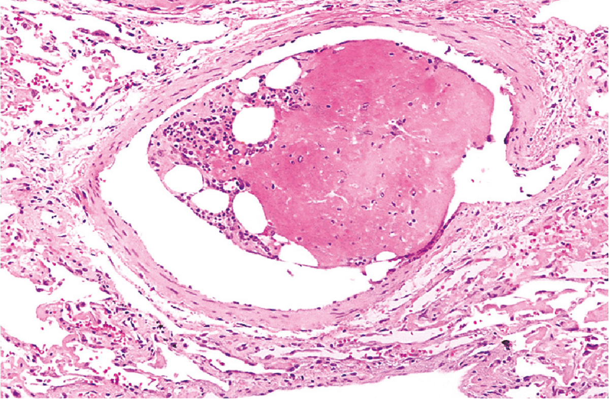
Bone marrow embolus in the pulmonary circulation. The cellular elements on the left are hematopoietic cells; the cleared vacuoles represent marrow fat. The red area on the right is an early organizing thrombus. - Robbins & Kumar Pathologic Basis of Disease
Microscopic findings:
- Fat emboli in pulmonary vasculature
- Alveolar edema, transudate, and exudate
- Special stains required: because solvents used in paraffin embedding dissolve lipids, demonstration of fat microglobules requires frozen sections and fat stains (e.g., Oil Red O, Sudan III)
- In the brain: small perivascular hemorrhages in the white matter around fat-containing vessels (may appear grossly normal in the early stages)
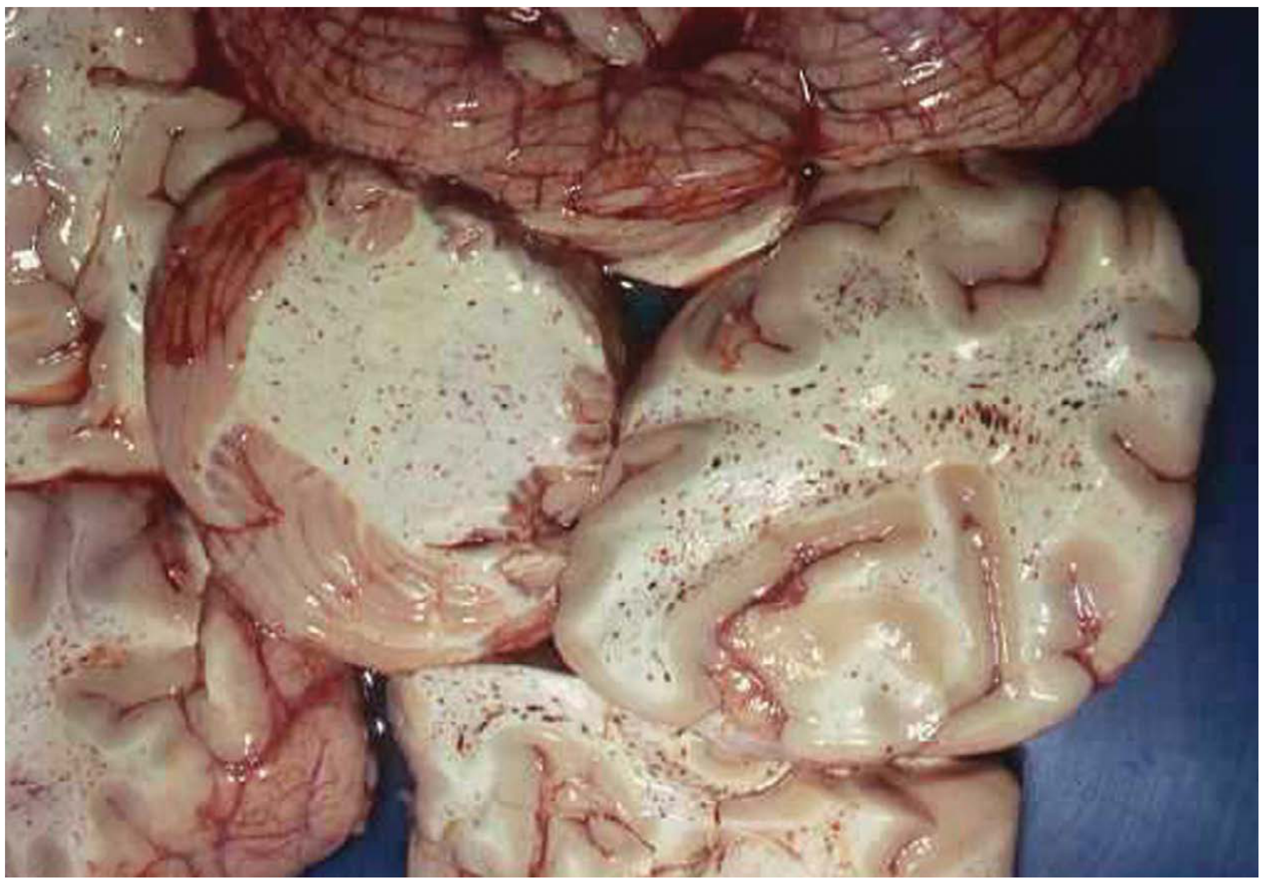
Petechial hemorrhages of the brain caused by fat embolism in a 21-year-old female with a right femur fracture. - DiMaio's Forensic Pathology
Diagnosis
FES is a clinical diagnosis - no single laboratory test is diagnostic.
Gurd's Diagnostic Criteria
Requires 1 major criterion + 4 minor/laboratory signs:
| Gurd's Criteria | |
|---|---|
| Major | Petechiae (axillary/subconjunctival), Hypoxemia (PaO₂ <60 mmHg), Depressed consciousness, Pulmonary edema |
| Minor | Tachycardia, Fever, Retinal fat emboli, Fat globules in sputum, Fat globules in urine |
| Laboratory | Microglobulinemia (required for diagnosis), Thrombocytopenia, Anemia, Elevated ESR |
Schonfeld Fat Embolism Index
An alternative point-based scoring system incorporating petechiae, diffuse alveolar infiltrates, hypoxemia, fever, tachycardia, and confusion - a score ≥5 is diagnostic.
Investigations
- Chest X-ray / CT: bilateral infiltrates ("snowstorm" appearance), can progress to ARDS pattern
- ABG: hypoxemia
- CBC: thrombocytopenia, anemia
- Serum lipase: elevated
- Urine/sputum: fat globules (not specific - seen with fractures alone)
- BAL: lipid-laden macrophages (not specific)
- MRI brain: high signal lesions in white matter ("starfield" pattern on DWI)
Important: Fat in serum and lipid-laden macrophages in BAL are seen in the majority of long-bone fracture patients regardless of FES - these findings are not diagnostic in isolation.
Management
Management is primarily supportive - no specific therapy has proven effective.
| Aspect | Approach |
|---|---|
| Respiratory support | Supplemental O₂, CPAP, or mechanical ventilation as needed (early intubation if deteriorating) |
| Hemodynamic support | Fluid resuscitation, vasopressors if needed |
| Fracture fixation | Early stabilization reduces fat release into circulation |
| Corticosteroids | Controversial - some evidence (including one RCT with high-dose methylprednisolone in 64 patients) that prophylactic steroids reduce FES incidence after long-bone fractures, but controlled data remain sparse |
| Treatments NOT proven effective | IV ethanol, albumin, dextran, heparin |
With meticulous supportive care, survival is now the rule in most cases. Severe FES with ARDS remains life-threatening.
Bone Cement Implantation Syndrome (BCIS) - Related Entity
BCIS is a syndrome of hypoxemia, hypotension, and altered consciousness during arthroplasty with cementation. It overlaps extensively with FES - the underlying mechanism involves embolization of fat and debris exacerbated by high intramedullary pressure from cement insertion, triggering endothelial damage, histamine release, and complement activation.
Recent Literature (2024-2025)
Recent reviews have continued to highlight FES as underdiagnosed in trauma patients, with a 2025 review in Cureus (PMID 41356881) noting evolving perspectives on diagnosis and care, and a 2024 review in J Trauma Acute Care Surg (PMID 39213184) emphasizing what trauma surgeons need to know about FES. A 2025 case series (PMID 40179158) documented FES occurring after both fractures and liposuction, reinforcing awareness beyond the classic trauma setting.
Key takeaways:
- FES = classic triad of respiratory distress + neurological dysfunction + petechial rash, onset 24-72 hours post-injury
- Diagnosis is clinical (Gurd's criteria); no test is pathognomonic
- Petechiae are absent in 50-80% of cases - do not use absence to rule out
- Requires frozen sections + fat stains on histology (paraffin processing dissolves fat)
- Treatment is supportive; early fracture fixation is the best preventive measure
- Corticosteroid prophylaxis remains debated
Sources: Robbins & Kumar Pathologic Basis of Disease; Murray & Nadel's Textbook of Respiratory Medicine; Fishman's Pulmonary Diseases and Disorders; Barash, Cullen, and Stoelting's Clinical Anesthesia 9e; DiMaio's Forensic Pathology 3e; Goldman-Cecil Medicine
This is a shared conversation. Sign in to Orris to start your own chat.