
Molecular testing of hpv / detection in cervical cytology and tissue 10 marks md pathology question
Reading File
Now let me search the medical library for HPV molecular testing content in parallel.
Finding Sources
Finding Sources
Finding Sources
Reading File
Reading File
Finding Sources
Excellent content found. Now let me read the key sections in parallel:
Reading File
Reading File
Reading File
Excellent. Let me get more content from the Tietz block which has very rich information on HPV molecular testing:
Reading File
Reading File
Finding Sources
Reading File
Reading File
Finding Sources
I now have comprehensive information. Let me also fetch the ISH image and search for relevant images:
Searching Images
HPV in situ hybridization cervical tissue
Finding Sources
I now have more than enough content to write a comprehensive 10-mark MD Pathology answer. Let me fetch the ISH panel image that was referenced in the Scott-Brown's textbook:
Loading Image
I now have all the content needed to write a thorough MD Pathology 10-mark answer. Here is the full answer:
Molecular Testing of HPV / Detection in Cervical Cytology and Tissue
1. Introduction
Human papillomavirus (HPV) is the causative agent of virtually all cervical carcinomas, and persistent high-risk HPV (hrHPV) infection is the central step in cervical oncogenesis. Over 200 HPV genotypes exist; 12 are classified as Group 1 carcinogens by IARC (types 16, 18, 31, 33, 35, 39, 45, 51, 52, 56, 58, 59). HPV 16 and 18 together account for approximately 70% of cervical cancers. Laboratory detection of HPV has two main purposes: (a) screening/risk stratification in cervical cytology and (b) confirming HPV status in tissue biopsies.
2. Cytologic Detection - Pap Smear and Morphological Clues
Since its introduction in 1941, cervical cytology (Pap smear/liquid-based cytology) has been the traditional cornerstone of screening. The transitional zone of the cervix (transformation zone, squamocolumnar junction) is sampled using a brush.
Bethesda Classification of cytologic abnormalities:
- LSIL (Low-grade squamous intraepithelial lesion) - corresponds to CIN 1; shows koilocytic atypia (perinuclear halo, nuclear enlargement, nuclear irregularity) - the classic cytopathic effect (CPE) of productive HPV infection.
- HSIL (High-grade squamous intraepithelial lesion) - corresponds to CIN 2/3.
- ASC-US (Atypical squamous cells of undetermined significance) - does not fulfil all criteria for LSIL/HSIL; reflex HPV testing is indicated.
- ASC-H - cannot exclude HSIL.
Histologic correlates (CIN grading on biopsy):
- CIN 1: Viral CPE (koilocytosis) + dysplasia confined to lower 1/3 of epithelium.
- CIN 2: Dysplasia in lower 2/3 of epithelium.
- CIN 3 (CIS): Full-thickness dysplasia.
3. Molecular Tests for HPV Detection
Molecular detection of HPV has emerged as a method to augment cytology and serves as primary screening or co-testing. Assays vary by:
- Nucleic acid target: DNA vs. mRNA (E6/E7 transcripts)
- Technology: nucleic acid amplification (PCR, TMA), signal amplification (hybrid capture, invader chemistry), or direct detection (ISH)
- Genotyping capability: broad high-risk group vs. individual genotype identification
A. Signal Amplification Methods
1. Hybrid Capture 2 (HC2) - Digene/Qiagen
- One of the earliest FDA-approved HPV assays.
- Principle: Uses RNA probes complementary to HPV DNA. RNA:DNA hybrids are captured by antibodies on a microplate and detected by chemiluminescence.
- Detects 18 high-risk genotypes as a group, without individual differentiation.
- Limitation: probe-based (not PCR-based), lacks an internal specimen adequacy control - inadequate specimens may falsely read as negative; slightly lower sensitivity compared to PCR-based assays.
- Approved collection media: SurePath and ThinPrep.
2. Cervista HPV HR / 16-18 (Hologic) - Invader Chemistry
- Uses the Third Wave Invader assay technology (isothermal, signal amplification).
- Cervista HR detects 14 hrHPV types without differentiation.
- Cervista 16/18 specifically reports genotypes 16 and 18 if present.
- Approved for ThinPrep specimens only.
B. Nucleic Acid Amplification Tests (NAATs) - DNA-Based
3. Cobas HPV Test (Roche) - Real-time PCR
- Targets HPV L1 gene and uses PCR with fluorescent probes.
- Detects 14 high-risk genotypes; specifically differentiates HPV 16 and HPV 18 (others reported as "HR HPV detected").
- Approved for primary screening (first FDA-approved for this indication in the USA).
- Approved for SurePath and ThinPrep.
4. BD Onclarity HPV Assay (BD Diagnostics) - Real-time PCR
- Targets E6 and E7 genes; detects 17 hrHPV genotypes.
- Specifically differentiates HPV 16, 18, and 45 - the three highest-risk genotypes.
- Also approved for primary screening.
- Approved for SurePath only.
C. mRNA-Based Assay - Transcription Mediated Amplification (TMA)
5. Aptima HPV Assay (Hologic)
- Detects E6/E7 oncogene mRNA transcripts rather than DNA.
- Key advantage: mRNA is produced only during active (transcriptionally active) infection. Large amounts of E6/E7 mRNA are not present during viral latency, so this assay is more specific for clinically relevant, biologically active infection.
- Detects 14 hrHPV types; does not differentiate them individually.
- A paired Aptima HPV 16 18/45 Genotype Assay is performed reflex on positive samples to differentiate HPV 16 and 18/45.
- Approved for ThinPrep specimens only.
Summary Table: FDA-Cleared HPV Molecular Assays
| Assay | Target | Genotyping | Primary Screening | Medium |
|---|---|---|---|---|
| Cervista HPV HR/16-18 (Hologic) | DNA | 14 HR (HPV16/18 separately) | No | ThinPrep |
| Digene HC2 (Qiagen) | DNA | 18 HR, no differentiation | No | SurePath + ThinPrep |
| Cobas HPV (Roche) | DNA | 14 HR; 16 and 18 individually | Yes | SurePath + ThinPrep |
| Aptima HPV (Hologic) | mRNA | 14 HR; 16, 18/45 by reflex | No | ThinPrep |
| BD Onclarity (BD) | DNA | 17 HR; 16, 18, 45 individually | Yes | SurePath |
(Tietz Textbook of Laboratory Medicine, 7th Edition)
4. HPV Detection in Tissue Biopsies
A. In Situ Hybridization (ISH) - DNA ISH
- A labeled DNA probe with a sequence complementary to the target HPV gene (typically the L1 gene) is applied to formalin-fixed, paraffin-embedded (FFPE) tissue sections.
- The probe hybridizes to complementary HPV DNA within the tumor cell nucleus.
- The chromogen label allows visualization by light microscopy - a positive result appears as a blue/brown dot in the nucleus.
- Staining patterns:
- Punctate (dot-like) pattern: suggests integrated HPV DNA.
- Diffuse pattern: suggests episomal (non-integrated) HPV.
- Commercial kits are available for high-risk HPV genotypes in FFPE tissue.
- Allows precise localization of HPV DNA to the host cell nucleus.
- Limitation: does not prove transcriptional activity (biological activity) of the virus.
B. Polymerase Chain Reaction (PCR) on Tissue
- Highly sensitive; can amplify a single copy of the target sequence.
- After DNA denaturation, specific primers bind to the E6 or E7 gene (or the L1 gene for consensus PCR) and amplify the target.
- Consensus PCR: detects presence of HPV DNA broadly.
- Real-time PCR (qPCR): quantifies viral load.
- Can be performed on FFPE tissue, though best results are on fresh tissue.
- Limitation: detects DNA only - cannot confirm active viral transcription.
C. Tests to Determine Biological Activity (Transcriptional Activity)
These are functionally the most important in tissue diagnosis:
1. Quantitative Reverse Transcription PCR (qRT-PCR)
- Directly detects E6 and E7 mRNA transcripts.
- cDNA is first synthesized from mRNA (reverse transcription), then amplified by PCR using E6/E7-specific primers.
- Current gold standard for confirming HPV biological activity (oncogenic transcription).
- Costly, labor-intensive, traditionally restricted to fresh/frozen tissue.
2. RNA In Situ Hybridization (RNA-ISH) / Chromogenic ISH (CISH)
- Detects E6/E7 mRNA transcripts directly in FFPE tissue without requiring reverse transcription.
- Commercial kits available with probes for E6/E7 mRNA of multiple hrHPV genotypes.
- Applied to FFPE sections - easily usable in routine practice.
- Sensitivity 97%, specificity 93% compared to qRT-PCR gold standard.
- Anticipated to become the routine standard for HPV testing in FFPE biopsies.
D. Surrogate Markers - p16INK4a Immunohistochemistry (IHC)
- Mechanism: HPV E7 oncoprotein degrades the retinoblastoma (RB) protein via the ubiquitin-proteasome system. Loss of RB leads to loss of E2F repression, which in turn drives overexpression of p16INK4a (a cyclin-dependent kinase inhibitor). Therefore, strong diffuse p16 overexpression is a surrogate marker for oncogenic HPV activity.
- Positive result: strong, diffuse block-positive nuclear and cytoplasmic staining in tumor cells.
- Highly sensitive, readily applicable to FFPE sections.
- Limitation: not 100% specific - p16 overexpression can occur via non-HPV mechanisms (e.g., methylation of RB).
- Used in conjunction with ISH for definitive HPV status determination (see image below).
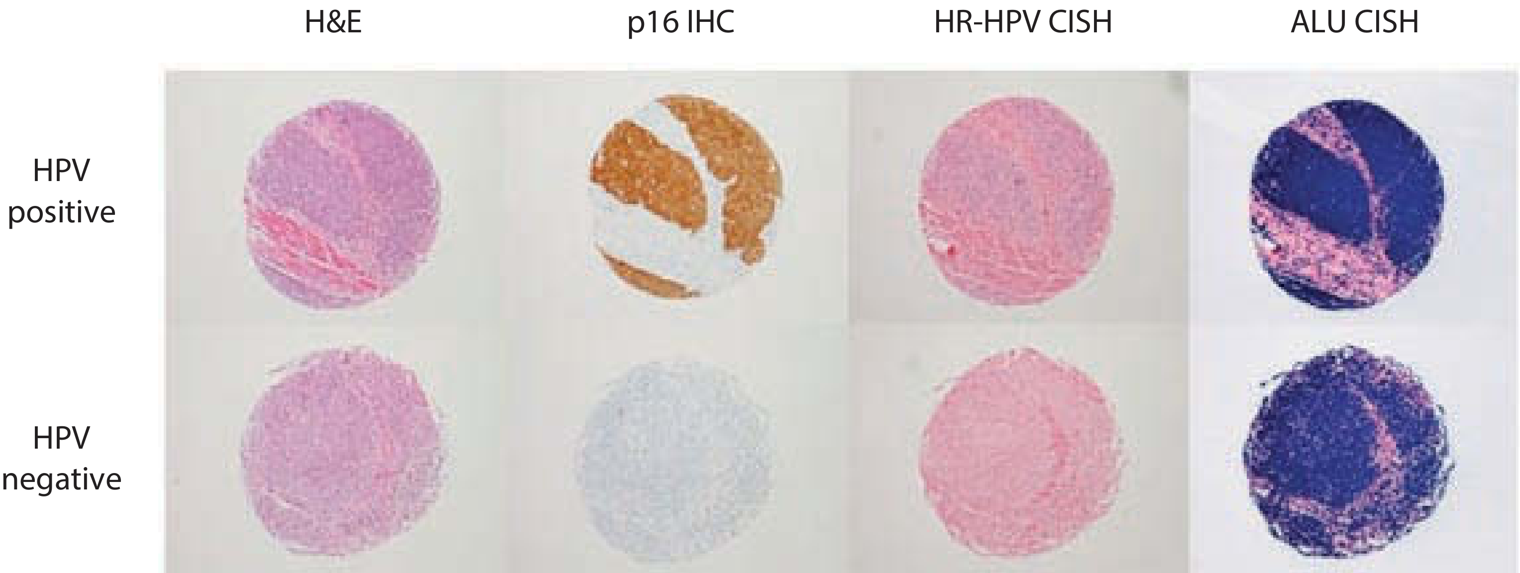
Figure: HPV positive (top row) shows strong p16 IHC positivity (brown) and HR-HPV CISH signal; HPV negative (bottom row) shows no p16 staining and no CISH signal. (Scott-Brown's Otorhinolaryngology, p. 258)
5. Screening Algorithms (ASCCP Guidelines)
- Ages 21-65: Cytology alone every 3 years is acceptable.
- Ages 30-65 (preferred): "Co-testing" with cytology + hrHPV molecular test every 5 years.
- This is based on meta-analysis data supporting high sensitivity of molecular HPV testing for early carcinoma detection.
- HPV 16/18 positive + cytology negative: Immediate colposcopy (higher risk ~11.4% for CIN 2+).
- Other hrHPV positive + cytology negative: Retest at 1 year.
- Reflex testing: ASC-US cytology triggers immediate hrHPV testing; HPV positivity triggers colposcopy.
- Primary HPV screening: Cobas and BD Onclarity are FDA-approved; genotyping for 16/18 drives management.
The ATHENA study confirmed that HPV 16/18 positivity confers nearly double the risk for CIN 2+ compared to other hrHPV types (11.4% vs 6.1%).
6. HPV Serology
HPV serology (antibody testing) is of no clinical diagnostic utility for individual patient management, though it is used in epidemiological and vaccine research settings. - Henry's Clinical Diagnosis and Management by Laboratory Methods
7. Key Points for MD Pathology Exam
| Feature | DNA ISH | PCR | qRT-PCR | RNA-ISH | p16 IHC |
|---|---|---|---|---|---|
| Detects | HPV DNA | HPV DNA | E6/E7 mRNA | E6/E7 mRNA | Surrogate marker |
| Tissue | FFPE | FFPE / fresh | Fresh/frozen (ideally) | FFPE | FFPE |
| Gold standard? | No | No | Yes (activity) | Near equivalent | No |
| Routine use | Yes | Research | Limited | Emerging standard | Yes |
| Specificity for active infection | Low | Low | High | High | Moderate |
| Sensitivity | Moderate | High | High | 97% | High |
Summary
Molecular HPV testing in cervical cytology uses signal amplification (HC2), PCR-based DNA assays (Cobas, Onclarity), or mRNA assays (Aptima) to risk-stratify patients, with genotyping of HPV 16/18 guiding management intensity. In tissue, ISH localizes HPV DNA to the nucleus in FFPE sections, PCR offers high sensitivity, and qRT-PCR is the gold standard for confirming biological (transcriptional) activity. p16 IHC serves as a widely-used surrogate for oncogenic HPV infection. RNA-ISH for E6/E7 mRNA in FFPE tissue is emerging as the preferred routine method for confirming biologically active HPV in tissue sections.
Sources: Tietz Textbook of Laboratory Medicine, 7th Edition; Henry's Clinical Diagnosis and Management by Laboratory Methods; Scott-Brown's Otorhinolaryngology Head & Neck Surgery; Harrison's Principles of Internal Medicine 22E
This is a shared conversation. Sign in to Orris to start your own chat.