
Appendicitis
appendicitis anatomy diagram appendix location
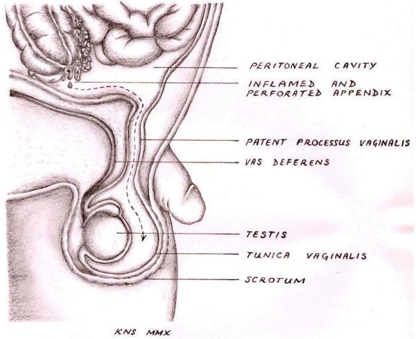
This medical anatomical diagram illustrates the pathophysiology of a scrotal abscess occurring as a rare complication of acute appendicitis. The sagittal view depicts the anatomical relationship between the peritoneal cavity and the scrotum. Key labeled structures include an 'inflamed and perforated appendix' situated superiorly, which is shown shedding purulent material. A dashed arrow traces the pathological pathway of the infection inferiorly through a 'patent processus vaginalis,' an embryonic developmental remnant. The diagram shows this channel passing adjacent to the 'vas deferens' and terminating in the 'scrotum,' where the 'testis' and 'tunica vaginalis' are located. This illustration serves to explain how intra-abdominal infection can track extra-abdominally into the scrotal sac when the processus vaginalis fails to obliterate. The diagram is intended for intermediate to advanced medical education, focusing on surgical anatomy, embryology, and atypical presentations of appendicitis.
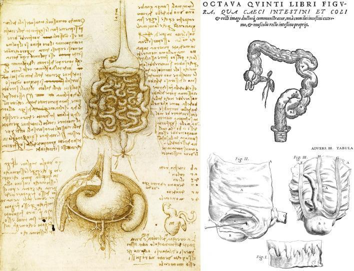
This composite image presents a historical comparison of anatomical drawings focusing on the vermiform appendix and gastrointestinal structures. On the left is a pen-and-ink anatomical diagram by Leonardo da Vinci (c. 1508), depicting the stomach with internal rugae, the esophagus, and the densely coiled small intestines. Below the stomach, a schematic representation of the cecum and appendix is visible, accompanied by mirror-writing notes. The upper right section features a woodcut by Andreas Vesalius showing a more morphologically accurate colon, highlighting the haustra (pouches) and taeniae coli (longitudinal bands), with a clear depiction of the cecum and the vermiform appendix. The lower right contains precise etchings by Giovanni Battista Morgagni, illustrating detailed observational studies of the cecal-appendiceal junction, including variations in appendiceal position and luminal characteristics. These illustrations represent the evolution of anatomical understanding in gastroenterology, moving from schematic representations to detailed structural observations relevant to the history of appendicitis diagnosis and surgical anatomy.
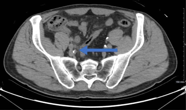
An axial non-contrast-enhanced computed tomography (CT) scan of the lower abdomen and pelvis at the level of the iliac wings. The diagnostic image shows cross-sectional anatomy including the sacrum, iliac bones, and abdominal wall musculature. A prominent blue arrow indicates the location of the appendix in the right lower quadrant, which appears mildly thickened with subtle surrounding fat stranding, suggestive of early or resolving inflammatory changes (appendicitis). Multiple high-attenuation (radiopaque) foci are visible within the pelvic cavity, consistent with documented renal calculi or vascular calcifications. The surrounding mesenteric fat and bowel loops are visible, providing a baseline for assessing intra-abdominal inflammation. This imaging is utilized in surgical and radiological education to demonstrate the CT presentation of suspected appendiceal pathology and its anatomical relationships within the pelvic inlet.
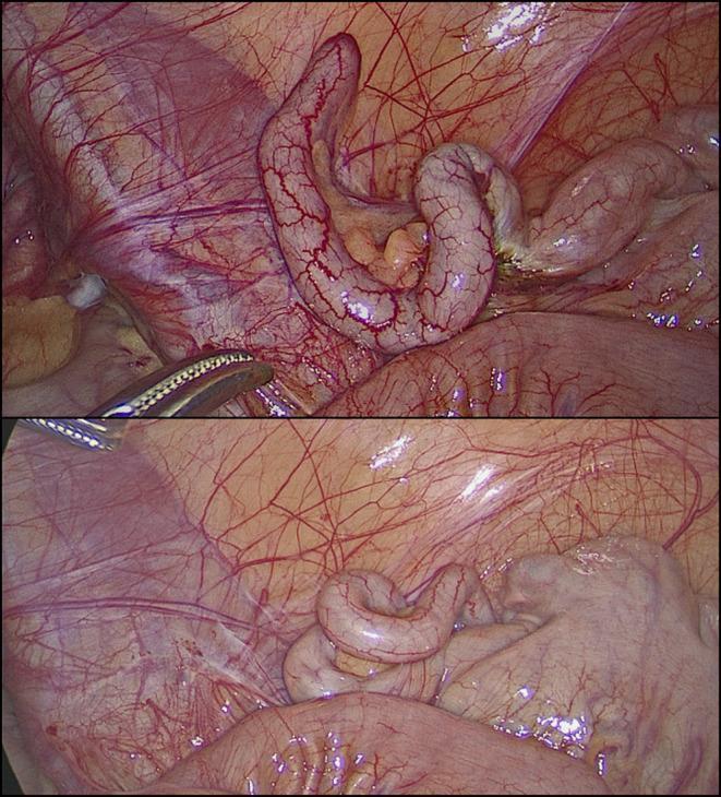
This clinical photograph consists of two laparoscopic views showing a macroscopically normal vermiform appendix during a diagnostic laparoscopy. The appendix appears as an elongated, tubular, and slightly coiled structure with a smooth, pale pink serosal surface. Fine, branching subserosal blood vessels are clearly visible across its length, indicating normal vascularity without the congestion or purulent exudate typical of acute appendicitis. In the upper frame, a laparoscopic grasper is visible, gently manipulating the appendix to facilitate inspection. The surrounding anatomy includes the mesoappendix containing adipose tissue and the glistening peritoneal lining of the right iliac fossa. There are no visual signs of inflammation, perforation, or abscess formation. This image serves as a reference for a 'grossly negative' appendix in the context of clinical suspicion of appendicitis, where symptoms may actually stem from non-inflammatory conditions such as follicular lymphoid hyperplasia or obstructive fecaliths.
"appendicitis"[MeSH Terms] AND "diagnosis OR treatment"
"appendicitis"[MeSH Terms] AND "diagnosis"[MeSH Terms]
Appendicitis
Overview
Pathophysiology
Clinical Features
Classic Presentation
- Abdominal pain - begins as vague, crampy, periumbilical pain (visceral/referred) that migrates over 12-24 hours to the right lower quadrant (RLQ) as parietal peritoneum becomes involved
- Anorexia (nearly universal)
- Nausea and vomiting (usually follow pain onset)
- Low-grade fever (develops later; high fever suggests perforation)
McBurney's Point
Examination Signs
| Sign | Maneuver | Mechanism |
|---|---|---|
| Rovsing sign | Palpation of LLQ causes RLQ pain | Referred peritoneal irritation |
| Psoas sign | Extension of right hip causes pain | Inflamed appendix overlies psoas |
| Obturator sign | Internal rotation of right hip causes pain | Pelvic appendix near obturator internus |
| Rebound tenderness | Pain on release of deep pressure | Parietal peritonitis |
Note: These signs indicate localized peritonitis rather than being specific for appendicitis. They are of limited sensitivity/specificity and should not be relied on exclusively, particularly in children. - Sabiston Textbook of Surgery, p. 2039
Atypical Presentations
- Retroperitoneal appendix: flank or back pain (mimics renal colic)
- Pelvic appendix: suprapubic pain (mimics UTI)
- Pregnancy: pain may not localize to RLQ, especially after first trimester; appendix displacement is less than traditionally taught (moves in <25% of third-trimester patients)
- Young children (<5 years): vague symptoms, high perforation rate (>50% at surgery)
- Elderly: blunted presentation, higher morbidity
Imaging
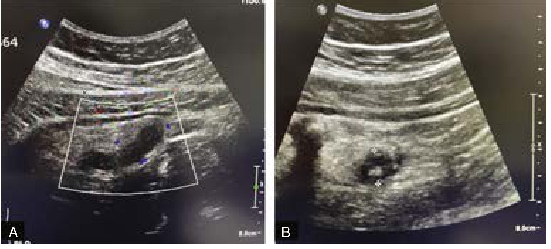
CT Findings in Appendicitis
- Dilated appendix (>6 mm diameter)
- Appendiceal wall thickening and enhancement
- Periappendiceal fat stranding
- Appendicolith (echogenic/calcified focus)
- Periappendiceal abscess (if perforated)
Laboratory Tests
| Test | Finding | Notes |
|---|---|---|
| WBC | >10,000/µL in ~90% of cases | Normal in 10%; doesn't distinguish simple vs. perforated |
| CRP | Often elevated | >10 mg/L more useful in children <6 years |
| Urinalysis | Mild sterile pyuria possible | Proximity of appendix to ureter/bladder |
| Beta-hCG | Mandatory in reproductive-age females | Excludes ectopic pregnancy |
| Procalcitonin | Elevated in complicated appendicitis | Useful adjunct |
Clinical Scoring
Modified Alvarado Score (MANTRELS)
| Feature | Points |
|---|---|
| Migration of pain to RLQ | 1 |
| Anorexia | 1 |
| Nausea/vomiting | 1 |
| RLQ tenderness | 2 |
| Rebound tenderness | 1 |
| Fever (>37.3°C) | 1 |
| WBC >10,000/mm³ | 2 |
| Total | 9 |
- 1-4: Low risk (but only 72% sensitive - do not rely on this alone)
- 5-6: Possible appendicitis
- 7-9: Probable appendicitis
Differential Diagnosis
Management
Surgical (Standard of Care)
- Preoperative antibiotics (e.g., cefotetan 2 g IV, or cefoxitin 2 g IV, or ticarcillin-clavulanic acid) reduce infectious complications
- Perforation increases mortality from 0.0002% to ~3% and morbidity from 3% to 47%
Nonoperative Management (Antibiotics Alone)
- ~80-90% initial success rate with broad-spectrum antibiotics
- ~40% recurrence rate at medium-term follow-up
- Patients with a fecalith are generally not candidates (higher failure rate)
- Many surgeons view it as a bridge to elective interval appendectomy rather than a definitive cure
Recent evidence update (2025): A Lancet Gastroenterol Hepatol individual patient data meta-analysis (PMID 39827891) confirmed antibiotics are a viable alternative to appendectomy in adults with uncomplicated appendicitis, but ~40% require appendectomy within 5 years. A 2026 JAMA Pediatrics meta-analysis (PMID 41046476) supports nonoperative management for pediatric uncomplicated appendicitis, though recurrence and failure rates remain important considerations.
Perforated Appendicitis with Abscess
- If >5 days of symptoms: percutaneous drain (if abscess accessible) + IV antibiotics, followed by interval appendectomy at 6-8 weeks
- IV antibiotics duration guided by clinical response (fever resolution, WBC normalization, return of bowel function)
Special Populations
Children
- Most common acute surgical problem in children
- Younger children (<5 years): >50% already perforated at surgery due to atypical/delayed presentation
- Prefer ultrasound and MRI over CT for imaging (radiation risk)
- Perforation rates inversely related to age - Current Surgical Therapy 14e, p. 1500
Pregnancy
- Most common surgical emergency in pregnancy; incidence same as non-pregnant women
- Delays in diagnosis lead to higher perforation rates - nearly 2x increase in sepsis, septic shock, and other complications
- Pain localization to RLQ persists in most cases even in the third trimester
- Imaging: ultrasound first, MRI if inconclusive - Rosen's Emergency Medicine
Elderly
- Presentation often blunted (less fever, less leukocytosis)
- Higher perforation rates at presentation
- Higher overall morbidity and mortality
Complications
| Complication | Notes |
|---|---|
| Perforation | Occurs if untreated; higher risk in extremes of age |
| Peritonitis | Diffuse (perforated) or localized (walled-off) |
| Appendiceal abscess | Walled-off perforation, may be drained percutaneously |
| Pylephlebitis | Septic thrombophlebitis of portal vein - rare but serious |
| Wound infection | More common after open/perforated cases |
| Post-appendectomy ileus | More prolonged in perforated cases |
Make a quick reference poster comparing appendicitis vs. mimics
A clean, professional medical quick reference poster titled "Appendicitis vs. Mimics" for clinical use. The poster has a dark navy blue header with white bold title text. Below are two main sections side by side: left side shows "Acute Appendicitis" in a red-orange highlighted box with key features listed: RLQ pain (migrates from periumbilical), anorexia, nausea/vomiting, low-grade fever, McBurney point tenderness, rebound tenderness, WBC >10,000, CRP elevated. Right side has a comparison grid table with 6 rows for the main mimics: Ovarian Torsion, Ectopic Pregnancy, Mesenteric Adenitis, Ureteral Colic, Crohn's/Terminal Ileitis, and PID/Salpingitis. Each mimic row has columns for Key Symptoms, Differentiating Features, and Preferred Test. Color-coded rows alternate light blue and white. At the bottom a yellow footer shows: "Imaging: CT preferred (adults) | Ultrasound/MRI preferred (pregnancy, children)". Small red warning box: "Perforation risk: highest in children <5 yrs and elderly - act early." Clean sans-serif fonts, organized grid layout, medical infographic style, white background for the body content area.
Error calling model 'gemini-3-pro-image' (RESOURCE_EXHAUSTED): 429 RESOURCE_EXHAUSTED. {'error': {'code': 429, 'message': 'Resource has been exhausted (e.g. check quota).', 'status': 'RESOURCE_EXHAUSTED'}}
We encountered an error while processing your request. Please try again or reach out to us for help.